

Pulmonary embolism
Encyclopedia
Pulmonary embolism is a blockage of the main artery of the lung
or one of its branches by a substance that has travelled from elsewhere in the body through the bloodstream (embolism
). Usually this is due to embolism of a thrombus
(blood clot) from the deep veins in the legs
, a process termed venous thromboembolism. A small proportion is due to the embolization of air
, fat
, talc in drugs of intravenous drug abusers or amniotic fluid
. The obstruction of the blood flow through the lung
s and the resultant pressure on the right ventricle
of the heart leads to the symptoms and signs of PE. The risk of PE is increased in various situations, such as cancer
or prolonged bed rest
.
Symptoms of pulmonary embolism include difficulty breathing
, chest pain on inspiration, and palpitation
s. Clinical signs include low blood oxygen saturation
and cyanosis
, rapid breathing
, and a rapid heart rate
. Severe cases of PE can lead to collapse
, abnormally low blood pressure, and sudden death
.
Diagnosis is based on these clinical findings in combination with laboratory tests (such as the D-dimer
test) and imaging studies, usually CT pulmonary angiography. Treatment is typically with anticoagulant
medication, including heparin
and warfarin
. Severe cases may require thrombolysis
with drugs such as tissue plasminogen activator
(tPA) or may require surgical intervention via pulmonary thrombectomy
.
(shortness of breath), tachypnea
(rapid breathing), chest pain
of a "pleuritic" nature (worsened by breathing), cough
and hemoptysis
(coughing up blood). More severe cases can include signs such as cyanosis
(blue discoloration, usually of the lips and fingers), collapse
, and circulatory instability due to decreased blood flow through the lungs and into the left side of the heart. About 15% of all cases of sudden death
are attributable to PE.
On physical examination, the lungs are usually normal. Occasionally, a pleural friction rub
may be audible over the affected area of the lung (mostly in PE with infarct) . A pleural effusion
is sometimes present that is transudative, detectable by decreased percussion note, audible breath sounds and vocal resonance. Strain on the right ventricle may be detected as a left parasternal heave, a loud pulmonary component of the second heart sound
, and raised jugular venous pressure
. A low-grade fever
may be present, particularly if there is associated pulmonary hemorrhage or infarction.
More rarely, inability of the right ventricle to remove fluid from the tissues leads to fluid accumulation in the legs (peripheral edema), congestion of the liver with mild jaundice
and tenderness, and ascites
(fluid in the abdominal cavity).
The development of thrombosis is classically due to a group of causes named Virchow's triad
(alterations in blood flow, factors in the vessel wall and factors affecting the properties of the blood). Often, more than one risk factor is present.
) cannot be definitively differentiated from other causes of chest pain and shortness of breath. The decision to do medical imaging is usually based on clinical grounds, i.e. the medical history
, symptoms and findings on physical examination
, followed by an assessment of clinical probability.
The most commonly used method to predict clinical probability, the Wells score, is a clinical prediction rule
, whose use is complicated by multiple versions being available. In 1995, Wells et al. initially developed a prediction rule (based on a literature search) to predict the likelihood of PE, based on clinical criteria. The prediction rule was revised in 1998 This prediction rule was further revised when simplified during a validation by Wells et al. in 2000. In the 2000 publication, Wells proposed two different scoring systems using cutoffs of 2 or 4 with the same prediction rule. In 2001, Wells published results using the more conservative cutoff of 2 to create three categories. An additional version, the "modified extended version", using the more recent cutoff of 2 but including findings from Wells's initial studies were proposed. Most recently, a further study reverted to Wells's earlier use of a cutoff of 4 points to create only two categories.
There are additional prediction rules for PE, such as the Geneva rule
. More importantly, the use of any rule is associated with reduction in recurrent thromboembolism.
The Wells score:
Traditional interpretation
Alternate interpretation
level (shown in a blood test
) is enough to exclude the possibility of thrombotic PE. This has been corroborated by a recent systematic review
of studies of patients with low pre-test probability
(PTP) of PE and negative D-dimer
results that found the three month risk of thromboembolic events in patients excluded in this manner was 0.14%, with 95% confidence intervals
from 0.05 to 0.41%, though this review was limited by its use of only one randomized-controlled clinical trial
, the remainder of studies being prospective cohorts
. D-dimer is highly sensitive but not very specific (specificity around 50%). In other words, a positive D-dimer is not synonymous with PE, but a negative D-dimer is, with a good degree of certainty, an indication of absence of a PE.
When a PE is being suspected, a number of blood test
s are done, in order to exclude important secondary causes of PE. This includes a full blood count, clotting status
(PT
, aPTT, TT), and some screening tests (erythrocyte sedimentation rate
, renal function
, liver enzymes, electrolyte
s). If one of these is abnormal, further investigations might be warranted.
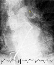

 The gold standard
for diagnosing pulmonary embolism (PE) is pulmonary angiography
. Pulmonary angiography is used less often due to wider acceptance of CT scans, which are non-invasive. CT pulmonary angiography is the recommended first line diagnostic imaging test in most people.
Non-invasive imaging
CT pulmonary angiography
(CTPA) is a pulmonary angiogram obtained using computed tomography
(CT) with radiocontrast
rather than right heart catheterization. Its advantages are clinical equivalence, its non-invasive nature, its greater availability to patients, and the possibility of identifying other lung disorders from the differential diagnosis
in case there is no pulmonary embolism. Assessing the accuracy of CT pulmonary angiography is hindered by the rapid changes in the number of rows of detectors available in multidetector CT (MDCT) machines. According to a cohort study
, single-slice spiral CT may help diagnose detection among patients with suspected pulmonary embolism . In this study, the sensitivity was 69% and specificity was 84%. In this study which had a prevalence of detection was 32%, the positive predictive value
of 67.0% and negative predictive value
of 85.2% (click here to adjust these results for patients at higher or lower risk of detection). However, this study's results may be biased due to possible incorporation bias, since the CT scan was the final diagnostic tool in patients with pulmonary embolism. The authors noted that a negative single slice CT scan is insufficient to rule out pulmonary embolism on its own. A separate study with a mixture of 4 slice and 16 slice scanners reported a sensitivity of 83% and a specificity of 96%. This study noted that additional testing is necessary when the clinical probability is inconsistent with the imaging results. CTPA is non-inferior to VQ scanning, and identifies more emboli (without necessarily improving the outcome) compared to VQ scanning.
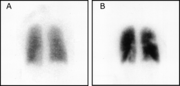
Ventilation/perfusion scan
(or V/Q scan or lung scintigraphy
), which shows that some areas of the lung are being ventilated
but not perfused
with blood (due to obstruction by a clot). This type of examination is used less often because of the more widespread availability of CT technology, however, it may be useful in patients who have an allergy to iodinated contrast
or in pregnancy
due to lower radiation exposure than CT.
Low probability diagnostic tests/non-diagnostic tests
Tests that are frequently done that are not sensitive for PE, but can be diagnostic.
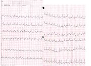 An electrocardiogram
(ECG) is routinely done on patients with chest pain to quickly diagnose myocardial infarction
s (heart attacks). An ECG may show signs of right heart strain or acute cor pulmonale
in cases of large PEs - the classic signs are a large S wave in lead I, a large Q wave in lead III and an inverted T wave
in lead III ("S1Q3T3"). This is occasionally (up to 20%) present, but may also occur in other acute lung conditions and has therefore limited diagnostic value. The most commonly seen signs in the ECG is sinus tachycardia
, right axis deviation and right bundle branch block
. Sinus tachycardia was however still only found in 8 - 69% of people with PE.
, an indication that the pulmonary artery
is severely obstructed and the heart is unable to match the pressure. Some studies (see below) suggest that this finding may be an indication for thrombolysis
. Not every patient with a (suspected) pulmonary embolism requires an echocardiogram, but elevations in cardiac troponins
or brain natriuretic peptide
may indicate heart strain and warrant an echocardiogram.
The specific appearance of the right ventricle on echocardiography is referred to as the McConnell's sign. This is the finding of akinesia of the mid-free wall but normal motion of the apex. This phenomenon has a 77% sensitivity and a 94% specificity for the diagnosis of acute pulmonary embolism in the setting of right ventricular dysfunction.
Pulmonary Embolism Rule-out Criteria
The Pulmonary Embolism Rule-out Criteria, or PERC rule, helps assess people in whom pulmonary embolism is suspected, but unlikely. Unlike the Wells Score and Geneva score, which are clinical prediction rules intended to risk stratify patients with suspected PE, the PERC rule is designed to rule out risk of PE in patients when the physician has already stratified them into a low-risk category.
Patients in this low risk category without any of these criteria may undergo no further diagnostic testing for PE: Hypoxia - Sa02 <95%, unilateral leg swelling, hemoptysis, prior DVT or PE, recent surgery or trauma, age >50, hormone use, tachycardia. The rationale behind this decision is that further testing (specifically CT angiogram of the chest) may cause more harm (from radiation exposure and contrast dye) than the risk of PE. The PERC rule has a sensitivity of 97.4% and specificity of 21.9% with a false negative rate of 1.0% (16/1666).
or analgesia, are often required.
therapy is the mainstay of treatment. Heparin
, low molecular weight heparin
s (such as enoxaparin
and dalteparin
), or fondaparinux
is administered initially, while warfarin
, acenocoumarol
, or phenprocoumon
therapy is commenced (this may take several days, usually while the patient is in the hospital). Low molecular weight heparin
may reduce bleeding among patients with pulmonary embolism as compared to heparin according to a systematic review
of randomized controlled trial
s by the Cochrane Collaboration
. The relative risk reduction
was 40.0%. For patients at similar risk to those in this study (2.0% had bleeding when not treated with low molecular weight heparin), this leads to an absolute risk reduction
of 0.8%. 125.0 patients must be treated for one to benefit
.
It is possible to treat low risk patients (risk class I or class II) as outpatients. A randomised trial
of 344 patients (171 outpatients and 168 inpatients) found that outcomes were equivalent whether patients were treated in hospital or at home (there was one death at 90 days in each group). This confirms the findings of an earlier systematic review
of observational studies
.
Warfarin therapy often requires frequent dose adjustment and monitoring of the INR. In PE, INRs between 2.0 and 3.0 are generally considered ideal. If another episode of PE occurs under warfarin treatment, the INR window may be increased to e.g. 2.5-3.5 (unless there are contraindications) or anticoagulation may be changed to a different anticoagulant e.g. low molecular weight heparin
. In patients with an underlying malignancy, therapy with a course of low molecular weight heparin
may be favored over warfarin based on the results of the CLOT trial.
Similarly, pregnant women are often maintained on low molecular weight heparin to avoid the known teratogenic effects of warfarin, especially in the early stages of pregnancy.
People are usually admitted to hospital in the early stages of treatment, and tend to remain under inpatient care until INR has reached therapeutic levels. Increasingly, low-risk cases are managed on an outpatient basis in a fashion already common in the treatment of DVT.
Warfarin therapy is usually continued for 3–6 months, or "lifelong" if there have been previous DVTs or PEs, or none of the usual risk factors is present. An abnormal D-dimer
level at the end of treatment might signal the need for continued treatment among patients with a first unprovoked pulmonary embolus.
, the enzymatic destruction of the clot with medication. It is the best available medical treatment in this situation and is supported by clinical guidelines.
The use of thrombolysis in non-massive PEs is still debated. The aim of the therapy is to dissolve the clot, but there is an attendant risk of bleeding or stroke. The main indication for thrombolysis is in submassive PE where right ventricular dysfunction can be demonstrated on echocardiography
, and the presence of visible thrombus in the atrium.
) is uncommon and has largely been abandoned because of poor long-term outcomes. However, recently, it has gone through a resurgence with the revision of the surgical technique and is thought to benefit selected patients.
Chronic pulmonary embolism leading to pulmonary hypertension
(known as chronic thromboembolic hypertension) is treated with a surgical procedure known as a pulmonary thromboendarterectomy
.
and/or ineffective, or to prevent new emboli from entering the pulmonary artery and combining with an existing blockage, an inferior vena cava filter
may be implanted.
from untreated PE is said to be 26%. This figure comes from a trial published in 1960 by Barrit and Jordan, which compared anticoagulation against placebo for the management of PE. Barritt and Jordan performed their study in the Bristol Royal Infirmary
in 1957. This study is the only placebo controlled trial ever to examine the place of anticoagulants in the treatment of PE, the results of which were so convincing that the trial has never been repeated as to do so would be considered unethical. That said, the reported mortality rate of 26% in the placebo group is probably an overstatement, given that the technology of the day may have detected only severe PEs.
Prognosis depends on the amount of lung that is affected and on the co-existence of other medical conditions; chronic embolisation to the lung can lead to pulmonary hypertension
. After a massive PE, the embolus must be resolved somehow if the patient is to survive. In thrombotic PE, the blood clot may be broken down by fibrinolysis
, or it may be organized and recanalized so that a new channel forms through the clot. Blood flow is restored most rapidly in the first day or two after a PE. Improvement slows thereafter and some deficits may be permanent. There is controversy over whether or not small subsegmental PEs need to be treated at all and some evidence exists that patients with subsegmental PEs may do well without treatment.
Once anticoagulation is stopped, the risk of a fatal pulmonary embolism is 0.5% per year.
therapy, a further search for underlying conditions is undertaken. This will include testing ("thrombophilia screen") for Factor V Leiden mutation, antiphospholipid antibodies, protein C and S and antithrombin levels, and later prothrombin mutation, MTHFR mutation, Factor VIII concentration and rarer inherited coagulation
abnormalities.
Pulmonary artery
The pulmonary arteries carry deoxygenated blood from the heart to the lungs. They are the only arteries that carry deoxygenated blood....
or one of its branches by a substance that has travelled from elsewhere in the body through the bloodstream (embolism
Embolism
In medicine, an embolism is the event of lodging of an embolus into a narrow capillary vessel of an arterial bed which causes a blockage in a distant part of the body.Embolization is...
). Usually this is due to embolism of a thrombus
Thrombus
A thrombus , or blood clot, is the final product of the blood coagulation step in hemostasis. It is achieved via the aggregation of platelets that form a platelet plug, and the activation of the humoral coagulation system...
(blood clot) from the deep veins in the legs
Deep vein thrombosis
Deep vein thrombosis is the formation of a blood clot in a deep vein. Deep vein thrombosis commonly affects the leg veins or the deep veins of the pelvis. Occasionally the veins of the arm are affected...
, a process termed venous thromboembolism. A small proportion is due to the embolization of air
Air embolism
An air embolism, or more generally gas embolism, is a pathological condition caused by gas bubbles in a vascular system. The most common context is a human body, in which case it refers to gas bubbles in the bloodstream...
, fat
Fat embolism
A fat embolism is a type of embolism that is often caused by physical trauma such as fracture of long bones, soft tissue trauma and burns.-Presentation:...
, talc in drugs of intravenous drug abusers or amniotic fluid
Amniotic fluid embolism
Amniotic fluid embolism is a rare and incompletely understood obstetric emergency in which amniotic fluid, fetal cells, hair, or other debris enters the mother's blood stream via the placental bed of the uterus and triggers an allergic reaction. This reaction then results in cardiorespiratory ...
. The obstruction of the blood flow through the lung
Lung
The lung is the essential respiration organ in many air-breathing animals, including most tetrapods, a few fish and a few snails. In mammals and the more complex life forms, the two lungs are located near the backbone on either side of the heart...
s and the resultant pressure on the right ventricle
Right ventricle
The right ventricle is one of four chambers in the human heart. It receives deoxygenated blood from the right atrium via the tricuspid valve, and pumps it into the pulmonary artery via the pulmonary valve and pulmonary trunk....
of the heart leads to the symptoms and signs of PE. The risk of PE is increased in various situations, such as cancer
Cancer
Cancer , known medically as a malignant neoplasm, is a large group of different diseases, all involving unregulated cell growth. In cancer, cells divide and grow uncontrollably, forming malignant tumors, and invade nearby parts of the body. The cancer may also spread to more distant parts of the...
or prolonged bed rest
Bed rest
Bed rest is a medical treatment involving a period of consistent recumbence in bed. It is used as a treatment for an illness or medical condition, especially when prescribed or chosen rather than resulting from severe prostration or imminent death...
.
Symptoms of pulmonary embolism include difficulty breathing
Dyspnea
Dyspnea , shortness of breath , or air hunger, is the subjective symptom of breathlessness.It is a normal symptom of heavy exertion but becomes pathological if it occurs in unexpected situations...
, chest pain on inspiration, and palpitation
Palpitation
A palpitation is an abnormality of heartbeat that causes a conscious awareness of its beating, whether it is too slow, too fast, irregular, or at its normal frequency. The word may also refer to this sensation itself...
s. Clinical signs include low blood oxygen saturation
Oxygen saturation
Oxygen saturation or dissolved oxygen is a relative measure of the amount of oxygen that is dissolved or carried in a given medium. It can be measured with a dissolved oxygen probe such as an oxygen sensor or an optode in liquid media, usually water.It has particular significance in medicine and...
and cyanosis
Cyanosis
Cyanosis is the appearance of a blue or purple coloration of the skin or mucous membranes due to the tissues near the skin surface being low on oxygen. The onset of cyanosis is 2.5 g/dL of deoxyhemoglobin. The bluish color is more readily apparent in those with high hemoglobin counts than it is...
, rapid breathing
Tachypnea
Tachypnea means rapid breathing. Any rate between 12-20 breaths per minute is normal. Tachypnea is a respiration rate greater than 20 breaths per minute. - Distinction from other breathing terms :...
, and a rapid heart rate
Tachycardia
Tachycardia comes from the Greek words tachys and kardia . Tachycardia typically refers to a heart rate that exceeds the normal range for a resting heart rate...
. Severe cases of PE can lead to collapse
Collapse (medical)
Collapse is a sudden and often unannounced loss of postural tone , often but not necessarily accompanied by loss of consciousness.If the episode was accompanied by a loss of consciousness, the term syncope is used. The main causes are cardiac , seizures or a psychological cause...
, abnormally low blood pressure, and sudden death
Cardiac arrest
Cardiac arrest, is the cessation of normal circulation of the blood due to failure of the heart to contract effectively...
.
Diagnosis is based on these clinical findings in combination with laboratory tests (such as the D-dimer
D-dimer
D-dimer is a fibrin degradation product , a small protein fragment present in the blood after a blood clot is degraded by fibrinolysis. It is so named because it contains two crosslinked D fragments of the fibrinogen protein....
test) and imaging studies, usually CT pulmonary angiography. Treatment is typically with anticoagulant
Anticoagulant
An anticoagulant is a substance that prevents coagulation of blood. A group of pharmaceuticals called anticoagulants can be used in vivo as a medication for thrombotic disorders. Some anticoagulants are used in medical equipment, such as test tubes, blood transfusion bags, and renal dialysis...
medication, including heparin
Heparin
Heparin , also known as unfractionated heparin, a highly sulfated glycosaminoglycan, is widely used as an injectable anticoagulant, and has the highest negative charge density of any known biological molecule...
and warfarin
Warfarin
Warfarin is an anticoagulant. It is most likely to be the drug popularly referred to as a "blood thinner," yet this is a misnomer, since it does not affect the thickness or viscosity of blood...
. Severe cases may require thrombolysis
Thrombolysis
Thrombolysis is the breakdown of blood clots by pharmacological means. It is colloquially referred to as clot busting for this reason...
with drugs such as tissue plasminogen activator
Tissue plasminogen activator
Tissue plasminogen activator is a protein involved in the breakdown of blood clots. It is a serine protease found on endothelial cells, the cells that line the blood vessels. As an enzyme, it catalyzes the conversion of plasminogen to plasmin, the major enzyme responsible for clot breakdown...
(tPA) or may require surgical intervention via pulmonary thrombectomy
Pulmonary thrombectomy
In thoracic surgery, a pulmonary thrombectomy, is an emergency procedure that removes clotted blood from the pulmonary arteries.Mechanical thrombectomies can be surgical or percutaneous ....
.
Signs and symptoms
Symptoms of PE are sudden-onset dyspneaDyspnea
Dyspnea , shortness of breath , or air hunger, is the subjective symptom of breathlessness.It is a normal symptom of heavy exertion but becomes pathological if it occurs in unexpected situations...
(shortness of breath), tachypnea
Tachypnea
Tachypnea means rapid breathing. Any rate between 12-20 breaths per minute is normal. Tachypnea is a respiration rate greater than 20 breaths per minute. - Distinction from other breathing terms :...
(rapid breathing), chest pain
Chest pain
Chest pain may be a symptom of a number of serious conditions and is generally considered a medical emergency. Even though it may be determined that the pain is non-cardiac in origin, this is often a diagnosis of exclusion made after ruling out more serious causes of the pain.-Differential...
of a "pleuritic" nature (worsened by breathing), cough
Cough
A cough is a sudden and often repetitively occurring reflex which helps to clear the large breathing passages from secretions, irritants, foreign particles and microbes...
and hemoptysis
Hemoptysis
Hemoptysis or haemoptysis is the expectoration of blood or of blood-stained sputum from the bronchi, larynx, trachea, or lungs Hemoptysis or haemoptysis is the expectoration (coughing up) of blood or of blood-stained sputum from the bronchi, larynx, trachea, or lungs Hemoptysis or haemoptysis ...
(coughing up blood). More severe cases can include signs such as cyanosis
Cyanosis
Cyanosis is the appearance of a blue or purple coloration of the skin or mucous membranes due to the tissues near the skin surface being low on oxygen. The onset of cyanosis is 2.5 g/dL of deoxyhemoglobin. The bluish color is more readily apparent in those with high hemoglobin counts than it is...
(blue discoloration, usually of the lips and fingers), collapse
Collapse (medical)
Collapse is a sudden and often unannounced loss of postural tone , often but not necessarily accompanied by loss of consciousness.If the episode was accompanied by a loss of consciousness, the term syncope is used. The main causes are cardiac , seizures or a psychological cause...
, and circulatory instability due to decreased blood flow through the lungs and into the left side of the heart. About 15% of all cases of sudden death
Sudden Cardiac Death
Sudden cardiac death is natural death from cardiac causes, heralded by abrupt loss of consciousness within one hour of the onset of acute symptoms. Other forms of sudden death may be noncardiac in origin...
are attributable to PE.
On physical examination, the lungs are usually normal. Occasionally, a pleural friction rub
Pleural friction rub
A pleural friction rub, or simply pleural rub, is a medical sign, audible by listening to the internal sounds of the body, usually using a stethoscope on the lungs, that is used in the diagnosis of pleurisy and other conditions affecting the chest cavity....
may be audible over the affected area of the lung (mostly in PE with infarct) . A pleural effusion
Pleural effusion
Pleural effusion is excess fluid that accumulates between the two pleural layers, the fluid-filled space that surrounds the lungs. Excessive amounts of such fluid can impair breathing by limiting the expansion of the lungs during ventilation.-Pathophysiology:...
is sometimes present that is transudative, detectable by decreased percussion note, audible breath sounds and vocal resonance. Strain on the right ventricle may be detected as a left parasternal heave, a loud pulmonary component of the second heart sound
Heart sounds
Heart sounds, or heartbeats, are the noises generated by the beating heart and the resultant flow of blood through it...
, and raised jugular venous pressure
Jugular venous pressure
The jugular venous pressure is the indirectly observed pressure over the venous system...
. A low-grade fever
Fever
Fever is a common medical sign characterized by an elevation of temperature above the normal range of due to an increase in the body temperature regulatory set-point. This increase in set-point triggers increased muscle tone and shivering.As a person's temperature increases, there is, in...
may be present, particularly if there is associated pulmonary hemorrhage or infarction.
More rarely, inability of the right ventricle to remove fluid from the tissues leads to fluid accumulation in the legs (peripheral edema), congestion of the liver with mild jaundice
Jaundice
Jaundice is a yellowish pigmentation of the skin, the conjunctival membranes over the sclerae , and other mucous membranes caused by hyperbilirubinemia . This hyperbilirubinemia subsequently causes increased levels of bilirubin in the extracellular fluid...
and tenderness, and ascites
Ascites
Ascites is a gastroenterological term for an accumulation of fluid in the peritoneal cavity.The medical condition is also known as peritoneal cavity fluid, peritoneal fluid excess, hydroperitoneum or more archaically as abdominal dropsy. Although most commonly due to cirrhosis and severe liver...
(fluid in the abdominal cavity).
Risk factors
The most common sources of embolism are proximal leg deep venous thrombosis (DVTs) or pelvic vein thromboses. Any risk factor for DVT also increases the risk that the venous clot will dislodge and migrate to the lung circulation, which happens in up to 15% of all DVTs. The conditions are generally regarded as a continuum termed venous thromboembolism (VTE).The development of thrombosis is classically due to a group of causes named Virchow's triad
Virchow's triad
Virchow's triad describes the three broad categories of factors that are thought to contribute to thrombosis.*Hypercoagulability*Hemodynamic changes *Endothelial injury/dysfunction...
(alterations in blood flow, factors in the vessel wall and factors affecting the properties of the blood). Often, more than one risk factor is present.
- Alterations in blood flow: immobilization (after surgery, injuryPhysical traumaTrauma refers to "a body wound or shock produced by sudden physical injury, as from violence or accident." It can also be described as "a physical wound or injury, such as a fracture or blow." Major trauma can result in secondary complications such as circulatory shock, respiratory failure and death...
or long-distance air travel), pregnancyPregnancyPregnancy refers to the fertilization and development of one or more offspring, known as a fetus or embryo, in a woman's uterus. In a pregnancy, there can be multiple gestations, as in the case of twins or triplets...
(also procoagulant), obesityObesityObesity is a medical condition in which excess body fat has accumulated to the extent that it may have an adverse effect on health, leading to reduced life expectancy and/or increased health problems...
(also procoagulant), cancerCancerCancer , known medically as a malignant neoplasm, is a large group of different diseases, all involving unregulated cell growth. In cancer, cells divide and grow uncontrollably, forming malignant tumors, and invade nearby parts of the body. The cancer may also spread to more distant parts of the...
(also procoagulant) - Factors in the vessel wall: of limited direct relevance in VTE
- Factors affecting the properties of the blood (procoagulant state):
- EstrogenEstrogenEstrogens , oestrogens , or œstrogens, are a group of compounds named for their importance in the estrous cycle of humans and other animals. They are the primary female sex hormones. Natural estrogens are steroid hormones, while some synthetic ones are non-steroidal...
-containing hormonal contraceptionHormonal contraceptionHormonal contraception refers to birth control methods that act on the endocrine system. Almost all methods are composed of steroid hormones, although in India one selective estrogen receptor modulator is marketed as a contraceptive. The original hormonal method—the combined oral contraceptive... - Genetic thrombophilia (factor V LeidenFactor V LeidenFactor V Leiden is the name given to a variant of human factor V that causes a hypercoagulability disorder. In this disorder the Leiden variant of factor V cannot be inactivated by activated protein C. Factor V Leiden is the most common hereditary hypercoagulability disorder amongst Eurasians...
, prothrombin mutation G20210A, protein C deficiencyProtein C deficiencyProtein C deficiency is a rare genetic trait that predisposes to thrombotic disease. It was first described in 1981. The disease belongs to a group of genetic disorders known as thrombophilias. The prevalence of protein C deficiency has been estimated to about 0.2% to 0.5% of the general population...
, protein S deficiencyProtein S deficiencyProtein S deficiency is a disorder associated with increased risk of venous thrombosis. Protein S, a vitamin K-dependent physiological anticoagulant, acts as a nonenzymatic cofactor to activated protein C in the proteolytic degradation of factor Va and factor VIIIa...
, antithrombinAntithrombinAntithrombin is a small protein molecule that inactivates several enzymes of the coagulation system. Antithrombin is a glycoprotein produced by the liver and consists of 432 amino acids. It contains three disulfide bonds and a total of four possible glycosylation sites...
deficiency, hyperhomocysteinemiaHyperhomocysteinemiaHyperhomocysteinemia or hyperhomocysteinaemia is a medical condition characterized by an abnormally large level of homocysteine in the blood....
and plasminogen/fibrinolysisFibrinolysisFibrinolysis is a process that prevents blood clots from growing and becoming problematic. This process has two types: primary fibrinolysis and secondary fibrinolysis...
disorders) - Acquired thrombophilia (antiphospholipid syndromeAntiphospholipid syndromeAntiphospholipid syndrome or antiphospholipid antibody syndrome , often also Hughes syndrome, is an autoimmune, hypercoagulable state caused by antibodies against cell-membrane phospholipids that provokes blood clots in both arteries and veins as well as pregnancy-related complications such as...
, nephrotic syndromeNephrotic syndromeNephrotic syndrome is a nonspecific disorder in which the kidneys are damaged, causing them to leak large amounts of protein from the blood into the urine....
, paroxysmal nocturnal hemoglobinuriaParoxysmal nocturnal hemoglobinuriaParoxysmal nocturnal hemoglobinuria , sometimes referred to as Marchiafava-Micheli syndrome, is a rare, acquired, potentially life-threatening disease of the blood characterised by complement-induced intravascular hemolytic anemia , red urine and thrombosis...
) - CancerCancerCancer , known medically as a malignant neoplasm, is a large group of different diseases, all involving unregulated cell growth. In cancer, cells divide and grow uncontrollably, forming malignant tumors, and invade nearby parts of the body. The cancer may also spread to more distant parts of the...
(due to secretion of pro-coagulants)
- Estrogen
Diagnosis
The diagnosis of PE is based primarily on validated clinical criteria combined with selective testing because the typical clinical presentation (shortness of breath, chest painChest pain
Chest pain may be a symptom of a number of serious conditions and is generally considered a medical emergency. Even though it may be determined that the pain is non-cardiac in origin, this is often a diagnosis of exclusion made after ruling out more serious causes of the pain.-Differential...
) cannot be definitively differentiated from other causes of chest pain and shortness of breath. The decision to do medical imaging is usually based on clinical grounds, i.e. the medical history
Medical history
The medical history or anamnesis of a patient is information gained by a physician by asking specific questions, either of the patient or of other people who know the person and can give suitable information , with the aim of obtaining information useful in formulating a diagnosis and providing...
, symptoms and findings on physical examination
Physical examination
Physical examination or clinical examination is the process by which a doctor investigates the body of a patient for signs of disease. It generally follows the taking of the medical history — an account of the symptoms as experienced by the patient...
, followed by an assessment of clinical probability.
The most commonly used method to predict clinical probability, the Wells score, is a clinical prediction rule
Clinical prediction rule
A clinical prediction rule is type of medical research study in which researchers try to identify the best combination of medical sign, symptoms, and other findings in predicting the probability of a specific disease or outcome....
, whose use is complicated by multiple versions being available. In 1995, Wells et al. initially developed a prediction rule (based on a literature search) to predict the likelihood of PE, based on clinical criteria. The prediction rule was revised in 1998 This prediction rule was further revised when simplified during a validation by Wells et al. in 2000. In the 2000 publication, Wells proposed two different scoring systems using cutoffs of 2 or 4 with the same prediction rule. In 2001, Wells published results using the more conservative cutoff of 2 to create three categories. An additional version, the "modified extended version", using the more recent cutoff of 2 but including findings from Wells's initial studies were proposed. Most recently, a further study reverted to Wells's earlier use of a cutoff of 4 points to create only two categories.
There are additional prediction rules for PE, such as the Geneva rule
Geneva score
The Geneva score is a clinical prediction rule used in determining the pre-test probability of pulmonary embolism based on a patient's risk factors and clinical findings. It has been shown to be as accurate as the Wells Score, and is less reliant on the experience of the doctor applying the rule....
. More importantly, the use of any rule is associated with reduction in recurrent thromboembolism.
The Wells score:
- clinically suspected DVT - 3.0 points
- alternative diagnosis is less likely than PE - 3.0 points
- tachycardiaTachycardiaTachycardia comes from the Greek words tachys and kardia . Tachycardia typically refers to a heart rate that exceeds the normal range for a resting heart rate...
- 1.5 points - immobilization/surgery in previous four weeks - 1.5 points
- history of DVT or PE - 1.5 points
- hemoptysisHemoptysisHemoptysis or haemoptysis is the expectoration of blood or of blood-stained sputum from the bronchi, larynx, trachea, or lungs Hemoptysis or haemoptysis is the expectoration (coughing up) of blood or of blood-stained sputum from the bronchi, larynx, trachea, or lungs Hemoptysis or haemoptysis ...
- 1.0 points - malignancy (treatment for within 6 months, palliative) - 1.0 points
Traditional interpretation
- Score >6.0 - High (probability 59% based on pooled data)
- Score 2.0 to 6.0 - Moderate (probability 29% based on pooled data)
- Score <2.0 - Low (probability 15% based on pooled data)
Alternate interpretation
- Score > 4 - PE likely. Consider diagnostic imaging.
- Score 4 or less - PE unlikely. Consider D-dimerD-dimerD-dimer is a fibrin degradation product , a small protein fragment present in the blood after a blood clot is degraded by fibrinolysis. It is so named because it contains two crosslinked D fragments of the fibrinogen protein....
to rule out PE.
Blood tests
Early primary research has shown that in low/moderate suspicion of PE, a normal D-dimerD-dimer
D-dimer is a fibrin degradation product , a small protein fragment present in the blood after a blood clot is degraded by fibrinolysis. It is so named because it contains two crosslinked D fragments of the fibrinogen protein....
level (shown in a blood test
Blood test
A blood test is a laboratory analysis performed on a blood sample that is usually extracted from a vein in the arm using a needle, or via fingerprick....
) is enough to exclude the possibility of thrombotic PE. This has been corroborated by a recent systematic review
Systematic review
A systematic review is a literature review focused on a research question that tries to identify, appraise, select and synthesize all high quality research evidence relevant to that question. Systematic reviews of high-quality randomized controlled trials are crucial to evidence-based medicine...
of studies of patients with low pre-test probability
Clinical utility of diagnostic tests
The clinical utility of a diagnostic test is its capacity to rule diagnosis in and/or out and to make a decision possible to adopt or to reject a therapeutic action. It can be integrated into clinical prediction rules for specific diseases or outcomes....
(PTP) of PE and negative D-dimer
D-dimer
D-dimer is a fibrin degradation product , a small protein fragment present in the blood after a blood clot is degraded by fibrinolysis. It is so named because it contains two crosslinked D fragments of the fibrinogen protein....
results that found the three month risk of thromboembolic events in patients excluded in this manner was 0.14%, with 95% confidence intervals
Confidence interval
In statistics, a confidence interval is a particular kind of interval estimate of a population parameter and is used to indicate the reliability of an estimate. It is an observed interval , in principle different from sample to sample, that frequently includes the parameter of interest, if the...
from 0.05 to 0.41%, though this review was limited by its use of only one randomized-controlled clinical trial
Randomized controlled trial
A randomized controlled trial is a type of scientific experiment - a form of clinical trial - most commonly used in testing the safety and efficacy or effectiveness of healthcare services or health technologies A randomized controlled trial (RCT) is a type of scientific experiment - a form of...
, the remainder of studies being prospective cohorts
Prospective cohort study
A prospective cohort study is a cohort study that follows over time a group of similar individuals who differ with respect to certain factors under study, to determine how these factors affect rates of a certain outcome...
. D-dimer is highly sensitive but not very specific (specificity around 50%). In other words, a positive D-dimer is not synonymous with PE, but a negative D-dimer is, with a good degree of certainty, an indication of absence of a PE.
When a PE is being suspected, a number of blood test
Blood test
A blood test is a laboratory analysis performed on a blood sample that is usually extracted from a vein in the arm using a needle, or via fingerprick....
s are done, in order to exclude important secondary causes of PE. This includes a full blood count, clotting status
Coagulation
Coagulation is a complex process by which blood forms clots. It is an important part of hemostasis, the cessation of blood loss from a damaged vessel, wherein a damaged blood vessel wall is covered by a platelet and fibrin-containing clot to stop bleeding and begin repair of the damaged vessel...
(PT
Prothrombin time
The prothrombin time and its derived measures of prothrombin ratio and international normalized ratio are measures of the extrinsic pathway of coagulation. This test is also called "ProTime INR" and "INR PT". They are used to determine the clotting tendency of blood, in the measure of warfarin...
, aPTT, TT), and some screening tests (erythrocyte sedimentation rate
Erythrocyte sedimentation rate
The erythrocyte sedimentation rate , also called a sedimentation rate or Biernacki Reaction, is the rate at which red blood cells sediment in a period of 1 hour...
, renal function
Renal function
Renal function, in nephrology, is an indication of the state of the kidney and its role in renal physiology. Glomerular filtration rate describes the flow rate of filtered fluid through the kidney...
, liver enzymes, electrolyte
Electrolyte
In chemistry, an electrolyte is any substance containing free ions that make the substance electrically conductive. The most typical electrolyte is an ionic solution, but molten electrolytes and solid electrolytes are also possible....
s). If one of these is abnormal, further investigations might be warranted.
Imaging
Gold standard (test)
In medicine and statistics, gold standard test refers to a diagnostic test or benchmark that is the best available under reasonable conditions. It does not have to be necessarily the best possible test for the condition in absolute terms...
for diagnosing pulmonary embolism (PE) is pulmonary angiography
Pulmonary angiography
Pulmonary angiography is a cardiological medical procedure. Pulmonary blood vessels are x-rayed to detect arteriovenous malformations....
. Pulmonary angiography is used less often due to wider acceptance of CT scans, which are non-invasive. CT pulmonary angiography is the recommended first line diagnostic imaging test in most people.
Non-invasive imaging
CT pulmonary angiography
CT pulmonary angiogram
CT pulmonary angiogram is a medical diagnostic test that employs computed tomography to obtain an image of the pulmonary arteries. Its main use is to diagnose pulmonary embolism...
(CTPA) is a pulmonary angiogram obtained using computed tomography
Computed tomography
X-ray computed tomography or Computer tomography , is a medical imaging method employing tomography created by computer processing...
(CT) with radiocontrast
Radiocontrast
Radiocontrast agents are a type of medical contrast medium used to improve the visibility of internal bodily structures in an X-ray based imaging techniques such as computed tomography or radiography...
rather than right heart catheterization. Its advantages are clinical equivalence, its non-invasive nature, its greater availability to patients, and the possibility of identifying other lung disorders from the differential diagnosis
Differential diagnosis
A differential diagnosis is a systematic diagnostic method used to identify the presence of an entity where multiple alternatives are possible , and may also refer to any of the included candidate alternatives A differential diagnosis (sometimes abbreviated DDx, ddx, DD, D/Dx, or ΔΔ) is a...
in case there is no pulmonary embolism. Assessing the accuracy of CT pulmonary angiography is hindered by the rapid changes in the number of rows of detectors available in multidetector CT (MDCT) machines. According to a cohort study
Cohort study
A cohort study or panel study is a form of longitudinal study used in medicine, social science, actuarial science, and ecology. It is an analysis of risk factors and follows a group of people who do not have the disease, and uses correlations to determine the absolute risk of subject contraction...
, single-slice spiral CT may help diagnose detection among patients with suspected pulmonary embolism . In this study, the sensitivity was 69% and specificity was 84%. In this study which had a prevalence of detection was 32%, the positive predictive value
Positive predictive value
In statistics and diagnostic testing, the positive predictive value, or precision rate is the proportion of subjects with positive test results who are correctly diagnosed. It is a critical measure of the performance of a diagnostic method, as it reflects the probability that a positive test...
of 67.0% and negative predictive value
Negative predictive value
In statistics and diagnostic testing, the negative predictive value is a summary statistic used to describe the performance of a diagnostic testing procedure. It is defined as the proportion of subjects with a negative test result who are correctly diagnosed. A high NPV means that when the test...
of 85.2% (click here to adjust these results for patients at higher or lower risk of detection). However, this study's results may be biased due to possible incorporation bias, since the CT scan was the final diagnostic tool in patients with pulmonary embolism. The authors noted that a negative single slice CT scan is insufficient to rule out pulmonary embolism on its own. A separate study with a mixture of 4 slice and 16 slice scanners reported a sensitivity of 83% and a specificity of 96%. This study noted that additional testing is necessary when the clinical probability is inconsistent with the imaging results. CTPA is non-inferior to VQ scanning, and identifies more emboli (without necessarily improving the outcome) compared to VQ scanning.
Ventilation/perfusion scan
Ventilation/perfusion scan
A ventilation/perfusion lung scan, also called a V/Q lung scan, is a type of medical imaging using scintigraphy and medical isotopes to evaluate the circulation of air and blood within a patient's lungs, in order to determine the ventilation/perfusion ratio...
(or V/Q scan or lung scintigraphy
Scintigraphy
Scintigraphy is a form of diagnostic test used in nuclear medicine, wherein radioisotopes are taken internally, and the emitted radiation is captured by external detectors to form two-dimensional images...
), which shows that some areas of the lung are being ventilated
Ventilation (physiology)
In respiratory physiology, ventilation is the rate at which gas enters or leaves the lung. It is categorized under the following definitions:-Sample values:...
but not perfused
Perfusion
In physiology, perfusion is the process of nutritive delivery of arterial blood to a capillary bed in the biological tissue. The word is derived from the French verb "perfuser" meaning to "pour over or through."...
with blood (due to obstruction by a clot). This type of examination is used less often because of the more widespread availability of CT technology, however, it may be useful in patients who have an allergy to iodinated contrast
Iodinated contrast
Iodinated contrast is a form of intravenous radiocontrast containing iodine, which enhances the visibility of vascular structures and organs during radiographic procedures...
or in pregnancy
Pregnancy
Pregnancy refers to the fertilization and development of one or more offspring, known as a fetus or embryo, in a woman's uterus. In a pregnancy, there can be multiple gestations, as in the case of twins or triplets...
due to lower radiation exposure than CT.
Low probability diagnostic tests/non-diagnostic tests
Tests that are frequently done that are not sensitive for PE, but can be diagnostic.
- Chest X-rayChest X-rayIn medicine, a chest radiograph, commonly called a chest X-ray , is a projection radiograph of the chest used to diagnose conditions affecting the chest, its contents, and nearby structures...
s are often done on patients with shortness of breath to help rule-out other causes, such as congestive heart failureCongestive heart failureHeart failure often called congestive heart failure is generally defined as the inability of the heart to supply sufficient blood flow to meet the needs of the body. Heart failure can cause a number of symptoms including shortness of breath, leg swelling, and exercise intolerance. The condition...
and rib fractureRib fractureA rib fracture is a break or fracture in one or more of the bones making up the rib cage.The first rib is rarely fractured because of its protected position behind the clavicle . However, if it is broken serious damage can occur to the brachial plexus of nerves and the subclavian vessels...
. Chest X-rays in PE are rarely normal, but usually lack signRadiologic signA radiologic sign is an "objective" indication of some medical fact or quality that is detected by a physician during examination of a radiograph .-Examples:*Football sign*Hampton's hump*Omental cake*Pneumatosis intestinalis...
s that suggest the diagnosis of PE (e.g. Westermark signWestermark signIn chest radiography, the Westermark Sign, is a sign that represents a focus of oligemia seen distal to a pulmonary embolus...
, Hampton's hump). - Ultrasonography of the legs, also known as leg doppler, in search of deep venous thrombosis (DVT). The presence of DVT, as shown on ultrasonography of the legs, is in itself enough to warrant anticoagulation, without requiring the V/Q or spiral CT scans (because of the strong association between DVT and PE). This may be valid approach in pregnancyPregnancyPregnancy refers to the fertilization and development of one or more offspring, known as a fetus or embryo, in a woman's uterus. In a pregnancy, there can be multiple gestations, as in the case of twins or triplets...
, in which the other modalities would increase the risk of birth defects in the unborn child. However, a negative scan does not rule out PE, and low-radiation dose scanning may be required if the mother is deemed at high risk of having pulmonary embolism.
Electrocardiogram
Electrocardiogram
Electrocardiography is a transthoracic interpretation of the electrical activity of the heart over a period of time, as detected by electrodes attached to the outer surface of the skin and recorded by a device external to the body...
(ECG) is routinely done on patients with chest pain to quickly diagnose myocardial infarction
Myocardial infarction
Myocardial infarction or acute myocardial infarction , commonly known as a heart attack, results from the interruption of blood supply to a part of the heart, causing heart cells to die...
s (heart attacks). An ECG may show signs of right heart strain or acute cor pulmonale
Cor pulmonale
Cor pulmonale or pulmonary heart disease is enlargement of the right ventricle of the heart as a response to increased resistance or high blood pressure in the lungs ....
in cases of large PEs - the classic signs are a large S wave in lead I, a large Q wave in lead III and an inverted T wave
T wave
In electrocardiography, the T wave represents the repolarization of the ventricles. The interval from the beginning of the QRS complex to the apex of the T wave is referred to as the absolute refractory period. The last half of the T wave is referred to as the relative refractory period...
in lead III ("S1Q3T3"). This is occasionally (up to 20%) present, but may also occur in other acute lung conditions and has therefore limited diagnostic value. The most commonly seen signs in the ECG is sinus tachycardia
Sinus tachycardia
Sinus tachycardia is a heart rhythm with elevated rate of impulses originating from the sinoatrial node, defined as a rate greater than 100 beats/min in an average adult. The normal heart rate in the average adult ranges from 60–100 beats/min...
, right axis deviation and right bundle branch block
Right bundle branch block
A right bundle branch block is a defect in the heart's electrical conduction system.During a right bundle branch block, the right ventricle is not directly activated by impulses travelling through the right bundle branch. The left ventricle however, is still normally activated by the left bundle...
. Sinus tachycardia was however still only found in 8 - 69% of people with PE.
Echocardiography
In massive and submassive PE, dysfunction of the right side of the heart can be seen on echocardiographyEchocardiography
An echocardiogram, often referred to in the medical community as a cardiac ECHO or simply an ECHO, is a sonogram of the heart . Also known as a cardiac ultrasound, it uses standard ultrasound techniques to image two-dimensional slices of the heart...
, an indication that the pulmonary artery
Pulmonary artery
The pulmonary arteries carry deoxygenated blood from the heart to the lungs. They are the only arteries that carry deoxygenated blood....
is severely obstructed and the heart is unable to match the pressure. Some studies (see below) suggest that this finding may be an indication for thrombolysis
Thrombolysis
Thrombolysis is the breakdown of blood clots by pharmacological means. It is colloquially referred to as clot busting for this reason...
. Not every patient with a (suspected) pulmonary embolism requires an echocardiogram, but elevations in cardiac troponins
Troponin
400px|thumb|right|alt = Colored dice with checkered background|Ribbon representation of the human cardiac troponin core complex in the calcium-saturated form...
or brain natriuretic peptide
Brain natriuretic peptide
Brain natriuretic peptide , now known as B-type natriuretic peptide or GC-B, is a 32 amino acid polypeptide secreted by the ventricles of the heart in response to excessive stretching of heart muscle cells...
may indicate heart strain and warrant an echocardiogram.
The specific appearance of the right ventricle on echocardiography is referred to as the McConnell's sign. This is the finding of akinesia of the mid-free wall but normal motion of the apex. This phenomenon has a 77% sensitivity and a 94% specificity for the diagnosis of acute pulmonary embolism in the setting of right ventricular dysfunction.
Algorithms
Recent recommendations for a diagnostic algorithm have been published by the PIOPED investigators; however, these recommendations do not reflect research using 64 slice MDCT. These investigators recommended:- Low clinical probability. If negative D-dimer, PE is excluded. If positive D-dimer, obtain MDCT and based treatment on results.
- Moderate clinical probability. If negative D-dimer, PE is excluded. However, the authors were not concerned that a negative MDCT with negative D-dimer in this setting has an 5% probability of being false. Presumably, the 5% error rate will fall as 64 slice MDCT is more commonly used. If positive D-dimer, obtain MDCT and based treatment on results.
- High clinical probability. Proceed to MDCT. If positive, treat, if negative, additional tests are needed to exclude PE.
Pulmonary Embolism Rule-out Criteria
The Pulmonary Embolism Rule-out Criteria, or PERC rule, helps assess people in whom pulmonary embolism is suspected, but unlikely. Unlike the Wells Score and Geneva score, which are clinical prediction rules intended to risk stratify patients with suspected PE, the PERC rule is designed to rule out risk of PE in patients when the physician has already stratified them into a low-risk category.
Patients in this low risk category without any of these criteria may undergo no further diagnostic testing for PE: Hypoxia - Sa02 <95%, unilateral leg swelling, hemoptysis, prior DVT or PE, recent surgery or trauma, age >50, hormone use, tachycardia. The rationale behind this decision is that further testing (specifically CT angiogram of the chest) may cause more harm (from radiation exposure and contrast dye) than the risk of PE. The PERC rule has a sensitivity of 97.4% and specificity of 21.9% with a false negative rate of 1.0% (16/1666).
Treatment
In most cases, anticoagulant therapy is the mainstay of treatment. Acutely, supportive treatments, such as oxygenOxygen therapy
Oxygen therapy is the administration of oxygen as a medical intervention, which can be for a variety of purposes in both chronic and acute patient care...
or analgesia, are often required.
Anticoagulation
In most cases, anticoagulantAnticoagulant
An anticoagulant is a substance that prevents coagulation of blood. A group of pharmaceuticals called anticoagulants can be used in vivo as a medication for thrombotic disorders. Some anticoagulants are used in medical equipment, such as test tubes, blood transfusion bags, and renal dialysis...
therapy is the mainstay of treatment. Heparin
Heparin
Heparin , also known as unfractionated heparin, a highly sulfated glycosaminoglycan, is widely used as an injectable anticoagulant, and has the highest negative charge density of any known biological molecule...
, low molecular weight heparin
Low molecular weight heparin
In medicine, low-molecular-weight heparin is a class of medication used as an anticoagulant in diseases that feature thrombosis, as well as for prophylaxis in situations that lead to a high risk of thrombosis....
s (such as enoxaparin
Enoxaparin
Enoxaparin is a low molecular weight heparin marketed under the trade names Lovenox and Clexane, among others. It is an anticoagulant used to prevent and treat deep vein thrombosis or pulmonary embolism, and is given as a subcutaneous injection...
and dalteparin
Dalteparin
Dalteparin is a low molecular weight heparin. It is marketed as Fragmin by Pfizer Inc. Like other low molecular weight heparins, dalteparin is used for prophylaxis or treatment of deep vein thrombosis and pulmonary embolism....
), or fondaparinux
Fondaparinux
Fondaparinux is an anticoagulant medication chemically related to low molecular weight heparins. It is marketed by GlaxoSmithKline.-Structure and mechanism:Fondaparinux is a synthetic pentasaccharide Factor Xa inhibitor...
is administered initially, while warfarin
Warfarin
Warfarin is an anticoagulant. It is most likely to be the drug popularly referred to as a "blood thinner," yet this is a misnomer, since it does not affect the thickness or viscosity of blood...
, acenocoumarol
Acenocoumarol
Acenocoumarol is an anticoagulant that functions as a vitamin K antagonist . It is a derivative of coumarin and is marketed under the brand names Sintrom and Sinthrome.- Dosage :...
, or phenprocoumon
Phenprocoumon
Phenprocoumon is an anticoagulant drug, a derivative of coumarin. It is a vitamin K antagonist that inhibits coagulation by blocking synthesis of coagulation factors II, VII, IX and X...
therapy is commenced (this may take several days, usually while the patient is in the hospital). Low molecular weight heparin
Low molecular weight heparin
In medicine, low-molecular-weight heparin is a class of medication used as an anticoagulant in diseases that feature thrombosis, as well as for prophylaxis in situations that lead to a high risk of thrombosis....
may reduce bleeding among patients with pulmonary embolism as compared to heparin according to a systematic review
Systematic review
A systematic review is a literature review focused on a research question that tries to identify, appraise, select and synthesize all high quality research evidence relevant to that question. Systematic reviews of high-quality randomized controlled trials are crucial to evidence-based medicine...
of randomized controlled trial
Randomized controlled trial
A randomized controlled trial is a type of scientific experiment - a form of clinical trial - most commonly used in testing the safety and efficacy or effectiveness of healthcare services or health technologies A randomized controlled trial (RCT) is a type of scientific experiment - a form of...
s by the Cochrane Collaboration
Cochrane Collaboration
The Cochrane Collaboration is a group of over 28,000 volunteers in more than 100 countries who review the effects of health care interventions tested in biomedical randomized controlled trials. A few more recent reviews have also studied the results of non-randomized, observational studies...
. The relative risk reduction
Relative risk reduction
In epidemiology, the relative risk reduction is a measure calculated by dividing the absolute risk reduction by the control event rate.The relative risk reduction can be more useful than the absolute risk reduction in determining an appropriate treatment plan, because it accounts not only for the...
was 40.0%. For patients at similar risk to those in this study (2.0% had bleeding when not treated with low molecular weight heparin), this leads to an absolute risk reduction
Absolute risk reduction
In epidemiology, the absolute risk reduction or risk difference is the decrease in risk of a given activity or treatment in relation to a control activity or treatment. It is the inverse of the number needed to treat....
of 0.8%. 125.0 patients must be treated for one to benefit
Number needed to treat
The number needed to treat is an epidemiological measure used in assessing the effectiveness of a health-care intervention, typically a treatment with medication. The NNT is the average number of patients who need to be treated to prevent one additional bad outcome...
.
It is possible to treat low risk patients (risk class I or class II) as outpatients. A randomised trial
Clinical trial
Clinical trials are a set of procedures in medical research and drug development that are conducted to allow safety and efficacy data to be collected for health interventions...
of 344 patients (171 outpatients and 168 inpatients) found that outcomes were equivalent whether patients were treated in hospital or at home (there was one death at 90 days in each group). This confirms the findings of an earlier systematic review
Systematic review
A systematic review is a literature review focused on a research question that tries to identify, appraise, select and synthesize all high quality research evidence relevant to that question. Systematic reviews of high-quality randomized controlled trials are crucial to evidence-based medicine...
of observational studies
Observational study
In epidemiology and statistics, an observational study draws inferences about the possible effect of a treatment on subjects, where the assignment of subjects into a treated group versus a control group is outside the control of the investigator...
.
Warfarin therapy often requires frequent dose adjustment and monitoring of the INR. In PE, INRs between 2.0 and 3.0 are generally considered ideal. If another episode of PE occurs under warfarin treatment, the INR window may be increased to e.g. 2.5-3.5 (unless there are contraindications) or anticoagulation may be changed to a different anticoagulant e.g. low molecular weight heparin
Low molecular weight heparin
In medicine, low-molecular-weight heparin is a class of medication used as an anticoagulant in diseases that feature thrombosis, as well as for prophylaxis in situations that lead to a high risk of thrombosis....
. In patients with an underlying malignancy, therapy with a course of low molecular weight heparin
Low molecular weight heparin
In medicine, low-molecular-weight heparin is a class of medication used as an anticoagulant in diseases that feature thrombosis, as well as for prophylaxis in situations that lead to a high risk of thrombosis....
may be favored over warfarin based on the results of the CLOT trial.
Similarly, pregnant women are often maintained on low molecular weight heparin to avoid the known teratogenic effects of warfarin, especially in the early stages of pregnancy.
People are usually admitted to hospital in the early stages of treatment, and tend to remain under inpatient care until INR has reached therapeutic levels. Increasingly, low-risk cases are managed on an outpatient basis in a fashion already common in the treatment of DVT.
Warfarin therapy is usually continued for 3–6 months, or "lifelong" if there have been previous DVTs or PEs, or none of the usual risk factors is present. An abnormal D-dimer
D-dimer
D-dimer is a fibrin degradation product , a small protein fragment present in the blood after a blood clot is degraded by fibrinolysis. It is so named because it contains two crosslinked D fragments of the fibrinogen protein....
level at the end of treatment might signal the need for continued treatment among patients with a first unprovoked pulmonary embolus.
Thrombolysis
Massive PE causing hemodynamic instability (shock and/or hypotension, defined as a systolic blood pressure <90 mmHg or a pressure drop of 40 mmHg for>15 min if not caused by new-onset arrhythmia, hypovolemia or sepsis) is an indication for thrombolysisThrombolysis
Thrombolysis is the breakdown of blood clots by pharmacological means. It is colloquially referred to as clot busting for this reason...
, the enzymatic destruction of the clot with medication. It is the best available medical treatment in this situation and is supported by clinical guidelines.
The use of thrombolysis in non-massive PEs is still debated. The aim of the therapy is to dissolve the clot, but there is an attendant risk of bleeding or stroke. The main indication for thrombolysis is in submassive PE where right ventricular dysfunction can be demonstrated on echocardiography
Echocardiography
An echocardiogram, often referred to in the medical community as a cardiac ECHO or simply an ECHO, is a sonogram of the heart . Also known as a cardiac ultrasound, it uses standard ultrasound techniques to image two-dimensional slices of the heart...
, and the presence of visible thrombus in the atrium.
Surgery
Surgical management of acute pulmonary embolism (pulmonary thrombectomyPulmonary thrombectomy
In thoracic surgery, a pulmonary thrombectomy, is an emergency procedure that removes clotted blood from the pulmonary arteries.Mechanical thrombectomies can be surgical or percutaneous ....
) is uncommon and has largely been abandoned because of poor long-term outcomes. However, recently, it has gone through a resurgence with the revision of the surgical technique and is thought to benefit selected patients.
Chronic pulmonary embolism leading to pulmonary hypertension
Pulmonary hypertension
In medicine, pulmonary hypertension is an increase in blood pressure in the pulmonary artery, pulmonary vein, or pulmonary capillaries, together known as the lung vasculature, leading to shortness of breath, dizziness, fainting, and other symptoms, all of which are exacerbated by exertion...
(known as chronic thromboembolic hypertension) is treated with a surgical procedure known as a pulmonary thromboendarterectomy
Pulmonary thromboendarterectomy
In thoracic surgery, a pulmonary thromboendarterectomy, PTE, is an operation that removes organized clotted blood from the pulmonary arteries.-Indication:...
.
Inferior vena cava filter
If anticoagulant therapy is contraindicatedContraindication
In medicine, a contraindication is a condition or factor that serves as a reason to withhold a certain medical treatment.Some contraindications are absolute, meaning that there are no reasonable circumstances for undertaking a course of action...
and/or ineffective, or to prevent new emboli from entering the pulmonary artery and combining with an existing blockage, an inferior vena cava filter
Inferior vena cava filter
An inferior vena cava filter, also IVC filter or Greenfield Filter a type of vascular filter, is a medical device that is implanted by interventional radiologists or vascular surgeons into the inferior vena cava to prevent fatal pulmonary emboli .IVC filters are used in case of contraindication to...
may be implanted.
Prognosis
MortalityMortality rate
Mortality rate is a measure of the number of deaths in a population, scaled to the size of that population, per unit time...
from untreated PE is said to be 26%. This figure comes from a trial published in 1960 by Barrit and Jordan, which compared anticoagulation against placebo for the management of PE. Barritt and Jordan performed their study in the Bristol Royal Infirmary
Bristol Royal Infirmary
The Bristol Royal Infirmary, also known as the BRI, is a large teaching hospital situated in the centre of Bristol, England. It has links with the medical faculty of the nearby University of Bristol, and the Faculty of Health and Social Care at the University of the West of England, also in...
in 1957. This study is the only placebo controlled trial ever to examine the place of anticoagulants in the treatment of PE, the results of which were so convincing that the trial has never been repeated as to do so would be considered unethical. That said, the reported mortality rate of 26% in the placebo group is probably an overstatement, given that the technology of the day may have detected only severe PEs.
Prognosis depends on the amount of lung that is affected and on the co-existence of other medical conditions; chronic embolisation to the lung can lead to pulmonary hypertension
Pulmonary hypertension
In medicine, pulmonary hypertension is an increase in blood pressure in the pulmonary artery, pulmonary vein, or pulmonary capillaries, together known as the lung vasculature, leading to shortness of breath, dizziness, fainting, and other symptoms, all of which are exacerbated by exertion...
. After a massive PE, the embolus must be resolved somehow if the patient is to survive. In thrombotic PE, the blood clot may be broken down by fibrinolysis
Fibrinolysis
Fibrinolysis is a process that prevents blood clots from growing and becoming problematic. This process has two types: primary fibrinolysis and secondary fibrinolysis...
, or it may be organized and recanalized so that a new channel forms through the clot. Blood flow is restored most rapidly in the first day or two after a PE. Improvement slows thereafter and some deficits may be permanent. There is controversy over whether or not small subsegmental PEs need to be treated at all and some evidence exists that patients with subsegmental PEs may do well without treatment.
Once anticoagulation is stopped, the risk of a fatal pulmonary embolism is 0.5% per year.
Predicting mortality
The PESI and Geneva prediction rules can estimate mortality and so may guide selection of patients who can be considered for outpatient therapy.Underlying causes
After a first PE, the search for secondary causes is usually brief. Only when a second PE occurs, and especially when this happens while still under anticoagulantAnticoagulant
An anticoagulant is a substance that prevents coagulation of blood. A group of pharmaceuticals called anticoagulants can be used in vivo as a medication for thrombotic disorders. Some anticoagulants are used in medical equipment, such as test tubes, blood transfusion bags, and renal dialysis...
therapy, a further search for underlying conditions is undertaken. This will include testing ("thrombophilia screen") for Factor V Leiden mutation, antiphospholipid antibodies, protein C and S and antithrombin levels, and later prothrombin mutation, MTHFR mutation, Factor VIII concentration and rarer inherited coagulation
Coagulation
Coagulation is a complex process by which blood forms clots. It is an important part of hemostasis, the cessation of blood loss from a damaged vessel, wherein a damaged blood vessel wall is covered by a platelet and fibrin-containing clot to stop bleeding and begin repair of the damaged vessel...
abnormalities.