
Right bundle branch block
Encyclopedia
A right bundle branch block (RBBB) is a defect in the heart's electrical conduction system
.
During a right bundle branch block, the right ventricle
is not directly activated by impulses travelling through the right bundle branch. The left ventricle however, is still normally activated by the left bundle branch. These impulses are then able to travel through the myocardium of the left ventricle to the right ventricle and depolarise the right ventricle this way. As conduction through myocardium is slower than conduction through the Bundle of His
-Purkinje fibres the QRS complex
is seen to be widened. The QRS complex often shows an extra deflection which reflects the rapid depolarisation of the left ventricle followed by the slower depolarisation of the right ventricle.
RBBB usually has pathological cause, although it is seen in healthy individuals.
The T wave should be deflected opposite the terminal deflection of the QRS complex. This is known as appropriate T wave discordance with bundle branch block. A concordant T wave may suggest ischemia or myocardial infarction.
A mnemonic to remember the ECG changes is WiLLiaM MaRRoW, i.e. with LBBB there is a W in V1 and a M in V6 and with a RBBB there is a M in lead V1 and a W in lead V6
Prevalence of RBBB increases with age.
ICD 9 : 426.4
Electrical conduction system of the heart
The normal intrinsic electrical conduction of the heart allows electrical propagation to be transmitted from the Sinoatrial Node through both atria and forward to the Atrioventricular Node. Normal/baseline physiology allows further propagation from the AV node to the ventricle or Purkinje Fibers...
.
During a right bundle branch block, the right ventricle
Ventricle (heart)
In the heart, a ventricle is one of two large chambers that collect and expel blood received from an atrium towards the peripheral beds within the body and lungs. The Atria primes the Pump...
is not directly activated by impulses travelling through the right bundle branch. The left ventricle however, is still normally activated by the left bundle branch. These impulses are then able to travel through the myocardium of the left ventricle to the right ventricle and depolarise the right ventricle this way. As conduction through myocardium is slower than conduction through the Bundle of His
Bundle of His
The bundle of His, known as the AV bundle or atrioventricular bundle, is a collection of heart muscle cells specialized for electrical conduction that transmits the electrical impulses from the AV node to the point of the apex of the fascicular branches...
-Purkinje fibres the QRS complex
QRS complex
The QRS complex is a name for the combination of three of the graphical deflections seen on a typical electrocardiogram . It is usually the central and most visually obvious part of the tracing. It corresponds to the depolarization of the right and left ventricles of the human heart...
is seen to be widened. The QRS complex often shows an extra deflection which reflects the rapid depolarisation of the left ventricle followed by the slower depolarisation of the right ventricle.
RBBB usually has pathological cause, although it is seen in healthy individuals.
ECG diagnosis
The criteria to diagnose a right bundle branch block on the electrocardiogram:- The heart rhythm must originate above the ventricles (i.e. sinoatrial nodeSinoatrial nodeThe sinoatrial node is the impulse-generating tissue located in the right atrium of the heart, and thus the generator of normal sinus rhythm. It is a group of cells positioned on the wall of the right atrium, near the entrance of the superior vena cava...
, atriaAtriaAtria may refer to:*Atrium , an anatomical structure of the heart*Atrium , a large open space within a building*Atria or Alpha Trianguli Australis, a star in the constellation Triangulum Australe...
or atrioventricular nodeAtrioventricular nodeThe atrioventricular node is a part of the electrical control system of the heart that coordinates heart rate. It electrically connects atrial and ventricular chambers...
) to activate the conduction system at the correct point. - The QRS duration must be more than 100 ms (incomplete block) or more than 120 ms (complete block)
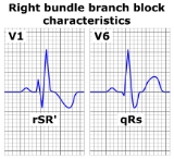
- There should be a terminal R wave in lead V1 (e.g. R, rR', rsR', rSR' or qR)
- There should be a slurred S wave in leads I and V6.
The T wave should be deflected opposite the terminal deflection of the QRS complex. This is known as appropriate T wave discordance with bundle branch block. A concordant T wave may suggest ischemia or myocardial infarction.
A mnemonic to remember the ECG changes is WiLLiaM MaRRoW, i.e. with LBBB there is a W in V1 and a M in V6 and with a RBBB there is a M in lead V1 and a W in lead V6
Prevalence of RBBB increases with age.
ICD 9 : 426.4