

Scintigraphy
Encyclopedia
Scintigraphy is a form of diagnostic test used in nuclear medicine
, wherein radioisotopes (here called radiopharmaceuticals) are taken internally, and the emitted radiation is captured by external detectors (gamma camera
s) to form two-dimensional images. In contrast, SPECT
and positron emission tomography
(PET) form 3-dimensional images, and are therefore classified as separate techniques to scintigraphy, although they also use gamma camera
s to detect internal radiation. Scintigraphy is unlike a diagnostic X-ray
where external radiation is passed through the body to form an image.
s. In cholescintigraphy, the injected radioactive chemical is taken up by the liver and secreted into the bile. The radiopharmaceutical then goes into the bile ducts, the gallbladder, and the intestines. The gamma camera is placed on the abdomen to picture these perfused organs. Other scintigraphic tests are done similarly.
 The most common indication for lung scintigraphy is to diagnose pulmonary embolism
, e.g. with a ventilation/perfusion scan
. Less common indications include evaluation of lung transplantation
, preoperative evaluation, evaluation of right-to-left shunt
s.
In the ventilation phase of a ventilation/perfusion scan, a gaseous radionuclide xenon
or technetium
DTPA in an aerosol form (or ideally using Technegas, a radioaerosol invented in Australia by Dr Bill Burch and Dr Richard Fawdry) is inhaled by the patient through a mouthpiece or mask that covers the nose and mouth. The perfusion phase of the test involves the intravenous injection of radioactive technetium macro aggregated albumin (Tc99m-MAA). A gamma camera acquires the images for both phases of the study.
to MDP, radioactivity can be transported and attached to bone via the hydroxyapatite for imaging. Any increased physiological function, such as a fracture in the bone, will usually mean increased concentration of the tracer.
-201 detected in cardiac tissues correlates with tissue blood supply. Viable cardiac cells have normal Na+/K+ ion exchange pumps
. Thallium binds the K+ pumps and is transported into the cells. Exercise or dipyridamole
induces widening (vasodilation
) of normal coronary arteries. This produces coronary steal from areas where arteries are maximally dilated. Areas of infarct or ischemic tissue
will remain "cold". Pre- and post-stress thallium may indicate areas that will benefit from myocardial revascularization
. Redistribution indicates the existence of coronary steal and the presence of ischemic coronary artery disease.
is used to detect parathyroid adenoma
s.
or Technetium-99m is generally used, and for this purpose the iodide isotope does not need to be attached to another protein or molecule, because thyroid tissue takes up free iodide actively.
s, indium white blood cell scan
s, Iobenguane
scan (MIBG) and octreotide scan
s. The MIBG scan detects adrenergic tissue and thus can be used to identify the location of tumor
s such as phaeochromocytomas and neuroblastoma
s.
and Urea breath test
, use radioisotopes but are not used to produce a specific image.
Nuclear medicine
In nuclear medicine procedures, elemental radionuclides are combined with other elements to form chemical compounds, or else combined with existing pharmaceutical compounds, to form radiopharmaceuticals. These radiopharmaceuticals, once administered to the patient, can localize to specific organs...
, wherein radioisotopes (here called radiopharmaceuticals) are taken internally, and the emitted radiation is captured by external detectors (gamma camera
Gamma camera
A gamma camera, also called a scintillation camera or Anger camera, is a device used to image gamma radiation emitting radioisotopes, a technique known as scintigraphy...
s) to form two-dimensional images. In contrast, SPECT
Single photon emission computed tomography
Single-photon emission computed tomography is a nuclear medicine tomographic imaging technique using gamma rays. It is very similar to conventional nuclear medicine planar imaging using a gamma camera. However, it is able to provide true 3D information...
and positron emission tomography
Positron emission tomography
Positron emission tomography is nuclear medicine imaging technique that produces a three-dimensional image or picture of functional processes in the body. The system detects pairs of gamma rays emitted indirectly by a positron-emitting radionuclide , which is introduced into the body on a...
(PET) form 3-dimensional images, and are therefore classified as separate techniques to scintigraphy, although they also use gamma camera
Gamma camera
A gamma camera, also called a scintillation camera or Anger camera, is a device used to image gamma radiation emitting radioisotopes, a technique known as scintigraphy...
s to detect internal radiation. Scintigraphy is unlike a diagnostic X-ray
X-ray
X-radiation is a form of electromagnetic radiation. X-rays have a wavelength in the range of 0.01 to 10 nanometers, corresponding to frequencies in the range 30 petahertz to 30 exahertz and energies in the range 120 eV to 120 keV. They are shorter in wavelength than UV rays and longer than gamma...
where external radiation is passed through the body to form an image.
Biliary system (Cholescintigraphy)
Scintigraphy of the biliary system is called cholescintigraphy and is done to diagnose obstruction of the bile ducts by a gallstone (cholelithiasis), a tumor, or another cause. It can also diagnose gallbladder diseases, e.g. bile leaks of biliary fistulaBiliary fistula
A biliary fistula is a type of fistula where bile leaks from the bile ducts into outside areas.-Causes:It can occur as a complication following biliary trauma , either iatrogenic or a result of a penetrating injury.-Presentation:...
s. In cholescintigraphy, the injected radioactive chemical is taken up by the liver and secreted into the bile. The radiopharmaceutical then goes into the bile ducts, the gallbladder, and the intestines. The gamma camera is placed on the abdomen to picture these perfused organs. Other scintigraphic tests are done similarly.
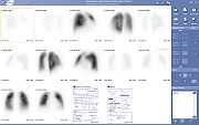
Lung scintigraphy
Pulmonary embolism
Pulmonary embolism is a blockage of the main artery of the lung or one of its branches by a substance that has travelled from elsewhere in the body through the bloodstream . Usually this is due to embolism of a thrombus from the deep veins in the legs, a process termed venous thromboembolism...
, e.g. with a ventilation/perfusion scan
Ventilation/perfusion scan
A ventilation/perfusion lung scan, also called a V/Q lung scan, is a type of medical imaging using scintigraphy and medical isotopes to evaluate the circulation of air and blood within a patient's lungs, in order to determine the ventilation/perfusion ratio...
. Less common indications include evaluation of lung transplantation
Lung transplantation
Lung transplantation, or pulmonary transplantation is a surgical procedure in which a patient's diseased lungs are partially or totally replaced by lungs which come from a donor...
, preoperative evaluation, evaluation of right-to-left shunt
Right-to-left shunt
A right-to-left shunt is a cardiac shunt which allows blood to flow from the right heart to the left heart. This terminology is used both for the abnormal state in humans and for normal physiological shunts in reptiles...
s.
In the ventilation phase of a ventilation/perfusion scan, a gaseous radionuclide xenon
Xenon
Xenon is a chemical element with the symbol Xe and atomic number 54. The element name is pronounced or . A colorless, heavy, odorless noble gas, xenon occurs in the Earth's atmosphere in trace amounts...
or technetium
Technetium
Technetium is the chemical element with atomic number 43 and symbol Tc. It is the lowest atomic number element without any stable isotopes; every form of it is radioactive. Nearly all technetium is produced synthetically and only minute amounts are found in nature...
DTPA in an aerosol form (or ideally using Technegas, a radioaerosol invented in Australia by Dr Bill Burch and Dr Richard Fawdry) is inhaled by the patient through a mouthpiece or mask that covers the nose and mouth. The perfusion phase of the test involves the intravenous injection of radioactive technetium macro aggregated albumin (Tc99m-MAA). A gamma camera acquires the images for both phases of the study.
Bone
For example, the ligand methylene-diphosphonate (MDP) can be preferentially taken up by bone. By chemically attaching technetium-99mTechnetium-99m
Technetium-99m is a metastable nuclear isomer of technetium-99, symbolized as 99mTc. The "m" indicates that this is a metastable nuclear isomer, i.e., that its half-life of 6 hours is considerably longer than most nuclear isomers that undergo gamma decay...
to MDP, radioactivity can be transported and attached to bone via the hydroxyapatite for imaging. Any increased physiological function, such as a fracture in the bone, will usually mean increased concentration of the tracer.
Heart
A thallium stress test is a form of scintigraphy, where the amount of thalliumThallium
Thallium is a chemical element with the symbol Tl and atomic number 81. This soft gray poor metal resembles tin but discolors when exposed to air. The two chemists William Crookes and Claude-Auguste Lamy discovered thallium independently in 1861 by the newly developed method of flame spectroscopy...
-201 detected in cardiac tissues correlates with tissue blood supply. Viable cardiac cells have normal Na+/K+ ion exchange pumps
Na+/K+-ATPase
Na+/K+-ATPase is an enzyme located in the plasma membrane in all animals.- Sodium-potassium pumps :Active transport is responsible for cells containing relatively high...
. Thallium binds the K+ pumps and is transported into the cells. Exercise or dipyridamole
Dipyridamole
Dipyridamole is a drug that inhibits thrombus formation when given chronically and causes vasodilation when given at high doses over a short time.-Mechanism and effects:...
induces widening (vasodilation
Vasodilation
Vasodilation refers to the widening of blood vessels resulting from relaxation of smooth muscle cells within the vessel walls, particularly in the large arteries, smaller arterioles and large veins. The process is essentially the opposite of vasoconstriction, or the narrowing of blood vessels. When...
) of normal coronary arteries. This produces coronary steal from areas where arteries are maximally dilated. Areas of infarct or ischemic tissue
Ischemia
In medicine, ischemia is a restriction in blood supply, generally due to factors in the blood vessels, with resultant damage or dysfunction of tissue. It may also be spelled ischaemia or ischæmia...
will remain "cold". Pre- and post-stress thallium may indicate areas that will benefit from myocardial revascularization
Revascularization
Revascularization is "a surgical procedure for the provision of a new, additional, or augmented blood supply to a body part or organ." The term derives from the prefix re-, in this case meaning "restoration" and vasculature, which refers to the circulatory structures of an organ.Revascularization...
. Redistribution indicates the existence of coronary steal and the presence of ischemic coronary artery disease.
Parathyroid
99mTC-sestamibiSestamibi
Technetium sestamibi is a pharmaceutical agent used in nuclear medicine imaging. The drug is a coordination complex of the radioisotope technetium-99m with the ligand methoxyisobutylisonitrile . The generic drug became available late September 2008...
is used to detect parathyroid adenoma
Parathyroid adenoma
A parathyroid adenoma is a benign tumor of the parathyroid gland. It can cause hyperparathyroidism.CT scans or sestamibi parathyroid scintigraphy can be used in detection.It can be associated with Cyclin D1 expression....
s.
Thyroid
To detect metastases/function of thyroid, the isotopes iodine-131Iodine-131
Iodine-131 , also called radioiodine , is an important radioisotope of iodine. It has a radioactive decay half-life of about eight days. Its uses are mostly medical and pharmaceutical...
or Technetium-99m is generally used, and for this purpose the iodide isotope does not need to be attached to another protein or molecule, because thyroid tissue takes up free iodide actively.
Full body
Examples are gallium scanGallium scan
A gallium scan or gallium 67 scan is a type of nuclear medicine study that uses a radioactive tracer to obtain images of a specific type of tissue, or disease state of tissue. Gallium salts like gallium citrate and gallium nitrate are used. The form of salt is not important, since it is the freely...
s, indium white blood cell scan
Indium white blood cell scan
The indium white blood cell scan, also called "indium leukocyte imaging," "indium-111 scan," or simply "indium scan," is a nuclear medicine procedure in which white blood cells are removed from the patient, tagged with the radioisotope Indium-111, and then injected intravenously into the patient...
s, Iobenguane
Iobenguane
Iobenguane, also known as metaiodobenzylguanidine or mIBG, or MIBG is a radiopharmaceutical, used in a scintigraphy method called MIBG scan...
scan (MIBG) and octreotide scan
Octreotide scan
An octreotide scan or octreoscan is a type of scintigraphy used to find carcinoid and other types of tumors and to localize sarcoidosis. Octreotide, a drug similar to somatostatin, is radiolabeled with indium-111, and is injected into a vein and travels through the bloodstream. The radioactive...
s. The MIBG scan detects adrenergic tissue and thus can be used to identify the location of tumor
Tumor
A tumor or tumour is commonly used as a synonym for a neoplasm that appears enlarged in size. Tumor is not synonymous with cancer...
s such as phaeochromocytomas and neuroblastoma
Neuroblastoma
Neuroblastoma is the most common extracranial solid cancer in childhood and the most common cancer in infancy, with an annual incidence of about 650 cases per year in the US , and 100 cases per year in the UK . Close to 50 percent of neuroblastoma cases occur in children younger than two years old...
s.
Function tests
Certain tests, such as the Schilling testSchilling test
The Schilling test is a medical investigation used for patients with vitamin B deficiency. The purpose of the test is to determine whether the patient has pernicious anemia.It is named for Robert F. Schilling.-Process:The Schilling test has multiple stages...
and Urea breath test
Urea breath test
The urea breath test is a rapid diagnostic procedure used to identify infections by Helicobacter pylori, a spiral bacterium implicated in gastritis, gastric ulcer, and peptic ulcer disease. It is based upon the ability of H. pylori to convert urea to ammonia and carbon dioxide...
, use radioisotopes but are not used to produce a specific image.