

Jugular venous pressure
Encyclopedia
The jugular venous pressure (JVP, sometimes referred to as jugular venous pulse) is the indirectly observed pressure over the venous system
. It can be useful in the differentiation of different forms of heart
and lung disease.
Classically three upward deflections and two downward deflections have been described.
 The patient is positioned under 45°, and the filling level of the jugular vein
determined. Visualize the internal jugular vein
when looking for the pulsation. In healthy people, the filling level of the jugular vein should be less than 3 centimetre
s vertical height above the sternal angle
. A pen-light can aid in discerning the jugular filling level by providing tangential light.
The JVP is easiest to observe if one looks along the surface of the sternocleidomastoid muscle
, as it is easier to appreciate the movement relative to the neck when looking from the side (as opposed to looking at the surface at a 90 degree angle). Like judging the movement of an automobile from a distance, it is easier to see the movement of an automobile when it is crossing one's path at 90 degrees (i.e. moving left to right or right to left), as opposed to coming toward one.
Pulse
s in the JVP are rather hard to observe, but trained cardiologists do try to discern these as signs of the state of the right atrium
.
The absence of 'a' waves may be seen in atrial fibrillation
.
"Cannon a waves" or increased amplitude 'a' waves, are associated with AV dissociation (third degree heart block
), when the atrium is contracting against a closed tricuspid valve.
In a prospective randomized study involving 86 patients who underwent right and left cardiac catheterization, the abdominojugular test was shown to correlate best with the pulmonary arterial wedge pressure. Furthermore, patients with a positive response had lower left ventricular ejection fractions and stroke volumes, higher left ventricular filling pressure, higher mean pulmonary arterial, and higher right atrial pressures.
The abdominojugular test, when done in a standardized fashion, correlates best with the pulmonary arterial wedge pressure, and therefore, is probably a reflection of an increased central blood volume. In the absence of isolated right ventricular failure, seen in some patients with right ventricular infarction, a positive abdominojugular test suggests a pulmonary artery wedge pressure of 15 mm Hg or greater.
. Another abnormality, "c-v waves", can be a sign of tricuspid regurgitation.
An elevated JVP is the classic sign of venous hypertension (e.g. right-sided heart failure). JVP elevation can be visualized as jugular venous distension, whereby the JVP is visualized at a level of the neck that is higher than normal. The paradoxical increase of the JVP with inspiration (instead of the expected decrease) is referred to as the Kussmaul sign, and indicates impaired filling of the right ventricle. The differential diagnosis of Kussmaul's sign includes constrictive pericarditis
, restrictive cardiomyopathy
, pericardial effusion
, and severe right-sided heart failure.
An important use of the jugular venous pressure is to assess the central venous pressure
in the absence of invasive measurements (e.g. with a central venous catheter
, which is a tube inserted in the neck veins). A 1996 systematic review
concluded that a high jugular venous pressure makes a high central venous pressure more likely, but does not significantly help confirm a low central venous pressure. The study also found that agreement between doctors on the jugular venous pressure can be poor.
Vein
In the circulatory system, veins are blood vessels that carry blood towards the heart. Most veins carry deoxygenated blood from the tissues back to the heart; exceptions are the pulmonary and umbilical veins, both of which carry oxygenated blood to the heart...
. It can be useful in the differentiation of different forms of heart
Heart disease
Heart disease, cardiac disease or cardiopathy is an umbrella term for a variety of diseases affecting the heart. , it is the leading cause of death in the United States, England, Canada and Wales, accounting for 25.4% of the total deaths in the United States.-Types:-Coronary heart disease:Coronary...
and lung disease.
Classically three upward deflections and two downward deflections have been described.
- The upward deflections are the "a" (atrial contraction), "c" (ventricular contraction and resulting bulging of tricuspid into the right atrium during isovolumetric systole) and "v" = atrial venous filling.
- The downward deflections of the wave are the "x" (the atrium relaxes and the tricuspid valve moves downward) and the "y" descent (filling of ventricle after tricuspid opening).
Visualization
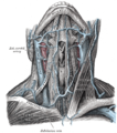
Jugular vein
The jugular veins are veins that bring deoxygenated blood from the head back to the heart via the superior vena cava.-Internal and external:There are two sets of jugular veins: external and internal....
determined. Visualize the internal jugular vein
Internal jugular vein
The two internal jugular veins collect the blood from the brain, the superficial parts of the face, and the neck.-Path:On both sides and at the base of the brain, the inferior petrosal sinus and the sigmoid sinus join to form the internal jugular vein...
when looking for the pulsation. In healthy people, the filling level of the jugular vein should be less than 3 centimetre
Centimetre
A centimetre is a unit of length in the metric system, equal to one hundredth of a metre, which is the SI base unit of length. Centi is the SI prefix for a factor of . Hence a centimetre can be written as or — meaning or respectively...
s vertical height above the sternal angle
Sternal angle
The sternal angle or 'angle of Louis', from the Latin angulus Ludovici is the anterior angle formed by the junction of the manubrium and the body of the sternum in the form of a secondary cartilaginous joint . This is also called the manubriosternal joint or Angle of Louis...
. A pen-light can aid in discerning the jugular filling level by providing tangential light.
The JVP is easiest to observe if one looks along the surface of the sternocleidomastoid muscle
Sternocleidomastoid muscle
In human anatomy, the sternocleidomastoid muscle , also known as sternomastoid and commonly abbreviated as SCM, is a paired muscle in the superficial layers of the anterior portion of the neck...
, as it is easier to appreciate the movement relative to the neck when looking from the side (as opposed to looking at the surface at a 90 degree angle). Like judging the movement of an automobile from a distance, it is easier to see the movement of an automobile when it is crossing one's path at 90 degrees (i.e. moving left to right or right to left), as opposed to coming toward one.
Pulse
Pulse
In medicine, one's pulse represents the tactile arterial palpation of the heartbeat by trained fingertips. The pulse may be palpated in any place that allows an artery to be compressed against a bone, such as at the neck , at the wrist , behind the knee , on the inside of the elbow , and near the...
s in the JVP are rather hard to observe, but trained cardiologists do try to discern these as signs of the state of the right atrium
Right atrium
The right atrium is one of four chambers in the hearts of mammals and archosaurs...
.
Differentiation from the carotid pulse
The JVP and carotid pulse can be differentiated several ways:- multiphasic - the JVP "beats" twice (in quick succession) in the cardiac cycleCardiac cycleThe cardiac cycle is a term referring to all or any of the events related to the flow or blood pressure that occurs from the beginning of one heartbeat to the beginning of the next. The frequency of the cardiac cycle is described by the heart rate. Each beat of the heart involves five major stages...
. In other words, there are two waves in the JVP for each contraction-relaxation cycle by the heart. The first beat represents that atrial contraction (termed a) and second beat represents venous filling of the right atrium against a closed tricuspid valve (termed v) and not the commonly mistaken 'ventricular contraction'. These wave forms may be altered by certain medical conditions therefore this is not always an accurate way to differentiate the JVP from the carotid pulse. The carotid artery only has one beat in the cardiac cycle. - non-palpable - the JVP cannot be palpated. If one feels a pulse in the neck, it is generally the common carotid arteryCommon carotid arteryIn human anatomy, the common carotid artery is an artery that supplies the head and neck with oxygenated blood; it divides in the neck to form the external and internal carotid arteries. - Structure :...
. - occludable - the JVP can be stopped by occluding the internal jugular veinInternal jugular veinThe two internal jugular veins collect the blood from the brain, the superficial parts of the face, and the neck.-Path:On both sides and at the base of the brain, the inferior petrosal sinus and the sigmoid sinus join to form the internal jugular vein...
by lightly pressing against the neck. It will fill from above. - varies with head-up-tilt (HUT) - the JVP varies with the angle of neck. If a person is standing, his JVP appears to be lower on the neck (or may not be seen at all because it is below the sternal angleSternal angleThe sternal angle or 'angle of Louis', from the Latin angulus Ludovici is the anterior angle formed by the junction of the manubrium and the body of the sternum in the form of a secondary cartilaginous joint . This is also called the manubriosternal joint or Angle of Louis...
). The carotid pulse's location does not vary with HUT. - varies with respiration - the JVP usually decreases with deep inspiration. Physiologically, this is a consequence of the Frank–Starling mechanism as inspiration decreases the thoracic pressure and increases blood movement into the heart (venous return), which a healthy heart moves into the pulmonary circulationPulmonary circulationPulmonary circulation is the half portion of the cardiovascular system which carries Oxygen-depleted Blood away from the heart, to the Lungs, and returns oxygenated blood back to the heart. Encyclopedic description and discovery of the pulmonary circulation is widely attributed to Doctor Ibn...
.
JVP waveform
The jugular venous pulsation has a biphasic waveform.- The 'a' wave corresponds to atrial contraction and ends synchronously with the carotid artery pulse. The peak of the 'a' wave demarcates the end of atrial systole.
- The 'c' wave occurs when the ventricles begin to contract causing the atrioventricular (AV) valves to bulge towards the atria.
- The 'x' descent follows the 'a' wave and represents atrial relaxation and rapid atrial filling due to low pressure. The 'x' ' (x prime) descent follows the 'c' wave and occurs as a result of the right ventricle pulling the tricuspid valve downward during ventricular systole. The 'x' ' (x prime) descent can be used as a measure of right ventricle contractility.
- The 'v' wave is seen when the tricuspid valve is closed and is caused by a pressure increase in the atrium as the venous return fills the atria - this occurs during and following the carotid pulse.
- The 'y' descent represents the rapid emptying of the atrium into the ventricle following the opening of the tricuspid valve.
The absence of 'a' waves may be seen in atrial fibrillation
Atrial fibrillation
Atrial fibrillation is the most common cardiac arrhythmia . It is a common cause of irregular heart beat, identified clinically by taking a pulse. Chaotic electrical activity in the two upper chambers of the heart result in the muscle fibrillating , instead of achieving coordinated contraction...
.
"Cannon a waves" or increased amplitude 'a' waves, are associated with AV dissociation (third degree heart block
Third degree heart block
-Presentation:Third-degree AV block, also known as complete heart block, is a medical condition in which the impulse generated in the SA node in the atrium does not propagate to the ventricles....
), when the atrium is contracting against a closed tricuspid valve.
Quantification
A classical method for quantifying the JVP was described by Borst & Molhuysen in 1952. It has since been modified in various ways. A venous arch may be used to measure the JVP more accurately.Abdominojugular test
The term hepatojugular reflux was previously used as it was thought that compression of the liver resulted in "reflux" of blood out the hepatic sinusoids into the great veins thereby elevating right atrial pressure and visualized as jugular venous distention. The exact physiologic mechanism of jugular venous distention with a positive test is much more complex and the commonly accepted term is now "Abdominojugular test."In a prospective randomized study involving 86 patients who underwent right and left cardiac catheterization, the abdominojugular test was shown to correlate best with the pulmonary arterial wedge pressure. Furthermore, patients with a positive response had lower left ventricular ejection fractions and stroke volumes, higher left ventricular filling pressure, higher mean pulmonary arterial, and higher right atrial pressures.
The abdominojugular test, when done in a standardized fashion, correlates best with the pulmonary arterial wedge pressure, and therefore, is probably a reflection of an increased central blood volume. In the absence of isolated right ventricular failure, seen in some patients with right ventricular infarction, a positive abdominojugular test suggests a pulmonary artery wedge pressure of 15 mm Hg or greater.
Interpretation
Certain wave form abnormalities, include "Cannon a-waves", which result when the atrium contracts against a closed tricuspid valve, due to complete heart block (3rd degree heart block), or even in ventricular tachycardiaVentricular tachycardia
Ventricular tachycardia is a tachycardia, or fast heart rhythm, that originates in one of the ventricles of the heart...
. Another abnormality, "c-v waves", can be a sign of tricuspid regurgitation.
An elevated JVP is the classic sign of venous hypertension (e.g. right-sided heart failure). JVP elevation can be visualized as jugular venous distension, whereby the JVP is visualized at a level of the neck that is higher than normal. The paradoxical increase of the JVP with inspiration (instead of the expected decrease) is referred to as the Kussmaul sign, and indicates impaired filling of the right ventricle. The differential diagnosis of Kussmaul's sign includes constrictive pericarditis
Constrictive pericarditis
In many cases, constrictive pericarditis is a late sequela, in other words a condition that is the consequence of a previous disease, of an inflammatory condition of the pericardium...
, restrictive cardiomyopathy
Restrictive cardiomyopathy
Restrictive cardiomyopathy is a form of cardiomyopathy in which the walls are rigid, and the heart is restricted from stretching and filling with blood properly....
, pericardial effusion
Pericardial effusion
Pericardial effusion is an abnormal accumulation of fluid in the pericardial cavity. Because of the limited amount of space in the pericardial cavity, fluid accumulation will lead to an increased intrapericardial pressure and this can negatively affect heart function...
, and severe right-sided heart failure.
- Raised JVP, normal waveform
- BradycardiaBradycardiaBradycardia , in the context of adult medicine, is the resting heart rate of under 60 beats per minute, though it is seldom symptomatic until the rate drops below 50 beat/min. It may cause cardiac arrest in some patients, because those with bradycardia may not be pumping enough oxygen to their heart...
- Fluid overload
- Heart Failure
- Bradycardia
- Raised JVP, absent pulsation
- Superior vena cava syndromeSuperior vena cava syndromeSuperior vena cava syndrome , or superior vena cava obstruction , is usually the result of the direct obstruction of the superior vena cava by malignancies such as compression of the vessel wall by right upper lobe tumors or thymoma and/or mediastinal lymphadenopathy. The most common malignancies...
- Superior vena cava syndrome
- Large 'a' wave (increased atrial contraction pressure)
- tricuspid stenosis
- Right heart failure
- Pulmonary hypertensionPulmonary hypertensionIn medicine, pulmonary hypertension is an increase in blood pressure in the pulmonary artery, pulmonary vein, or pulmonary capillaries, together known as the lung vasculature, leading to shortness of breath, dizziness, fainting, and other symptoms, all of which are exacerbated by exertion...
- Cannon 'a' wave (atria contracting against closed tricuspid valve)
- Atrial flutterAtrial flutterAtrial flutter is an abnormal heart rhythm that occurs in the atria of the heart. When it first occurs, it is usually associated with a fast heart rate or tachycardia , and falls into the category of supra-ventricular tachycardias. While this rhythm occurs most often in individuals with...
- Premature atrial rhythm (or tachycardia)
- third degree heart blockThird degree heart block-Presentation:Third-degree AV block, also known as complete heart block, is a medical condition in which the impulse generated in the SA node in the atrium does not propagate to the ventricles....
- Ventricular ectopics
- Ventricular tachycardia
- Atrial flutter
- Absent 'a' wave (no unifocal atrial depolarisation)
- atrial fibrillationAtrial fibrillationAtrial fibrillation is the most common cardiac arrhythmia . It is a common cause of irregular heart beat, identified clinically by taking a pulse. Chaotic electrical activity in the two upper chambers of the heart result in the muscle fibrillating , instead of achieving coordinated contraction...
- atrial fibrillation
- Large 'v' wave (c-v wave)
- Tricuspid regurgitation
- Slow 'y' descent
- Tricuspid stenosis
- Parodoxical JVP (Kussmaul's signKussmaul's signKussmaul's sign is the observation of a rise in jugular venous pressure on inspiration. It can be seen in some forms of heart disease and is usually indicative of limited right ventricular filling.-Background:...
: JVP rises with inspiration, drops with expiration)- Pericardial effusionPericardial effusionPericardial effusion is an abnormal accumulation of fluid in the pericardial cavity. Because of the limited amount of space in the pericardial cavity, fluid accumulation will lead to an increased intrapericardial pressure and this can negatively affect heart function...
- Constrictive pericarditisConstrictive pericarditisIn many cases, constrictive pericarditis is a late sequela, in other words a condition that is the consequence of a previous disease, of an inflammatory condition of the pericardium...
- Pericardial tamponade
- Pericardial effusion
An important use of the jugular venous pressure is to assess the central venous pressure
Central venous pressure
Central venous pressure describes the pressure of blood in the thoracic vena cava, near the right atrium of the heart...
in the absence of invasive measurements (e.g. with a central venous catheter
Central venous catheter
In medicine, a central venous catheter is a catheter placed into a large vein in the neck , chest or groin...
, which is a tube inserted in the neck veins). A 1996 systematic review
Systematic review
A systematic review is a literature review focused on a research question that tries to identify, appraise, select and synthesize all high quality research evidence relevant to that question. Systematic reviews of high-quality randomized controlled trials are crucial to evidence-based medicine...
concluded that a high jugular venous pressure makes a high central venous pressure more likely, but does not significantly help confirm a low central venous pressure. The study also found that agreement between doctors on the jugular venous pressure can be poor.
External links
- Clinical Examination page on JVP
- JVP (GPnotebook)
- Normal jugular vein waves - Merck Manual
- JVD bedside workup