

Computed tomography
Encyclopedia
X-ray computed tomography or Computer tomography (CT), is a medical imaging
method employing tomography
created by computer processing. Digital geometry processing
is used to generate a three-dimensional
image of the inside of an object from a large series of two-dimensional X-ray
images taken around a single axis of rotation.
CT produces a volume of data that can be manipulated, through a process known as "windowing", in order to demonstrate various bodily structures based on their ability to block the X-ray beam. Although historically the images generated were in the axial or transverse plane, perpendicular to the long axis of the body, modern scanners allow this volume of data to be reformatted in various planes or even as volumetric (3D) representations of structures. Although most common in medicine, CT is also used in other fields, such as nondestructive materials testing
. Another example is archaeological uses such as imaging the contents of sarcophagi.
Usage of CT has increased dramatically over the last two decades in many countries. An estimated 72 million scans were performed in the United States in 2007. It is estimated that 0.4% of current cancers in the United States are due to CTs performed in the past and that this may increase to as high as 1.5-2% with 2007 rates of CT usage; however, this estimate is disputed.
tomos (slice) and graphein (to write). Computed tomography was originally known as the "EMI scan" as it was developed at a research branch of EMI
, a company best known today for its music and recording business. It was later known as computed axial tomography (CAT or CT scan) and body section röntgenography.
Although the term "computed tomography" could be used to describe positron emission tomography
and single photon emission computed tomography
, in practice it usually refers to the computation of tomography from X-ray
images, especially in older medical literature and smaller medical facilities.
In MeSH
, "computed axial tomography" was used from 1977–79, but the current indexing explicitly includes "X-ray" in the title.
is spun around the central axis of the area being scanned. These are the dominant type of scanners on the market because of they have been manufactured longer and offer lower cost of production and purchase. The main limitation of this type is the bulk and intertia of the equipment (X-Ray tube assembly and Detector array on the opposite side of the circle) which limits the speed at which the equipment can spin.
Spinning electrons, commonly called EBT
, in which a large enough X-Ray tube
is constructed so that only the path of the electrons, traveling between the cathode and anode of the X-Ray tube are spun using deflection coils. This type has a major advantage in that sweep speeds can be much faster, imaging the heart which never stops moving. However, few of this machine design have been produced and sold because of the higher cost in building a much large X-Ray tube and the detector array, which is also stationary, much also be far larger, thus more expensive.
to supplement X-ray
s and medical ultrasonography
. It has more recently been used for preventive medicine
or screening
for disease, for example CT colonography for patients with a high risk of colon cancer, or full-motion heart scans for patients with high risk of heart disease. A number of institutions offer full-body scan
s for the general population.
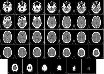 CT scanning of the head is typically used to detect infarction
, tumours, calcification
s, haemorrhage and bone trauma. Of the above, hypodense (dark) structures indicate infarction or tumours, hyperdense (bright) structures indicate calcifications and haemorrhage and bone trauma can be seen as disjunction in bone windows. Ambulances equipped with small bore multi-sliced CT scanners respond to cases involving stroke or head trauma.
parenchyma, that is, the internals of the lungs. It is particularly relevant here because normal two-dimensional X-rays do not show such defects. A variety of techniques are used, depending on the suspected abnormality. For evaluation of chronic interstitial processes (emphysema
, fibrosis
, and so forth), thin sections with high spatial frequency reconstructions are used; often scans are performed both in inspiration and expiration. This special technique is called high resolution CT. Therefore, it produces a sampling of the lung and not continuous images.
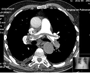 CT pulmonary angiogram
(CTPA) is a medical diagnostic test used to diagnose pulmonary embolism
(PE). It employs computed tomography to obtain an image of the pulmonary arteries
.
. First, CT completely eliminates the superimposition of images of structures outside the area of interest. Second, because of the inherent high-contrast resolution of CT, differences between tissues that differ in physical density by less than 1% can be distinguished. Finally, data from a single CT imaging procedure consisting of either multiple contiguous or one helical scan can be viewed as images in the axial, coronal, or sagittal planes, depending on the diagnostic task. This is referred to as multiplanar reformatted imaging.
CT is regarded as a moderate- to high-radiation
diagnostic technique. The improved resolution of CT has permitted the development of new investigations, which may have advantages; compared to conventional radiography, for example, CT angiography avoids the invasive insertion of a catheter. CT Colonography (also known as Virtual Colonoscopy or VC for short) may be as useful as a barium enema for detection of tumors, but may use a lower radiation dose. CT VC is increasingly being used in the UK as a diagnostic test for bowel cancer and can negate the need for a colonoscopy.
The radiation dose for a particular study depends on multiple factors: volume scanned, patient build, number and type of scan sequences, and desired resolution and image quality. In addition, two helical CT scanning parameters that can be adjusted easily and that have a profound effect on radiation dose are tube current and pitch. Computed tomography (CT) scan has been shown to be more accurate than radiographs in evaluating anterior interbody fusion but may still over-read the extent of fusion.
). CT scans of children have been estimated to produce non-negligible increases in the probability of lifetime cancer mortality, leading to calls for the use of reduced current settings for CT scans of children.
These calculations are based on similar radiation exposures experienced by those present during the atomic bomb explosions in Japan during the second world war and nuclear industry works. Estimated lifetime cancer mortality risks attributable to the radiation exposure from a CT in a 1-year-old are 0.18% (abdominal) and 0.07% (head) — an order of magnitude higher than for adults — although those figures still represent a small increase in cancer mortality over the background rate. In the United States, of approximately 600,000 abdominal and head CT examinations annually performed in children under the age of 15 years, a rough estimate is that 500 of these individuals might ultimately die from cancer attributable to the CT radiation. The additional risk is still low: 0.35% compared to the background risk of dying from cancer of 23%. However, if these statistics are extrapolated to the current number of CT scans, the additional rise in cancer mortality could be 1.5 to 2%. Furthermore, certain conditions can require children to be exposed to multiple CT scans.
In 2009, a number of studies that further defined the risk of cancer that may be caused by CT scans appeared. One study indicated that radiation by CT scans is often higher and more variable than cited, and each of the 19,500 CT scans that are daily performed in the US is equivalent to 30 to 442 chest X-rays in radiation. It has been estimated that CT radiation exposure will result in 29,000 new cancer cases just from the CT scans performed in 2007. The most common cancers caused by CT are thought to be lung cancer
, colon cancer, and leukemia
with younger people, and women more at risk. These conclusions, however, are criticized by the American College of Radiology
(ACR), which maintains that the life expectancy of CT scanned patients is not that of the general population and that the model of calculating cancer is based on total-body radiation exposure and thus faulty.
CT scans can be performed with different settings for lower exposure in children, although these techniques are often not employed. Surveys have suggested that, at the current time, many CT scans are performed unnecessarily. Ultrasound scanning or magnetic resonance imaging
are alternatives (for example, in appendicitis or brain imaging) without the risk of radiation exposure. Although CT scans come with an additional risk of cancer (it can be estimated that the radiation exposure from a full body scan is the same as standing 2.4 km away from the World War II
atomic bomb blasts in Japan), especially in children, the benefits that stem from their use outweigh the risk in many cases. Studies support informing parents of the risks of pediatric CT scanning.
For purposes of comparison, the average background exposure
in the UK is 1-3 mSv per year.
contrast agents
in order to provide superior image quality, there is a low but non-negligible level of risk associated with the contrast agents themselves. Many patients report nausea and discomfort, including warmth in the crotch, which mimics the sensation of wetting oneself. Certain patients may experience severe and potentially life-threatening allergic reactions
to the contrast dye.
The contrast agent may also induce kidney damage
. The risk of this is increased with patients who have preexisting renal insufficiency
, preexisting diabetes
, or reduced intravascular volume. In general, if a patient has normal kidney function, then the risks of contrast nephropathy are negligible. Patients with mild kidney impairment are usually advised to ensure full hydration for several hours before and after the injection. For moderate kidney failure, the use of iodinated contrast
should be avoided; this may mean using an alternative technique instead of CT, e.g., MRI. However, patients with severe renal failure requiring dialysis do not require special precautions, as their kidneys have so little function remaining that any further damage would not be noticeable and the dialysis will remove the contrast agent.
Streaks are often seen around materials that block most X-rays, such as metal or bone. These streaks can be caused by undersampling, photon starvation, motion, beam hardening, or scatter. This type of artifact commonly occurs in the posterior fossa of the brain, or if there are metal implants. The streaks can be reduced using newer reconstruction techniques.
This appears as "blurring" over sharp edges. It is due to the scanner being unable to differentiate between a small amount of high-density material (e.g., bone) and a larger amount of lower density (e.g., cartilage). The processor tries to average out the two densities or structures, and information is lost. This can be partially overcome by scanning using thinner slices.
Probably the most common mechanical artifact, the image of one or many "rings" appears within an image. This is usually due to a detector fault.
This appears as graining on the image and is caused by a low signal to noise ratio. This occurs more commonly when a thin slice thickness is used. It can also occur when the power supplied to the X-ray tube is insufficient to penetrate the anatomy.
This is seen as blurring and/or streaking, which is caused by movement of the object being imaged. Motion blurring might be reduced using a new technique called IFT (incompressible flow tomography).
Streaking appearances can occur when the detectors intersect the reconstruction plane. This can be reduced with filters or a reduction in pitch.
This can give a "cupped appearance". It occurs when there is more attenuation in the center of the object than around the edge. This is easily corrected by filtration and software.
who saw them from 1.8% to 25%. In the emergency department in the United States, CT or MRI imaging is done in 15% of people who present with injuries as of 2007 (up from 6% in 1998).
gas. These systems were in turn replaced by scintillation systems based on photodiode
s instead of photomultipliers and modern scintillation materials with more desirable characteristics. Many data scans are progressively taken as the object is gradually passed through the gantry.
Newer machines with faster computer systems and newer software strategies can process not only individual cross sections but continuously changing cross sections as the gantry, with the object to be imaged slowly and smoothly slid through the X-ray circle. These are called helical or spiral CT
machines. Their computer systems integrate the data of the moving individual slices to generate three dimensional volumetric information (3D-CT scan), in turn viewable from multiple different perspectives on attached CT workstation monitors. This type of data acquisition requires enormous processing power, as the data are arriving in a continuous stream and must be processed in real-time.
In conventional CT machines, an X-ray tube
and detector are physically rotated behind a circular shroud (see the image above right); in the electron beam tomography
(EBT), the tube is far larger and higher power to support the high temporal resolution. The electron beam is deflected in a hollow funnel-shaped vacuum chamber. X-rays are generated when the beam hits the stationary target. The detector is also stationary. This arrangement can result in very fast scans, but is extremely expensive.
CT is used in medicine
as a diagnostic tool and as a guide for interventional procedures. Sometimes contrast materials such as intravenous iodinated
contrast are used. This is useful to highlight structures such as blood vessels that otherwise would be difficult to delineate from their surroundings. Using contrast material can also help to obtain functional information about tissues.
Once the scan data has been acquired, the data must be processed using a form of tomographic reconstruction
, which produces a series of cross-sectional images. The most common technique in general use is filtered back projection, which is straightforward to implement and can be computed rapidly. In terms of mathematics, this method is based on the Radon transform
. However, this is not the only technique available: the original EMI scanner solved the tomographic reconstruction problem by linear algebra
, but this approach was limited by its high computational complexity, especially given the computer technology available at the time. More recently, manufacturers have developed iterative physical model-based expectation-maximization techniques. These techniques are advantageous because they use an internal model of the scanner's physical properties and of the physical laws of X-ray interactions. By contrast, earlier methods have assumed a perfect scanner and highly simplified physics, which leads to a number of artifacts and reduced resolution - the result is images with improved resolution, reduced noise and fewer artifacts, as well as the ability to greatly reduce the radiation dose in certain circumstances. The disadvantage is a very high computational requirement, which is at the limits of practicality for current scan protocols.
Pixel
s in an image obtained by CT scanning are displayed in terms of relative radiodensity
. The pixel itself is displayed according to the mean attenuation of the tissue(s) that it corresponds to on a scale from +3071 (most attenuating) to -1024 (least attenuating) on the Hounsfield scale
. Pixel
is a two dimensional unit based on the matrix size and the field of view. When the CT slice thickness is also factored in, the unit is known as a Voxel
, which is a three-dimensional unit. The phenomenon that one part of the detector cannot differentiate between different tissues is called the "Partial Volume Effect". That means that a big amount of cartilage and a thin layer of compact bone can cause the same attenuation in a voxel as hyperdense cartilage alone. Water has an attenuation of 0 Hounsfield units (HU), while air is -1000 HU, cancellous bone is typically +400 HU, cranial bone can reach 2000 HU or more (os temporale) and can cause artifacts. The attenuation of metallic implants depends on atomic number of the element used: Titanium usually has an amount of +1000 HU, iron steel can completely extinguish the X-ray and is, therefore, responsible for well-known line-artifacts in computed tomograms. Artifacts are caused by abrupt transitions between low- and high-density materials, which results in data values that exceed the dynamic range of the processing electronics.
Contrast medium
s used for X-ray CT, as well as for plain film X-ray
, are called radiocontrast
s. Radiocontrasts for X-ray CT are, in general, iodine-based. Often, images are taken both with and without radiocontrast. CT images are called precontrast or native-phase images before any radiocontrast has been administrated, and postcontrast after radiocontrast administration.
or near isotropic, resolution, display of images does not need to be restricted to the conventional axial images. Instead, it is possible for a software program to build a volume by "stacking" the individual slices one on top of the other. The program may then display the volume in an alternative manner.
(MIP) or minimum-intensity projection (mIP), can be used to build the reconstructed slices.
MPR is frequently used for examining the spine. Axial images through the spine will only show one vertebral body at a time and cannot reliably show the intervertebral discs. By reformatting the volume, it becomes much easier to visualise the position of one vertebral body in relation to the others.
Modern software allows reconstruction in non-orthogonal (oblique) planes so that the optimal plane can be chosen to display an anatomical structure. This may be particularly useful for visualising the structure of the bronchi as these do not lie orthogonal to the direction of the scan.
For vascular imaging, curved-plane reconstruction can be performed. This allows bends in a vessel to be "straightened" so that the entire length can be visualised on one image, or a short series of images. Once a vessel has been "straightened" in this way, quantitative measurements of length and cross sectional area can be made, so that surgery or interventional treatment can be planned.
MIP reconstructions enhance areas of high radiodensity, and so are useful for angiographic studies. MIP reconstructions tend to enhance air spaces so are useful for assessing lung structure.
image processing algorithms and displayed on screen. Multiple models can be constructed from various thresholds, allowing different colors to represent each anatomical component such as bone, muscle, and cartilage. However, the interior structure of each element is not visible in this mode of operation.
, transparency and colors are used to allow a better representation of the volume to be shown in a single image. For example, the bones of the pelvis could be displayed as semi-transparent, so that, even at an oblique angle, one part of the image does not conceal another.
(industrial computed tomography) is a process which utilizes x-ray equipment to produce 3D representations of components both externally and internally. Industrial CT scanning has been utilized in many areas of industry for internal inspection of components. Some of the key uses for CT scanning have been flaw detection, failure analysis, metrology, assembly analysis and reverse engineering applications
 In the early 1900s, the Italian radiologist Alessandro Vallebona proposed a method to represent a single slice of the body on the radiographic film. This method was known as tomography
. The idea is based on simple principles of projective geometry
: moving synchronously and in opposite directions the X-ray tube and the film, which are connected together by a rod whose pivot point is the focus; the image created by the points on the focal plane appears sharper, while the images of the other points annihilate as noise. This is only marginally effective, as blurring occurs in only the "x" plane. There are also more complex devices that can move in more than one plane and perform more effective blurring.
In 1959, William Oldendorf, a UCLA neurologist and senior medical investigator at the West Los Angeles Veterans Administration hospital, conceived an idea for "scanning a head through a transmitted beam of X-rays, and being able to reconstruct the radiodensity patterns of a plane through the head" after watching an automated apparatus built to reject frost-bitten fruit by detecting dehydrated portions. In 1961, he built a prototype in which an X-ray source and a mechanically coupled detector rotated around the object to be imaged. By reconstructing the image, this instrument could get an X-ray picture of a nail surrounded by a circle of other nails, which made it impossible to X-ray from any single angle. In his landmark paper, published in 1961, he described the basic concept later used by Allan McLeod Cormack
to develop the mathematics behind computerized tomography. In October, 1963 Oldendorf received a U.S. patent for a "radiant energy apparatus for investigating selected areas of interior objects obscured by dense material," Oldendorf shared the 1975 Lasker award with Hounsfield for that discovery.
Tomography has been one of the pillars of radiologic diagnostics until the late 1970s, when the availability of minicomputers and of the transverse axial scanning method – this last due to the work of Godfrey Hounsfield
and South African-born Allan McLeod Cormack
– gradually supplanted it as the modality of CT. In terms of mathematics, the method is based upon the use of the Radon Transform
invented by Johann Radon
in 1917. But as Cormack remembered later, he had to find the solution himself since it was only in 1972 that he learned of the work of Radon, by chance.
The first commercially viable CT scanner was invented by Sir Godfrey Hounsfield
in Hayes
, United Kingdom
, at EMI
Central Research Laboratories using X-rays. Hounsfield conceived his idea in 1967. The first EMI-Scanner was installed in Atkinson Morley Hospital
in Wimbledon
, England, and the first patient brain-scan was done on 1 October 1971. It was publicly announced in 1972.
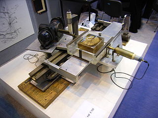
The original 1971 prototype took 160 parallel readings through 180 angles, each 1° apart, with each scan taking a little over 5 minutes. The images from these scans took 2.5 hours to be processed by algebraic reconstruction technique
s on a large computer. The scanner had a single photomultiplier detector, and operated on the Translate/Rotate principle.
It has been claimed that thanks to the success of The Beatles
, EMI
could fund research and build early models for medical use. The first production X-ray CT machine (in fact called the "EMI-Scanner") was limited to making tomographic sections of the brain, but acquired the image data in about 4 minutes (scanning two adjacent slices), and the computation time (using a Data General Nova
minicomputer) was about 7 minutes per picture. This scanner required the use of a water-filled Perspex tank with a pre-shaped rubber "head-cap" at the front, which enclosed the patient's head. The water-tank was used to reduce the dynamic range of the radiation reaching the detectors (between scanning outside the head compared with scanning through the bone of the skull). The images were relatively low resolution, being composed of a matrix of only 80 x 80 pixels.
In the U.S., the first installation was at the Mayo Clinic
. As a tribute to the impact of this system on medical imaging the Mayo Clinic has an EMI scanner on display in the Radiology Department. Allan McLeod Cormack
of Tufts University
in Massachusetts
independently invented a similar process, and both Hounsfield and Cormack shared the 1979 Nobel Prize in Medicine
.
The first CT system that could make images of any part of the body and did not require the "water tank" was the ACTA (Automatic Computerized Transverse Axial) scanner designed by Robert S. Ledley, DDS, at Georgetown University
. This machine had 30 photomultiplier tubes as detectors and completed a scan in only 9 translate/rotate cycles, much faster than the EMI-scanner. It used a DEC
PDP11/34
minicomputer both to operate the servo-mechanisms and to acquire and process the images. The Pfizer
drug company acquired the prototype from the university, along with rights to manufacture it. Pfizer then began making copies of the prototype, calling it the "200FS" (FS meaning Fast Scan), which were selling as fast as they could make them. This unit produced images in a 256×256 matrix, with much better definition than the EMI-Scanner's 80×80.
Since the first CT scanner, CT technology has vastly improved. Improvements in speed, slice count, and image quality have been the major focus primarily for cardiac imaging. Scanners now produce images much faster and with higher resolution enabling doctors to diagnose patients more accurately and perform medical procedures with greater precision. In the late 90's CT scanners have broke into two major groups, "Fixed CT" and "Portable CT". "Fixed CT Scanners" are large, require a dedicated power supply, electrical closet, HVAC cooling system, a separate workstation room, and a large lead lined room. "Fixed CT Scanners" can also be mounted inside large tractor trailers and driven from site to site and are known as "Mobile CT Scanners". "Portable CT Scanners" are light weight, small, and mounted on wheels. These scanners often have built-in lead shielding and run off of batteries or standard wall power.
Study for brain was quickly replaced by CT. A form of tomography can be performed by moving the X-ray source and detector during an exposure. Anatomy at the target level remains sharp, while structures at different levels are blurred. By varying the extent and path of motion, a variety of effects can be obtained, with variable depth of field
and different degrees of blurring of "out of plane" structures.
Although largely obsolete, conventional tomography is still used in specific situations such as dental imaging (orthopantomography) or in intravenous urography.
. However, because the image processing is digital, a series of slices at different depths and with different thicknesses can be reconstructed from the same acquisition, saving both time and radiation exposure.
Because the data acquired are incomplete, tomosynthesis is unable to offer the extremely narrow slice widths that CT offers. However, higher resolution detectors can be used, allowing very high in-plane resolution, even if the Z-axis resolution is poor. The primary interest in tomosynthesis is in breast imaging, as an extension to mammography
, where it may offer better detection rates with little extra increase in radiation exposure.
Reconstruction algorithms for tomosynthesis are significantly different from those of conventional CT because the conventional filtered back projection algorithm requires a complete set of data. Iterative algorithms based upon expectation maximization are most commonly used, but are extremely computationally intensive. Some manufacturers have produced practical systems using off-the-shelf GPUs
to perform the reconstruction.
Medical imaging
Medical imaging is the technique and process used to create images of the human body for clinical purposes or medical science...
method employing tomography
Tomography
Tomography refers to imaging by sections or sectioning, through the use of any kind of penetrating wave. A device used in tomography is called a tomograph, while the image produced is a tomogram. The method is used in radiology, archaeology, biology, geophysics, oceanography, materials science,...
created by computer processing. Digital geometry processing
Geometry Processing
Geometry processing, or mesh processing, is a fast-growing area of research that uses concepts from applied mathematics, computer science and engineering to design efficient algorithms for the acquisition, reconstruction, analysis, manipulation, simulation and transmission of complex 3D models...
is used to generate a three-dimensional
Three-dimensional space
Three-dimensional space is a geometric 3-parameters model of the physical universe in which we live. These three dimensions are commonly called length, width, and depth , although any three directions can be chosen, provided that they do not lie in the same plane.In physics and mathematics, a...
image of the inside of an object from a large series of two-dimensional X-ray
X-ray
X-radiation is a form of electromagnetic radiation. X-rays have a wavelength in the range of 0.01 to 10 nanometers, corresponding to frequencies in the range 30 petahertz to 30 exahertz and energies in the range 120 eV to 120 keV. They are shorter in wavelength than UV rays and longer than gamma...
images taken around a single axis of rotation.
CT produces a volume of data that can be manipulated, through a process known as "windowing", in order to demonstrate various bodily structures based on their ability to block the X-ray beam. Although historically the images generated were in the axial or transverse plane, perpendicular to the long axis of the body, modern scanners allow this volume of data to be reformatted in various planes or even as volumetric (3D) representations of structures. Although most common in medicine, CT is also used in other fields, such as nondestructive materials testing
Nondestructive testing
Nondestructive testing or Non-destructive testing is a wide group of analysis techniques used in science and industry to evaluate the properties of a material, component or system without causing damage....
. Another example is archaeological uses such as imaging the contents of sarcophagi.
Usage of CT has increased dramatically over the last two decades in many countries. An estimated 72 million scans were performed in the United States in 2007. It is estimated that 0.4% of current cancers in the United States are due to CTs performed in the past and that this may increase to as high as 1.5-2% with 2007 rates of CT usage; however, this estimate is disputed.
Terminology
The word "tomography" is derived from the GreekGreek language
Greek is an independent branch of the Indo-European family of languages. Native to the southern Balkans, it has the longest documented history of any Indo-European language, spanning 34 centuries of written records. Its writing system has been the Greek alphabet for the majority of its history;...
tomos (slice) and graphein (to write). Computed tomography was originally known as the "EMI scan" as it was developed at a research branch of EMI
EMI
The EMI Group, also known as EMI Music or simply EMI, is a multinational music company headquartered in London, United Kingdom. It is the fourth-largest business group and family of record labels in the recording industry and one of the "big four" record companies. EMI Group also has a major...
, a company best known today for its music and recording business. It was later known as computed axial tomography (CAT or CT scan) and body section röntgenography.
Although the term "computed tomography" could be used to describe positron emission tomography
Positron emission tomography
Positron emission tomography is nuclear medicine imaging technique that produces a three-dimensional image or picture of functional processes in the body. The system detects pairs of gamma rays emitted indirectly by a positron-emitting radionuclide , which is introduced into the body on a...
and single photon emission computed tomography
Single photon emission computed tomography
Single-photon emission computed tomography is a nuclear medicine tomographic imaging technique using gamma rays. It is very similar to conventional nuclear medicine planar imaging using a gamma camera. However, it is able to provide true 3D information...
, in practice it usually refers to the computation of tomography from X-ray
X-ray
X-radiation is a form of electromagnetic radiation. X-rays have a wavelength in the range of 0.01 to 10 nanometers, corresponding to frequencies in the range 30 petahertz to 30 exahertz and energies in the range 120 eV to 120 keV. They are shorter in wavelength than UV rays and longer than gamma...
images, especially in older medical literature and smaller medical facilities.
In MeSH
Medical Subject Headings
Medical Subject Headings is a comprehensive controlled vocabulary for the purpose of indexing journal articles and books in the life sciences; it can also serve as a thesaurus that facilitates searching...
, "computed axial tomography" was used from 1977–79, but the current indexing explicitly includes "X-ray" in the title.
Types of CT Machine
Spinning tube, commonly called spiral CT, in which an entire X-Ray tubeX-ray tube
An X-ray tube is a vacuum tube that produces X-rays. They are used in X-ray machines. X-rays are part of the electromagnetic spectrum, an ionizing radiation with wavelengths shorter than ultraviolet light...
is spun around the central axis of the area being scanned. These are the dominant type of scanners on the market because of they have been manufactured longer and offer lower cost of production and purchase. The main limitation of this type is the bulk and intertia of the equipment (X-Ray tube assembly and Detector array on the opposite side of the circle) which limits the speed at which the equipment can spin.
Spinning electrons, commonly called EBT
EBT
EBT can mean:* Electron beam tomography, a diagnostic technology* Electron beam texturing, a texturing technology* Electronic Benefit Transfer, an electronic payment system* EuroBillTracker, a website for tracking Euro banknotes...
, in which a large enough X-Ray tube
X-ray tube
An X-ray tube is a vacuum tube that produces X-rays. They are used in X-ray machines. X-rays are part of the electromagnetic spectrum, an ionizing radiation with wavelengths shorter than ultraviolet light...
is constructed so that only the path of the electrons, traveling between the cathode and anode of the X-Ray tube are spun using deflection coils. This type has a major advantage in that sweep speeds can be much faster, imaging the heart which never stops moving. However, few of this machine design have been produced and sold because of the higher cost in building a much large X-Ray tube and the detector array, which is also stationary, much also be far larger, thus more expensive.
Diagnostic use
Since its introduction in the 1970s, CT has become an important tool in medical imagingMedical imaging
Medical imaging is the technique and process used to create images of the human body for clinical purposes or medical science...
to supplement X-ray
X-ray
X-radiation is a form of electromagnetic radiation. X-rays have a wavelength in the range of 0.01 to 10 nanometers, corresponding to frequencies in the range 30 petahertz to 30 exahertz and energies in the range 120 eV to 120 keV. They are shorter in wavelength than UV rays and longer than gamma...
s and medical ultrasonography
Medical ultrasonography
Diagnostic sonography is an ultrasound-based diagnostic imaging technique used for visualizing subcutaneous body structures including tendons, muscles, joints, vessels and internal organs for possible pathology or lesions...
. It has more recently been used for preventive medicine
Preventive medicine
Preventive medicine or preventive care refers to measures taken to prevent diseases, rather than curing them or treating their symptoms...
or screening
Screening (medicine)
Screening, in medicine, is a strategy used in a population to detect a disease in individuals without signs or symptoms of that disease. Unlike what generally happens in medicine, screening tests are performed on persons without any clinical sign of disease....
for disease, for example CT colonography for patients with a high risk of colon cancer, or full-motion heart scans for patients with high risk of heart disease. A number of institutions offer full-body scan
Full-body scan
Full-body scan is a scan of the patient's entire body to support the diagnosis and treatment of illnesses. It may also be known as a full-body CT scan if computed tomography technology is used, though there are many types of medical imaging technology which can perform full-body scans .-Use in...
s for the general population.
Head
Infarction
In medicine, infarction refers to tissue death that is caused by a local lack of oxygen due to obstruction of the tissue's blood supply. The resulting lesion is referred to as an infarct.-Causes:...
, tumours, calcification
Calcification
Calcification is the process in which calcium salts build up in soft tissue, causing it to harden. Calcifications may be classified on whether there is mineral balance or not, and the location of the calcification.-Causes:...
s, haemorrhage and bone trauma. Of the above, hypodense (dark) structures indicate infarction or tumours, hyperdense (bright) structures indicate calcifications and haemorrhage and bone trauma can be seen as disjunction in bone windows. Ambulances equipped with small bore multi-sliced CT scanners respond to cases involving stroke or head trauma.
Lungs
CT can be used for detecting both acute and chronic changes in the lungHuman lung
The human lungs are the organs of respiration in humans. Humans have two lungs, with the left being divided into two lobes and the right into three lobes. Together, the lungs contain approximately of airways and 300 to 500 million alveoli, having a total surface area of about in...
parenchyma, that is, the internals of the lungs. It is particularly relevant here because normal two-dimensional X-rays do not show such defects. A variety of techniques are used, depending on the suspected abnormality. For evaluation of chronic interstitial processes (emphysema
Emphysema
Emphysema is a long-term, progressive disease of the lungs that primarily causes shortness of breath. In people with emphysema, the tissues necessary to support the physical shape and function of the lungs are destroyed. It is included in a group of diseases called chronic obstructive pulmonary...
, fibrosis
Fibrosis
Fibrosis is the formation of excess fibrous connective tissue in an organ or tissue in a reparative or reactive process. This is as opposed to formation of fibrous tissue as a normal constituent of an organ or tissue...
, and so forth), thin sections with high spatial frequency reconstructions are used; often scans are performed both in inspiration and expiration. This special technique is called high resolution CT. Therefore, it produces a sampling of the lung and not continuous images.
Pulmonary angiogram
CT pulmonary angiogram
CT pulmonary angiogram is a medical diagnostic test that employs computed tomography to obtain an image of the pulmonary arteries. Its main use is to diagnose pulmonary embolism...
(CTPA) is a medical diagnostic test used to diagnose pulmonary embolism
Pulmonary embolism
Pulmonary embolism is a blockage of the main artery of the lung or one of its branches by a substance that has travelled from elsewhere in the body through the bloodstream . Usually this is due to embolism of a thrombus from the deep veins in the legs, a process termed venous thromboembolism...
(PE). It employs computed tomography to obtain an image of the pulmonary arteries
Pulmonary artery
The pulmonary arteries carry deoxygenated blood from the heart to the lungs. They are the only arteries that carry deoxygenated blood....
.
Cardiac
With the advent of subsecond rotation combined with multi-slice CT (up to 320-slices), high resolution and high speed can be obtained at the same time, allowing excellent imaging of the coronary arteries (cardiac CT angiography).Abdominal and pelvic
CT is a sensitive method for diagnosis of abdominal diseases. It is used frequently to determine stage of cancer and to follow progress. It is also a useful test to investigate acute abdominal pain.Extremities
CT is often used to image complex fractures, especially ones around joints, because of its ability to reconstruct the area of interest in multiple planes. Fractures, ligamentous injuries and dislocations can easily be recognised with a 0.2 mm resolution.Advantages
There are several advantages that CT has over traditional 2D medical radiographyMedical radiography
Radiography is the use of ionizing electromagnetic radiation such as X-rays to view objects. Although not technically radiographic techniques, imaging modalities such as PET and MRI are sometimes grouped in radiography because the radiology department of hospitals handle all forms of imaging...
. First, CT completely eliminates the superimposition of images of structures outside the area of interest. Second, because of the inherent high-contrast resolution of CT, differences between tissues that differ in physical density by less than 1% can be distinguished. Finally, data from a single CT imaging procedure consisting of either multiple contiguous or one helical scan can be viewed as images in the axial, coronal, or sagittal planes, depending on the diagnostic task. This is referred to as multiplanar reformatted imaging.
CT is regarded as a moderate- to high-radiation
Radiation
In physics, radiation is a process in which energetic particles or energetic waves travel through a medium or space. There are two distinct types of radiation; ionizing and non-ionizing...
diagnostic technique. The improved resolution of CT has permitted the development of new investigations, which may have advantages; compared to conventional radiography, for example, CT angiography avoids the invasive insertion of a catheter. CT Colonography (also known as Virtual Colonoscopy or VC for short) may be as useful as a barium enema for detection of tumors, but may use a lower radiation dose. CT VC is increasingly being used in the UK as a diagnostic test for bowel cancer and can negate the need for a colonoscopy.
The radiation dose for a particular study depends on multiple factors: volume scanned, patient build, number and type of scan sequences, and desired resolution and image quality. In addition, two helical CT scanning parameters that can be adjusted easily and that have a profound effect on radiation dose are tube current and pitch. Computed tomography (CT) scan has been shown to be more accurate than radiographs in evaluating anterior interbody fusion but may still over-read the extent of fusion.
Adverse effects
The increased use of CT scans has been the greatest in two fields: screening of adults (screening CT of the lung in smokers, virtual colonoscopy, CT cardiac screening, and whole-body CT in asymptomatic patients) and CT imaging of children. Shortening of the scanning time to around 1 second, eliminating the strict need for the subject to remain still or be sedated, is one of the main reasons for the large increase in the pediatric population (especially for the diagnosis of appendicitisAppendicitis
Appendicitis is a condition characterized by inflammation of the appendix. It is classified as a medical emergency and many cases require removal of the inflamed appendix, either by laparotomy or laparoscopy. Untreated, mortality is high, mainly because of the risk of rupture leading to...
). CT scans of children have been estimated to produce non-negligible increases in the probability of lifetime cancer mortality, leading to calls for the use of reduced current settings for CT scans of children.
These calculations are based on similar radiation exposures experienced by those present during the atomic bomb explosions in Japan during the second world war and nuclear industry works. Estimated lifetime cancer mortality risks attributable to the radiation exposure from a CT in a 1-year-old are 0.18% (abdominal) and 0.07% (head) — an order of magnitude higher than for adults — although those figures still represent a small increase in cancer mortality over the background rate. In the United States, of approximately 600,000 abdominal and head CT examinations annually performed in children under the age of 15 years, a rough estimate is that 500 of these individuals might ultimately die from cancer attributable to the CT radiation. The additional risk is still low: 0.35% compared to the background risk of dying from cancer of 23%. However, if these statistics are extrapolated to the current number of CT scans, the additional rise in cancer mortality could be 1.5 to 2%. Furthermore, certain conditions can require children to be exposed to multiple CT scans.
In 2009, a number of studies that further defined the risk of cancer that may be caused by CT scans appeared. One study indicated that radiation by CT scans is often higher and more variable than cited, and each of the 19,500 CT scans that are daily performed in the US is equivalent to 30 to 442 chest X-rays in radiation. It has been estimated that CT radiation exposure will result in 29,000 new cancer cases just from the CT scans performed in 2007. The most common cancers caused by CT are thought to be lung cancer
Lung cancer
Lung cancer is a disease characterized by uncontrolled cell growth in tissues of the lung. If left untreated, this growth can spread beyond the lung in a process called metastasis into nearby tissue and, eventually, into other parts of the body. Most cancers that start in lung, known as primary...
, colon cancer, and leukemia
Leukemia
Leukemia or leukaemia is a type of cancer of the blood or bone marrow characterized by an abnormal increase of immature white blood cells called "blasts". Leukemia is a broad term covering a spectrum of diseases...
with younger people, and women more at risk. These conclusions, however, are criticized by the American College of Radiology
American College of Radiology
The American College of Radiology , founded in 1923, is a non-profit professional medical association composed of diagnostic radiologists, radiation oncologists, interventional radiologists, nuclear medicine physicians, and medical physicists. It is based in Reston, Virginia, with offices in...
(ACR), which maintains that the life expectancy of CT scanned patients is not that of the general population and that the model of calculating cancer is based on total-body radiation exposure and thus faulty.
CT scans can be performed with different settings for lower exposure in children, although these techniques are often not employed. Surveys have suggested that, at the current time, many CT scans are performed unnecessarily. Ultrasound scanning or magnetic resonance imaging
Magnetic resonance imaging
Magnetic resonance imaging , nuclear magnetic resonance imaging , or magnetic resonance tomography is a medical imaging technique used in radiology to visualize detailed internal structures...
are alternatives (for example, in appendicitis or brain imaging) without the risk of radiation exposure. Although CT scans come with an additional risk of cancer (it can be estimated that the radiation exposure from a full body scan is the same as standing 2.4 km away from the World War II
World War II
World War II, or the Second World War , was a global conflict lasting from 1939 to 1945, involving most of the world's nations—including all of the great powers—eventually forming two opposing military alliances: the Allies and the Axis...
atomic bomb blasts in Japan), especially in children, the benefits that stem from their use outweigh the risk in many cases. Studies support informing parents of the risks of pediatric CT scanning.
Typical scan dose
| Examination | Typical effective dose Effective dose Effective dose may refer to:*Effective dose the dose of pharmacologic agent which will have a therapeutic effect in some fraction of the population receiving the drug... (mSv Sievert The sievert is the International System of Units SI derived unit of dose equivalent radiation. It attempts to quantitatively evaluate the biological effects of ionizing radiation as opposed to just the absorbed dose of radiation energy, which is measured in gray... ) |
(millirem) |
|---|---|---|
| X-ray Personnel security screening scan | 0.00025 | 0.025 |
| Chest X-ray | 0.1 | 10 |
| Head CT | 1.5 | 150 |
| Screening mammography Mammography Mammography is the process of using low-energy-X-rays to examine the human breast and is used as a diagnostic and a screening tool.... |
3 | 300 |
| Abdomen CT | 5.3 | 530 |
| Chest CT | 5.8 | 580 |
| CT colonography (virtual colonoscopy Virtual colonoscopy Virtual colonoscopy is a medical imaging procedure which uses x-rays and computers to produce two- and three-dimensional images of the colon from the lowest part, the rectum, all the way to the lower end of the small intestine and display them on a screen... ) |
3.6–8.8 | 360–880 |
| Chest, abdomen and pelvis CT | 9.9 | 990 |
| Cardiac CT angiogram | 6.7-13 | 670–1300 |
| Barium enema Barium enema A lower gastrointestinal series, also called a barium enema, is a medical procedure used to examine and diagnose problems with the human colon . X-ray pictures are taken while barium sulfate fills the colon via the rectum.-Procedure:... |
15 | 1500 |
| Neonatal abdominal CT | 20 | 2000 |
For purposes of comparison, the average background exposure
Background radiation
Background radiation is the ionizing radiation constantly present in the natural environment of the Earth, which is emitted by natural and artificial sources.-Overview:Both Natural and human-made background radiation varies by location....
in the UK is 1-3 mSv per year.
Reactions to contrast agents
Because contrast CT scans rely on intravenously administeredIntravenous therapy
Intravenous therapy or IV therapy is the infusion of liquid substances directly into a vein. The word intravenous simply means "within a vein". Therapies administered intravenously are often called specialty pharmaceuticals...
contrast agents
Radiocontrast
Radiocontrast agents are a type of medical contrast medium used to improve the visibility of internal bodily structures in an X-ray based imaging techniques such as computed tomography or radiography...
in order to provide superior image quality, there is a low but non-negligible level of risk associated with the contrast agents themselves. Many patients report nausea and discomfort, including warmth in the crotch, which mimics the sensation of wetting oneself. Certain patients may experience severe and potentially life-threatening allergic reactions
Anaphylaxis
Anaphylaxis is defined as "a serious allergic reaction that is rapid in onset and may cause death". It typically results in a number of symptoms including throat swelling, an itchy rash, and low blood pressure...
to the contrast dye.
The contrast agent may also induce kidney damage
Nephropathy
Nephropathy refers to damage to or disease of the kidney. An older term for this is nephrosis.-Causes:Causes of nephropathy include administration of analgesics, xanthine oxidase deficiency, and long-term exposure to lead or its salts...
. The risk of this is increased with patients who have preexisting renal insufficiency
Renal failure
Renal failure or kidney failure describes a medical condition in which the kidneys fail to adequately filter toxins and waste products from the blood...
, preexisting diabetes
Diabetes mellitus
Diabetes mellitus, often simply referred to as diabetes, is a group of metabolic diseases in which a person has high blood sugar, either because the body does not produce enough insulin, or because cells do not respond to the insulin that is produced...
, or reduced intravascular volume. In general, if a patient has normal kidney function, then the risks of contrast nephropathy are negligible. Patients with mild kidney impairment are usually advised to ensure full hydration for several hours before and after the injection. For moderate kidney failure, the use of iodinated contrast
Iodinated contrast
Iodinated contrast is a form of intravenous radiocontrast containing iodine, which enhances the visibility of vascular structures and organs during radiographic procedures...
should be avoided; this may mean using an alternative technique instead of CT, e.g., MRI. However, patients with severe renal failure requiring dialysis do not require special precautions, as their kidneys have so little function remaining that any further damage would not be noticeable and the dialysis will remove the contrast agent.
Low-dose CT scan
An important issue within radiology today is how to reduce the radiation dose during CT examinations without compromising the image quality. In general, higher radiation doses result in higher-resolution images, while lower doses lead to increased image noise and unsharp images. Increased dosage raises the risk of radiation induced cancer — a four-phase abdominal CT gives the same radiation dose as 300 chest x-rays. Several methods that can reduce the exposure to ionizing radiation during a CT scan exist.- New software technology can significantly reduce the required radiation dose.
- Individualize the examination and adjust the radiation dose to the body type and body organ examined. Different body types and organs require different amounts of radiation.
- Prior to every CT examination, evaluate the appropriateness of the exam whether it is motivated or if another type of examination is more suitable. Higher resolution is not always suitable for any given scenario, such as detection of small pulmonary masses
Artifacts
Although CT is a relatively accurate test, it is liable to produce artifacts, such as the following:, Chapters 3 and 5- Streak artifact
Streaks are often seen around materials that block most X-rays, such as metal or bone. These streaks can be caused by undersampling, photon starvation, motion, beam hardening, or scatter. This type of artifact commonly occurs in the posterior fossa of the brain, or if there are metal implants. The streaks can be reduced using newer reconstruction techniques.
- Partial volume effect
This appears as "blurring" over sharp edges. It is due to the scanner being unable to differentiate between a small amount of high-density material (e.g., bone) and a larger amount of lower density (e.g., cartilage). The processor tries to average out the two densities or structures, and information is lost. This can be partially overcome by scanning using thinner slices.
- Ring artifact
Probably the most common mechanical artifact, the image of one or many "rings" appears within an image. This is usually due to a detector fault.
- Noise artifact
This appears as graining on the image and is caused by a low signal to noise ratio. This occurs more commonly when a thin slice thickness is used. It can also occur when the power supplied to the X-ray tube is insufficient to penetrate the anatomy.
- Motion artifact
This is seen as blurring and/or streaking, which is caused by movement of the object being imaged. Motion blurring might be reduced using a new technique called IFT (incompressible flow tomography).
- Windmill
Streaking appearances can occur when the detectors intersect the reconstruction plane. This can be reduced with filters or a reduction in pitch.
- Beam hardening
This can give a "cupped appearance". It occurs when there is more attenuation in the center of the object than around the edge. This is easily corrected by filtration and software.
Prevalence
Usage of CT has increased dramatically over the last two decades. An estimated 72 million scans were performed in the United States in 2007. In Calgary, Canada 12.1% of people who present to the emergency with an urgent complaint received a CT scan, most commonly either of the head or of the abdomen. The percentage who received CT, however, varied markedly by the emergency physicianEmergency physician
An emergency physician is a physician who works at an emergency department to care for acutely ill patients. The emergency physician is a specialist in advanced cardiac life support , trauma care such as fractures and soft tissue injuries, and management of other life-threatening situations.In...
who saw them from 1.8% to 25%. In the emergency department in the United States, CT or MRI imaging is done in 15% of people who present with injuries as of 2007 (up from 6% in 1998).
Process
X-ray slice data is generated using an X-ray source that rotates around the object; X-ray sensors are positioned on the opposite side of the circle from the X-ray source. The earliest sensors were scintillation detectors, with photomultiplier tubes excited by (typically) cesium iodide crystals. Cesium iodide was replaced during the 1980s by ion chambers containing high-pressure XenonXenon
Xenon is a chemical element with the symbol Xe and atomic number 54. The element name is pronounced or . A colorless, heavy, odorless noble gas, xenon occurs in the Earth's atmosphere in trace amounts...
gas. These systems were in turn replaced by scintillation systems based on photodiode
Photodiode
A photodiode is a type of photodetector capable of converting light into either current or voltage, depending upon the mode of operation.The common, traditional solar cell used to generateelectric solar power is a large area photodiode....
s instead of photomultipliers and modern scintillation materials with more desirable characteristics. Many data scans are progressively taken as the object is gradually passed through the gantry.
Newer machines with faster computer systems and newer software strategies can process not only individual cross sections but continuously changing cross sections as the gantry, with the object to be imaged slowly and smoothly slid through the X-ray circle. These are called helical or spiral CT
Helical cone beam computed tomography
Spiral computed tomography is a computed tomography, technology involving movement in a spiral pattern for the purpose of increasing resolution...
machines. Their computer systems integrate the data of the moving individual slices to generate three dimensional volumetric information (3D-CT scan), in turn viewable from multiple different perspectives on attached CT workstation monitors. This type of data acquisition requires enormous processing power, as the data are arriving in a continuous stream and must be processed in real-time.
In conventional CT machines, an X-ray tube
X-ray tube
An X-ray tube is a vacuum tube that produces X-rays. They are used in X-ray machines. X-rays are part of the electromagnetic spectrum, an ionizing radiation with wavelengths shorter than ultraviolet light...
and detector are physically rotated behind a circular shroud (see the image above right); in the electron beam tomography
Electron beam tomography
Electron beam tomography , now owned by the General Electric company , is a specific form of computed tomography in which the X-ray tube is not mechanically spun in order to rotate the source of X-ray photons...
(EBT), the tube is far larger and higher power to support the high temporal resolution. The electron beam is deflected in a hollow funnel-shaped vacuum chamber. X-rays are generated when the beam hits the stationary target. The detector is also stationary. This arrangement can result in very fast scans, but is extremely expensive.
CT is used in medicine
Medicine
Medicine is the science and art of healing. It encompasses a variety of health care practices evolved to maintain and restore health by the prevention and treatment of illness....
as a diagnostic tool and as a guide for interventional procedures. Sometimes contrast materials such as intravenous iodinated
Iodine
Iodine is a chemical element with the symbol I and atomic number 53. The name is pronounced , , or . The name is from the , meaning violet or purple, due to the color of elemental iodine vapor....
contrast are used. This is useful to highlight structures such as blood vessels that otherwise would be difficult to delineate from their surroundings. Using contrast material can also help to obtain functional information about tissues.
Once the scan data has been acquired, the data must be processed using a form of tomographic reconstruction
Tomographic reconstruction
The mathematical basis for tomographic imaging was laid down by Johann Radon. It is applied in computed tomography to obtain cross-sectional images of patients...
, which produces a series of cross-sectional images. The most common technique in general use is filtered back projection, which is straightforward to implement and can be computed rapidly. In terms of mathematics, this method is based on the Radon transform
Radon transform
thumb|right|Radon transform of the [[indicator function]] of two squares shown in the image below. Lighter regions indicate larger function values. Black indicates zero.thumb|right|Original function is equal to one on the white region and zero on the dark region....
. However, this is not the only technique available: the original EMI scanner solved the tomographic reconstruction problem by linear algebra
Linear algebra
Linear algebra is a branch of mathematics that studies vector spaces, also called linear spaces, along with linear functions that input one vector and output another. Such functions are called linear maps and can be represented by matrices if a basis is given. Thus matrix theory is often...
, but this approach was limited by its high computational complexity, especially given the computer technology available at the time. More recently, manufacturers have developed iterative physical model-based expectation-maximization techniques. These techniques are advantageous because they use an internal model of the scanner's physical properties and of the physical laws of X-ray interactions. By contrast, earlier methods have assumed a perfect scanner and highly simplified physics, which leads to a number of artifacts and reduced resolution - the result is images with improved resolution, reduced noise and fewer artifacts, as well as the ability to greatly reduce the radiation dose in certain circumstances. The disadvantage is a very high computational requirement, which is at the limits of practicality for current scan protocols.
Pixel
Pixel
In digital imaging, a pixel, or pel, is a single point in a raster image, or the smallest addressable screen element in a display device; it is the smallest unit of picture that can be represented or controlled....
s in an image obtained by CT scanning are displayed in terms of relative radiodensity
Radiodensity
Radiodensity refers to the relative inability of electromagnetic radiation, particularly X-rays, to pass through a particular material. Radiolucency indicates greater transparency or "transradiancy" to X-ray photons...
. The pixel itself is displayed according to the mean attenuation of the tissue(s) that it corresponds to on a scale from +3071 (most attenuating) to -1024 (least attenuating) on the Hounsfield scale
Hounsfield scale
The Hounsfield scale, named after Sir Godfrey Newbold Hounsfield, is a quantitative scale for describing radiodensity.-Definition:The Hounsfield unit scale is a linear transformation of the original linear attenuation coefficient measurement into one in which the radiodensity of distilled water at...
. Pixel
Pixel
In digital imaging, a pixel, or pel, is a single point in a raster image, or the smallest addressable screen element in a display device; it is the smallest unit of picture that can be represented or controlled....
is a two dimensional unit based on the matrix size and the field of view. When the CT slice thickness is also factored in, the unit is known as a Voxel
Voxel
A voxel is a volume element, representing a value on a regular grid in three dimensional space. This is analogous to a pixel, which represents 2D image data in a bitmap...
, which is a three-dimensional unit. The phenomenon that one part of the detector cannot differentiate between different tissues is called the "Partial Volume Effect". That means that a big amount of cartilage and a thin layer of compact bone can cause the same attenuation in a voxel as hyperdense cartilage alone. Water has an attenuation of 0 Hounsfield units (HU), while air is -1000 HU, cancellous bone is typically +400 HU, cranial bone can reach 2000 HU or more (os temporale) and can cause artifacts. The attenuation of metallic implants depends on atomic number of the element used: Titanium usually has an amount of +1000 HU, iron steel can completely extinguish the X-ray and is, therefore, responsible for well-known line-artifacts in computed tomograms. Artifacts are caused by abrupt transitions between low- and high-density materials, which results in data values that exceed the dynamic range of the processing electronics.
Contrast medium
Contrast medium
A medical contrast medium is a substance used to enhance the contrast of structures or fluids within the body in medical imaging...
s used for X-ray CT, as well as for plain film X-ray
Radiography
Radiography is the use of X-rays to view a non-uniformly composed material such as the human body. By using the physical properties of the ray an image can be developed which displays areas of different density and composition....
, are called radiocontrast
Radiocontrast
Radiocontrast agents are a type of medical contrast medium used to improve the visibility of internal bodily structures in an X-ray based imaging techniques such as computed tomography or radiography...
s. Radiocontrasts for X-ray CT are, in general, iodine-based. Often, images are taken both with and without radiocontrast. CT images are called precontrast or native-phase images before any radiocontrast has been administrated, and postcontrast after radiocontrast administration.
Three-dimensional reconstruction
Because contemporary CT scanners offer isotropicIsotropy
Isotropy is uniformity in all orientations; it is derived from the Greek iso and tropos . Precise definitions depend on the subject area. Exceptions, or inequalities, are frequently indicated by the prefix an, hence anisotropy. Anisotropy is also used to describe situations where properties vary...
or near isotropic, resolution, display of images does not need to be restricted to the conventional axial images. Instead, it is possible for a software program to build a volume by "stacking" the individual slices one on top of the other. The program may then display the volume in an alternative manner.
Multiplanar reconstruction
Multiplanar reconstruction (MPR) is the simplest method of reconstruction. A volume is built by stacking the axial slices. The software then cuts slices through the volume in a different plane (usually orthogonal). As an option, a special projection method, such as maximum-intensity projectionMaximum intensity projection
In scientific visualization, a maximum intensity projection is a volume rendering method for 3D data that projects in the visualization plane the voxels with maximum intensity that fall in the way of parallel rays traced from the viewpoint to the plane of projection...
(MIP) or minimum-intensity projection (mIP), can be used to build the reconstructed slices.
MPR is frequently used for examining the spine. Axial images through the spine will only show one vertebral body at a time and cannot reliably show the intervertebral discs. By reformatting the volume, it becomes much easier to visualise the position of one vertebral body in relation to the others.
Modern software allows reconstruction in non-orthogonal (oblique) planes so that the optimal plane can be chosen to display an anatomical structure. This may be particularly useful for visualising the structure of the bronchi as these do not lie orthogonal to the direction of the scan.
For vascular imaging, curved-plane reconstruction can be performed. This allows bends in a vessel to be "straightened" so that the entire length can be visualised on one image, or a short series of images. Once a vessel has been "straightened" in this way, quantitative measurements of length and cross sectional area can be made, so that surgery or interventional treatment can be planned.
MIP reconstructions enhance areas of high radiodensity, and so are useful for angiographic studies. MIP reconstructions tend to enhance air spaces so are useful for assessing lung structure.
3D rendering techniques
Surface rendering: A threshold value of radiodensity is set by the operator (e.g., a level that corresponds to bone). From this, a three-dimensional model can be constructed using edge detectionEdge detection
Edge detection is a fundamental tool in image processing and computer vision, particularly in the areas of feature detection and feature extraction, which aim at identifying points in a digital image at which the image brightness changes sharply or, more formally, has discontinuities...
image processing algorithms and displayed on screen. Multiple models can be constructed from various thresholds, allowing different colors to represent each anatomical component such as bone, muscle, and cartilage. However, the interior structure of each element is not visible in this mode of operation.
Volume rendering
Surface rendering is limited in that it will display only surfaces that meet a threshold density, and will display only the surface that is closest to the imaginary viewer. In volume renderingVolume rendering
In scientific visualization and computer graphics, volume rendering is a set of techniques used to display a 2D projection of a 3D discretely sampled data set.A typical 3D data set is a group of 2D slice images acquired by aCT, MRI, or MicroCT scanner....
, transparency and colors are used to allow a better representation of the volume to be shown in a single image. For example, the bones of the pelvis could be displayed as semi-transparent, so that, even at an oblique angle, one part of the image does not conceal another.
Image segmentation
Where different structures have similar radiodensity, it can become impossible to separate them simply by adjusting volume rendering parameters. The solution is called segmentation, a manual or automatic procedure that can remove the unwanted structures from the image.Industrial use
Industrial CT ScanningIndustrial CT Scanning
Industrial CT scanning is a process which uses X-ray equipment to produce three-dimensional representations of components both externally and internally. Industrial CT scanning has been used in many areas of industry for internal inspection of components...
(industrial computed tomography) is a process which utilizes x-ray equipment to produce 3D representations of components both externally and internally. Industrial CT scanning has been utilized in many areas of industry for internal inspection of components. Some of the key uses for CT scanning have been flaw detection, failure analysis, metrology, assembly analysis and reverse engineering applications
History
Tomography
Tomography refers to imaging by sections or sectioning, through the use of any kind of penetrating wave. A device used in tomography is called a tomograph, while the image produced is a tomogram. The method is used in radiology, archaeology, biology, geophysics, oceanography, materials science,...
. The idea is based on simple principles of projective geometry
Projective geometry
In mathematics, projective geometry is the study of geometric properties that are invariant under projective transformations. This means that, compared to elementary geometry, projective geometry has a different setting, projective space, and a selective set of basic geometric concepts...
: moving synchronously and in opposite directions the X-ray tube and the film, which are connected together by a rod whose pivot point is the focus; the image created by the points on the focal plane appears sharper, while the images of the other points annihilate as noise. This is only marginally effective, as blurring occurs in only the "x" plane. There are also more complex devices that can move in more than one plane and perform more effective blurring.
In 1959, William Oldendorf, a UCLA neurologist and senior medical investigator at the West Los Angeles Veterans Administration hospital, conceived an idea for "scanning a head through a transmitted beam of X-rays, and being able to reconstruct the radiodensity patterns of a plane through the head" after watching an automated apparatus built to reject frost-bitten fruit by detecting dehydrated portions. In 1961, he built a prototype in which an X-ray source and a mechanically coupled detector rotated around the object to be imaged. By reconstructing the image, this instrument could get an X-ray picture of a nail surrounded by a circle of other nails, which made it impossible to X-ray from any single angle. In his landmark paper, published in 1961, he described the basic concept later used by Allan McLeod Cormack
Allan McLeod Cormack
Allan MacLeod Cormack was a South African-born American physicist who won the 1979 Nobel Prize in Physiology or Medicine for his work on X-ray computed tomography ....
to develop the mathematics behind computerized tomography. In October, 1963 Oldendorf received a U.S. patent for a "radiant energy apparatus for investigating selected areas of interior objects obscured by dense material," Oldendorf shared the 1975 Lasker award with Hounsfield for that discovery.
Tomography has been one of the pillars of radiologic diagnostics until the late 1970s, when the availability of minicomputers and of the transverse axial scanning method – this last due to the work of Godfrey Hounsfield
Godfrey Hounsfield
Sir Godfrey Newbold Hounsfield CBE, FRS, was an English electrical engineer who shared the 1979 Nobel Prize for Physiology or Medicine with Allan McLeod Cormack for his part in developing the diagnostic technique of X-ray computed tomography .His name is immortalised in the Hounsfield scale, a...
and South African-born Allan McLeod Cormack
Allan McLeod Cormack
Allan MacLeod Cormack was a South African-born American physicist who won the 1979 Nobel Prize in Physiology or Medicine for his work on X-ray computed tomography ....
– gradually supplanted it as the modality of CT. In terms of mathematics, the method is based upon the use of the Radon Transform
Radon transform
thumb|right|Radon transform of the [[indicator function]] of two squares shown in the image below. Lighter regions indicate larger function values. Black indicates zero.thumb|right|Original function is equal to one on the white region and zero on the dark region....
invented by Johann Radon
Johann Radon
Johann Karl August Radon was an Austrian mathematician. His doctoral dissertation was on calculus of variations .- Life :...
in 1917. But as Cormack remembered later, he had to find the solution himself since it was only in 1972 that he learned of the work of Radon, by chance.
The first commercially viable CT scanner was invented by Sir Godfrey Hounsfield
Godfrey Hounsfield
Sir Godfrey Newbold Hounsfield CBE, FRS, was an English electrical engineer who shared the 1979 Nobel Prize for Physiology or Medicine with Allan McLeod Cormack for his part in developing the diagnostic technique of X-ray computed tomography .His name is immortalised in the Hounsfield scale, a...
in Hayes
Hayes, Hillingdon
Hayes is a town in the London Borough of Hillingdon, West London. It is a suburban development situated west of Charing Cross. Hayes was developed in the late 19th and 20th centuries as an industrial locality to which residential districts were later added in order to house factory workers...
, United Kingdom
United Kingdom
The United Kingdom of Great Britain and Northern IrelandIn the United Kingdom and Dependencies, other languages have been officially recognised as legitimate autochthonous languages under the European Charter for Regional or Minority Languages...
, at EMI
EMI
The EMI Group, also known as EMI Music or simply EMI, is a multinational music company headquartered in London, United Kingdom. It is the fourth-largest business group and family of record labels in the recording industry and one of the "big four" record companies. EMI Group also has a major...
Central Research Laboratories using X-rays. Hounsfield conceived his idea in 1967. The first EMI-Scanner was installed in Atkinson Morley Hospital
Atkinson Morley Hospital
Atkinson Morley Hospital was located at Copse Hill, Wimbledon, London , SW20, England from 1869 until 2003. The hospital was noted as one of the most advanced brain surgery centres in the world, and in particular for the first use of computed tomography on a human being in 1972 by Godfrey...
in Wimbledon
Wimbledon, London
Wimbledon is a district in the south west area of London, England, located south of Wandsworth, and east of Kingston upon Thames. It is situated within Greater London. It is home to the Wimbledon Tennis Championships and New Wimbledon Theatre, and contains Wimbledon Common, one of the largest areas...
, England, and the first patient brain-scan was done on 1 October 1971. It was publicly announced in 1972.
The original 1971 prototype took 160 parallel readings through 180 angles, each 1° apart, with each scan taking a little over 5 minutes. The images from these scans took 2.5 hours to be processed by algebraic reconstruction technique
Algebraic Reconstruction Technique
frame|right|Animated sequence of reconstruction steps, one iteration.The Algebraic Reconstruction Technique is an iterative algorithm for the reconstruction of a images from a series of angular projections , used in computed tomography...
s on a large computer. The scanner had a single photomultiplier detector, and operated on the Translate/Rotate principle.
It has been claimed that thanks to the success of The Beatles
The Beatles
The Beatles were an English rock band, active throughout the 1960s and one of the most commercially successful and critically acclaimed acts in the history of popular music. Formed in Liverpool, by 1962 the group consisted of John Lennon , Paul McCartney , George Harrison and Ringo Starr...
, EMI
EMI
The EMI Group, also known as EMI Music or simply EMI, is a multinational music company headquartered in London, United Kingdom. It is the fourth-largest business group and family of record labels in the recording industry and one of the "big four" record companies. EMI Group also has a major...
could fund research and build early models for medical use. The first production X-ray CT machine (in fact called the "EMI-Scanner") was limited to making tomographic sections of the brain, but acquired the image data in about 4 minutes (scanning two adjacent slices), and the computation time (using a Data General Nova
Data General Nova
The Data General Nova was a popular 16-bit minicomputer built by the American company Data General starting in 1969. The Nova was packaged into a single rack mount case and had enough power to do most simple computing tasks. The Nova became popular in science laboratories around the world, and...
minicomputer) was about 7 minutes per picture. This scanner required the use of a water-filled Perspex tank with a pre-shaped rubber "head-cap" at the front, which enclosed the patient's head. The water-tank was used to reduce the dynamic range of the radiation reaching the detectors (between scanning outside the head compared with scanning through the bone of the skull). The images were relatively low resolution, being composed of a matrix of only 80 x 80 pixels.
In the U.S., the first installation was at the Mayo Clinic
Mayo Clinic
Mayo Clinic is a not-for-profit medical practice and medical research group specializing in treating difficult patients . Patients are referred to Mayo Clinic from across the U.S. and the world, and it is known for innovative and effective treatments. Mayo Clinic is known for being at the top of...
. As a tribute to the impact of this system on medical imaging the Mayo Clinic has an EMI scanner on display in the Radiology Department. Allan McLeod Cormack
Allan McLeod Cormack
Allan MacLeod Cormack was a South African-born American physicist who won the 1979 Nobel Prize in Physiology or Medicine for his work on X-ray computed tomography ....
of Tufts University
Tufts University
Tufts University is a private research university located in Medford/Somerville, near Boston, Massachusetts. It is organized into ten schools, including two undergraduate programs and eight graduate divisions, on four campuses in Massachusetts and on the eastern border of France...
in Massachusetts
Massachusetts
The Commonwealth of Massachusetts is a state in the New England region of the northeastern United States of America. It is bordered by Rhode Island and Connecticut to the south, New York to the west, and Vermont and New Hampshire to the north; at its east lies the Atlantic Ocean. As of the 2010...
independently invented a similar process, and both Hounsfield and Cormack shared the 1979 Nobel Prize in Medicine
Nobel Prize in Physiology or Medicine
The Nobel Prize in Physiology or Medicine administered by the Nobel Foundation, is awarded once a year for outstanding discoveries in the field of life science and medicine. It is one of five Nobel Prizes established in 1895 by Swedish chemist Alfred Nobel, the inventor of dynamite, in his will...
.
The first CT system that could make images of any part of the body and did not require the "water tank" was the ACTA (Automatic Computerized Transverse Axial) scanner designed by Robert S. Ledley, DDS, at Georgetown University
Georgetown University
Georgetown University is a private, Jesuit, research university whose main campus is in the Georgetown neighborhood of Washington, D.C. Founded in 1789, it is the oldest Catholic university in the United States...
. This machine had 30 photomultiplier tubes as detectors and completed a scan in only 9 translate/rotate cycles, much faster than the EMI-scanner. It used a DEC
Digital Equipment Corporation
Digital Equipment Corporation was a major American company in the computer industry and a leading vendor of computer systems, software and peripherals from the 1960s to the 1990s...
PDP11/34
PDP-11
The PDP-11 was a series of 16-bit minicomputers sold by Digital Equipment Corporation from 1970 into the 1990s, one of a succession of products in the PDP series. The PDP-11 replaced the PDP-8 in many real-time applications, although both product lines lived in parallel for more than 10 years...
minicomputer both to operate the servo-mechanisms and to acquire and process the images. The Pfizer
Pfizer
Pfizer, Inc. is an American multinational pharmaceutical corporation. The company is based in New York City, New York with its research headquarters in Groton, Connecticut, United States...
drug company acquired the prototype from the university, along with rights to manufacture it. Pfizer then began making copies of the prototype, calling it the "200FS" (FS meaning Fast Scan), which were selling as fast as they could make them. This unit produced images in a 256×256 matrix, with much better definition than the EMI-Scanner's 80×80.
Since the first CT scanner, CT technology has vastly improved. Improvements in speed, slice count, and image quality have been the major focus primarily for cardiac imaging. Scanners now produce images much faster and with higher resolution enabling doctors to diagnose patients more accurately and perform medical procedures with greater precision. In the late 90's CT scanners have broke into two major groups, "Fixed CT" and "Portable CT". "Fixed CT Scanners" are large, require a dedicated power supply, electrical closet, HVAC cooling system, a separate workstation room, and a large lead lined room. "Fixed CT Scanners" can also be mounted inside large tractor trailers and driven from site to site and are known as "Mobile CT Scanners". "Portable CT Scanners" are light weight, small, and mounted on wheels. These scanners often have built-in lead shielding and run off of batteries or standard wall power.
Previous studies
PneumoencephalographyPneumoencephalography
Pneumoencephalography is a medical procedure in which most of the cerebrospinal fluid is drained from around the brain and replaced with air, oxygen, or helium to allow the structure of the brain to show up more clearly on an X-ray image...
Study for brain was quickly replaced by CT. A form of tomography can be performed by moving the X-ray source and detector during an exposure. Anatomy at the target level remains sharp, while structures at different levels are blurred. By varying the extent and path of motion, a variety of effects can be obtained, with variable depth of field
Depth of field
In optics, particularly as it relates to film and photography, depth of field is the distance between the nearest and farthest objects in a scene that appear acceptably sharp in an image...
and different degrees of blurring of "out of plane" structures.
Although largely obsolete, conventional tomography is still used in specific situations such as dental imaging (orthopantomography) or in intravenous urography.
Tomosynthesis
Digital tomosynthesis combines digital image capture and processing with simple tube/detector motion as used in conventional radiographic tomography. Although there are some similarities to CT, it is a separate technique. In CT, the source/detector makes a complete 360-degree rotation about the subject obtaining a complete set of data from which images may be reconstructed. In digital tomosynthesis, only a small rotation angle (e.g. 40 degrees) with a small number of discrete exposures (e.g., 10) are used. This incomplete set of data can be digitally processed to yield images similar to conventional tomography with a limited depth of fieldDepth of field
In optics, particularly as it relates to film and photography, depth of field is the distance between the nearest and farthest objects in a scene that appear acceptably sharp in an image...
. However, because the image processing is digital, a series of slices at different depths and with different thicknesses can be reconstructed from the same acquisition, saving both time and radiation exposure.
Because the data acquired are incomplete, tomosynthesis is unable to offer the extremely narrow slice widths that CT offers. However, higher resolution detectors can be used, allowing very high in-plane resolution, even if the Z-axis resolution is poor. The primary interest in tomosynthesis is in breast imaging, as an extension to mammography
Mammography
Mammography is the process of using low-energy-X-rays to examine the human breast and is used as a diagnostic and a screening tool....
, where it may offer better detection rates with little extra increase in radiation exposure.
Reconstruction algorithms for tomosynthesis are significantly different from those of conventional CT because the conventional filtered back projection algorithm requires a complete set of data. Iterative algorithms based upon expectation maximization are most commonly used, but are extremely computationally intensive. Some manufacturers have produced practical systems using off-the-shelf GPUs
Graphics processing unit
A graphics processing unit or GPU is a specialized circuit designed to rapidly manipulate and alter memory in such a way so as to accelerate the building of images in a frame buffer intended for output to a display...
to perform the reconstruction.
See also
- VirtopsyVirtopsyVirtopsy is a portmanteau of virtual and autopsy. It is a way of performing a non- or minimally-invasive autopsy by scanning a corpse. Virtopsy is a registered name of the research team in Bern, Switzerland.- Virtopsies in popular culture :...
- Xenon-enhanced CT scanningXenon-enhanced CT scanningXenon-enhanced CT scanning is a method of computed tomography used for neuroimaging in which the subject inhales xenon gas while CT images are made. The method can be used to assess changes in cerebral blood flow in the period shortly after a traumatic brain injury. The diffusion of the gas...
- X-ray microtomography
- MRI versus CT
External links
- CTisus: Information on body CT (Computed tomography) including a 3D vascular atlas with volume rendering, spiral CT protocols, and teaching files
- CT Cases, Imaging cases of medical interest - a free tool for specialists by MD Aris Babis (Athens, Greece) and Dimitris Sioutis (Bologna, Italy).
- Open-source computed tomography simulator with educational tracing displays
- idoimaging.com: Free software for viewing CT and other medical imaging files
- CT Artefacts by David Platten
- DigiMorph A library of 3D imagery based on CT scans of the internal and external structure of living and extinct plants and animals.
- MicroCT and calcified tissues A website dedicated to microCT in the microscopic analysis of calcified tissues.
- Radiation Risk Calculator Calculate cancer risk from CT scans and xrays.
- CT scanner video - gantry
- CT in your clinical practice by Gregory J. Kohs and Joel Legunn.
- Coronary CT angiography by Eugene Lin
- CT physics lecture excellent video lectures about physics in computed tomography
- Video documentary of patient getting a CT Scan
- Atlas of Radiological Images on atlas.mudr.org. Hundreds of imaging cases and thousands of radiological images sorted by method, topography, organs, with short description.