

Medical ultrasonography
Encyclopedia
Diagnostic sonography (ultrasonography) is an ultrasound
-based diagnostic imaging
technique used for visualizing subcutaneous body structures including tendons, muscles, joints, vessels and internal organs for possible pathology or lesion
s. Obstetric sonography
is commonly used during pregnancy
and is widely recognized by the public.
In physics, the term "ultrasound" applies to all sound
waves with a frequency above the audible range of human hearing, about 20 kHz. The frequencies used in diagnostic ultrasound are typically between 2 and 18 MHz.
 Typical diagnostic sonographic scanners operate in the frequency range of 2 to 18 megahertz, though frequencies up to 50–100 megahertz has been used experimentally in a technique known as biomicroscopy in special regions, such as the anterior chamber of eye. The choice of frequency is a trade-off between spatial resolution of the image and imaging depth: lower frequencies produce less resolution but image deeper into the body. Higher frequency sound waves have a smaller wavelength and thus are capable of reflecting or scattering from smaller structures. Higher frequency sound waves also have a larger attenuation coefficient and thus are more readily absorbed in tissue, limiting the depth of penetration of the sound wave into the body.
Sonography (ultrasonography) is widely used in medicine
. It is possible to perform both diagnosis
and therapeutic procedures, using ultrasound to guide interventional procedures (for instance biopsies
or drainage of fluid collections). Sonographer
s are medical professional
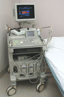
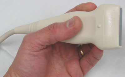
s who perform scans which are then typically interpreted by Radiologists, physicians who specialize in the application and interpretation of a wide variety of medical imaging modalities, or by Cardiologists in the case of cardiac ultrasonography (echocardiography). Sonographers typically use a hand-held probe (called a transducer) that is placed directly on and moved over the patient.
Sonography is effective for imaging soft tissues of the body. Superficial structures such as muscle
s, tendon
s, testes, breast
and the neonatal brain are imaged at a higher frequency
(7–18 MHz), which provides better axial and lateral resolution
. Deeper structures such as liver and kidney are imaged at a lower frequency 1–6 MHz with lower axial and lateral resolution but greater penetration.
Medical sonography is used in the study of many different systems:
Other types of uses include:
A general-purpose sonographic machine may be used for most imaging purposes. Usually specialty applications may be served only by use of a specialty transducer. Most ultrasound procedures are done using a transducer on the surface of the body, but improved diagnostic confidence is often possible if a transducer can be placed inside the body. For this purpose, specialty transducers, including endovaginal, endorectal, and transesophageal transducers are commonly employed. At the extreme of this, very small transducers can be mounted on small diameter catheters and placed into blood vessels to image the walls and disease of those vessels.
, and interpreting those echoes.
 A sound wave is typically produced by a piezoelectric transducer
encased in a housing which can take a number of forms. Strong, short electrical pulses from the ultrasound machine make the transducer ring at the desired frequency. The frequencies can be anywhere between 2 and 18 MHz. The sound is focused either by the shape of the transducer, a lens in front of the transducer, or a complex set of control pulses from the ultrasound scanner machine (Beamforming
). This focusing produces an arc-shaped sound wave from the face of the transducer. The wave travels into the body and comes into focus at a desired depth.
Older technology transducers focus their beam with physical lenses. Newer technology transducers use phased array
techniques to enable the sonographic machine to change the direction and depth of focus. Almost all piezoelectric transducers are made of ceramic
.
Materials on the face of the transducer enable the sound to be transmitted efficiently into the body (usually seeming to be a rubbery coating, a form of impedance matching
). In addition, a water-based gel is placed between the patient's skin and the probe.
The sound wave is partially reflected from the layers between different tissues. Specifically, sound is reflected anywhere there are density changes in the body: e.g. blood cells in blood plasma, small structures in organs, etc. Some of the reflections return to the transducer.
Once the ultrasonic scanner determines these three things, it can locate which pixel in the image to light up and to what intensity and at what hue
if frequency is processed (see redshift
for a natural mapping to hue).
Transforming the received signal into a digital image may be explained by using a blank spreadsheet as an analogy. First picture a long, flat transducer at the top of the sheet. Send pulses down the 'columns' of the spreadsheet (A, B, C, etc.). Listen at each column for any return echoes. When an echo is heard, note how long it took for the echo to return. The longer the wait, the deeper the row (1,2,3, etc.). The strength of the echo determines the brightness setting for that cell (white for a strong echo, black for a weak echo, and varying shades of grey for everything in between.) When all the echoes are recorded on the sheet, we have a greyscale image.
to capture and digitize the analog video signal. The captured signal can then be post-processed on the computer itself.
For computational details see also: Confocal laser scanning microscopy
, Radar
,
 Ultrasonography (sonography) uses a probe containing multiple acoustic transducer
s to send pulses of sound into a material. Whenever a sound wave encounters a material with a different density (acoustical impedance), part of the sound wave is reflected back to the probe and is detected as an echo. The time it takes for the echo
to travel back to the probe is measured and used to calculate the depth of the tissue interface causing the echo. The greater the difference between acoustic impedances, the larger the echo is. If the pulse hits gases or solids, the density difference is so great that most of the acoustic energy is reflected and it becomes impossible to see deeper.
The frequencies used for medical imaging are generally in the range of 1 to 18 MHz. Higher frequencies have a correspondingly smaller wavelength, and can be used to make sonograms with smaller details. However, the attenuation of the sound wave is increased at higher frequencies, so in order to have better penetration of deeper tissues, a lower frequency (3–5 MHz) is used.
Seeing deep into the body with sonography is very difficult. Some acoustic energy is lost every time an echo is formed, but most of it (approximately ) is lost from acoustic absorption.
The speed of sound varies as it travels through different materials, and is dependent on the acoustical impedance
of the material. However, the sonographic instrument assumes that the acoustic velocity is constant at 1540 m/s. An effect of this assumption is that in a real body with non-uniform tissues, the beam becomes somewhat de-focused and image resolution is reduced.
To generate a 2D-image, the ultrasonic beam is swept. A transducer may be swept mechanically by rotating or swinging. Or a 1D phased array
transducer may be use to sweep the beam electronically. The received data is processed and used to construct the image. The image is then a 2D representation of the slice into the body.
3D
images can be generated by acquiring a series of adjacent 2D images. Commonly a specialised probe that mechanically scans a conventional 2D-image transducer is used. However, since the mechanical scanning is slow, it is difficult to make 3D images of moving tissues. Recently, 2D phased array transducers that can sweep the beam in 3D have been developed. These can image faster and can even be used to make live 3D images of a beating heart.
Doppler
ultrasonography is used to study blood flow and muscle motion. The different detected speeds are represented in color for ease of interpretation, for example leaky heart valves: the leak shows up as a flash of unique color. Colors may alternatively be used to represent the amplitudes of the received echoes.
, many 2D planes are digitally added together to create a 3-dimensional image of the object. In contrast-enhanced ultrasound, microbubble contrast agents enhance the ultrasound waves, resulting in increased contrast.
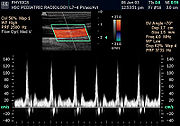
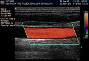
 Sonography can be enhanced with Doppler measurements, which employ the Doppler effect
to assess whether structures (usually blood) are moving towards or away from the probe, and its relative velocity. By calculating the frequency shift of a particular sample volume, for example flow in an artery or a jet of blood flow over a heart valve, its speed and direction can be determined and visualised. This is particularly useful in cardiovascular studies (sonography of the vascular system and heart) and essential in many areas such as determining reverse blood flow in the liver vasculature in portal hypertension
. The Doppler information is displayed graphically using spectral Doppler, or as an image using color Doppler (directional Doppler) or power Doppler (non directional Doppler). This Doppler shift falls in the audible range and is often presented audibly using stereo speakers: this produces a very distinctive, although synthetic, pulsating sound.
Most modern sonographic machines use pulsed Doppler to measure velocity. Pulsed wave machines transmit and receive series of pulses. The frequency shift of each pulse is ignored, however the relative phase changes of the pulses are used to obtain the frequency shift (since frequency is the rate of change of phase). The major advantages of pulsed Doppler over continuous wave is that distance information is obtained (the time between the transmitted and received pulses can be converted into a distance with knowledge of the speed of sound) and gain correction is applied. The disadvantage of pulsed Doppler is that the measurements can suffer from aliasing
. The terminology "Doppler ultrasound" or "Doppler sonography", has been accepted to apply to both pulsed and continuous Doppler systems despite the different mechanisms by which the velocity is measured.
It should be noted here that there are no standards for the display of color Doppler. Some laboratories show arteries as red and veins as blue, as medical illustrators usually show them, even though some vessels may have portions flowing towards and portions flowing away from the transducer. This results in the illogical appearance of a vessel being partly a vein and partly an artery. Other laboratories use red to indicate flow toward the transducer and blue away from the transducer. Still other laboratories prefer to display the sonographic Doppler color map more in accord with the prior published physics with the red shift
representing longer waves of echoes (scattered) from blood flowing away from the transducer; and with blue representing the shorter waves of echoes reflecting from blood flowing toward the transducer. Because of this confusion and lack of standards in the various laboratories, the sonographer must understand the underlying acoustic physics of color Doppler and the physiology of normal and abnormal blood flow in the human body (see Red shift
.
is known as contrast-enhanced ultrasound. This technique is currently used in echocardiography
, and may have future applications in molecular imaging and drug delivery.
and combines ultrasonography of the deep veins with venous compression. The technique can be used on deep veins of the upper and lower extremities, with some laboratories limiting the examination to the common femoral vein and the popliteal vein
, whereas other laboratories examine the deep veins from the inguinal region to the calf, including the calf veins.
Compression ultrasonography in B-mode has both high sensitivity and specificity
for detecting proximal deep vein thrombosis in symptomatic patients. The sensitivity lies somewhere between 90 to 100% for the diagnosis of symptomatic deep vein thrombosis, and the specificity ranges between 95% and 100%.
Diagnostic ultrasound studies of the fetus are generally considered to be safe during pregnancy. This diagnostic procedure should be performed only when there is a valid medical indication, and the lowest possible ultrasonic exposure setting should be used to gain the necessary diagnostic information under the "as low as reasonably achievable" or ALARA
principle.
World Health Organizations technical report series 875 (1998). supports that ultrasound is harmless:
"Diagnostic ultrasound is recognized as a safe, effective, and highly flexible imaging modality capable of providing clinically relevant information about most parts of the body in a rapid and cost-effective fashion". Although there is no evidence ultrasound could be harmful for the fetus, US Food and Drug Administration views promotion, selling, or leasing of ultrasound equipment for making "keepsake fetal videos" to be an unapproved use of a medical device.
Potential adverse ultrasound-related biological effects were recently reviewed and discussed on that journal's blog.
Currently New Mexico is the only state in the USA which regulates diagnostic medical sonographers. Certification examinations for sonographers are available in the US from three organizations: The American Registry of Diagnostic Medical Sonography,Cardiovascular Credentialing International and the American Registry of Radiological Technologists.
The primary regulated metrics are MI (Mechanical Index) a metric associated with the cavitation bio-effect, and TI (Thermal Index) a metric associated with the tissue heating bio-effect. The FDA requires that the machine not exceed limits that they have established. This requires self-regulation on the part of the manufacturer in terms of the calibration of the machine. The established limits are reasonably conservative so as to maintain diagnostic ultrasound as a safe imaging modality.
In India, lack of social security and consequent preference for a male child has popularized the use of ultrasound technology to identify and abort female fetuses. India's Antenatal (US: Prenatal) Diagnostic Techniques act makes use of ultrasound for sex selection illegal, but unscrupulous Indian doctors and would-be parents continue to discriminate against the girl child.
at the Naval Medical Research Institute, Bethesda, Maryland
in the late 1940s. English born and educated John Wild
(1914–2009) first used ultrasound to assess the thickness of bowel tissue as early as 1949: for his early work he has been described as the "father of medical ultrasound".
In 1962, after about two years of work, Joseph Holmes, William Wright, and Ralph Meyerdirk developed the first compound contact B-mode scanner. Their work had been supported by U.S. Public Health Services
and the University of Colorado
. Wright and Meyerdirk left the University to form Physionic Engineering Inc., which launched the first commercial hand-held articulated arm compound contact B-mode scanner in 1963. This was the start of the most popular design in the history of ultrasound scanners.
The first demonstration of color Doppler was by Geoff Stevenson, who was involved in the early developments and medical use of Doppler shifted ultrasonic energy.
by cardiologist
Inge Edler and Carl Hellmuth Hertz
, the son of Gustav Ludwig Hertz
, who was a graduate student at the department of nuclear physics
.
Edler had asked Hertz if it was possible to use radar
to look into the body, but Hertz said this was impossible. However, he said, it might be possible to use ultrasonography. Hertz was familiar with using ultrasonic
reflectoscopes for nondestructive materials testing
, and together they developed the idea of using this method in medicine.
The first successful measurement of heart activity was made on October 29, 1953 using a device borrowed from the ship construction company Kockums
in Malmö
. On December 16 the same year, the method was used to generate an echo-encephalogram (ultrasonic probe of the brain
). Edler and Hertz published their findings in 1954.
, Scotland
by Professor Ian Donald
and colleagues at the Glasgow Royal Maternity Hospital
(GRMH) led to the first diagnostic applications of the technique. Donald was an obstetrician
with a self-confessed "childish interest in machines, electronic and otherwise", who, having treated the wife of one of the company's directors, was invited to visit the Research Department of boilermakers Babcock & Wilcox
at Renfrew
, where he used their industrial ultrasound equipment to conduct experiments on various morbid anatomical specimens and assess their ultrasonic characteristics. Together with the medical physicist Tom Brown and fellow obstetrician Dr John MacVicar, Donald refined the equipment to enable differentiation of pathology in live volunteer patients. These findings were reported in The Lancet
on 7 June 1958 as "Investigation of Abdominal Masses by Pulsed Ultrasound" – possibly one of the most important papers ever published in the field of diagnostic medical imaging
.
At GRMH, Professor Donald and Dr James Willocks then refined their techniques to obstetric applications including fetal head measurement to assess the size and growth of the fetus. With the opening of the new Queen Mother's Hospital in Yorkhill
in 1964, it became possible to improve these methods even further. Dr Stuart Campbell
's pioneering work on
fetal cephalometry led to it acquiring long-term status as the definitive method of study of foetal growth. As the technical quality of the scans was further developed, it soon became possible to study pregnancy from start to finish and diagnose its many complications such as multiple pregnancy, fetal abnormality and placenta praevia
. Diagnostic ultrasound has since been imported into practically every other area of medicine.
Ultrasound
Ultrasound is cyclic sound pressure with a frequency greater than the upper limit of human hearing. Ultrasound is thus not separated from "normal" sound based on differences in physical properties, only the fact that humans cannot hear it. Although this limit varies from person to person, it is...
-based diagnostic imaging
Medical imaging
Medical imaging is the technique and process used to create images of the human body for clinical purposes or medical science...
technique used for visualizing subcutaneous body structures including tendons, muscles, joints, vessels and internal organs for possible pathology or lesion
Lesion
A lesion is any abnormality in the tissue of an organism , usually caused by disease or trauma. Lesion is derived from the Latin word laesio which means injury.- Types :...
s. Obstetric sonography
Obstetric ultrasonography
Obstetric sonography is the application of medical ultrasonography to obstetrics, in which sonography is used to visualize the embryo or foetus in its mother's uterus...
is commonly used during pregnancy
Pregnancy
Pregnancy refers to the fertilization and development of one or more offspring, known as a fetus or embryo, in a woman's uterus. In a pregnancy, there can be multiple gestations, as in the case of twins or triplets...
and is widely recognized by the public.
In physics, the term "ultrasound" applies to all sound
Sound
Sound is a mechanical wave that is an oscillation of pressure transmitted through a solid, liquid, or gas, composed of frequencies within the range of hearing and of a level sufficiently strong to be heard, or the sensation stimulated in organs of hearing by such vibrations.-Propagation of...
waves with a frequency above the audible range of human hearing, about 20 kHz. The frequencies used in diagnostic ultrasound are typically between 2 and 18 MHz.
Diagnostic applications
Sonography (ultrasonography) is widely used in medicine
Medicine
Medicine is the science and art of healing. It encompasses a variety of health care practices evolved to maintain and restore health by the prevention and treatment of illness....
. It is possible to perform both diagnosis
Medical diagnosis
Medical diagnosis refers both to the process of attempting to determine or identify a possible disease or disorder , and to the opinion reached by this process...
and therapeutic procedures, using ultrasound to guide interventional procedures (for instance biopsies
Biopsy
A biopsy is a medical test involving sampling of cells or tissues for examination. It is the medical removal of tissue from a living subject to determine the presence or extent of a disease. The tissue is generally examined under a microscope by a pathologist, and can also be analyzed chemically...
or drainage of fluid collections). Sonographer
Sonographer
Sonographers are diagnostic medical professionals who operate ultrasonic imaging devices to produce diagnostic images, scans, videos, or 3D volumes of anatomy and diagnostic data. Sonography requires specialized education and skills to view, analyze and modify the scan to optimize the information...
s are medical professional
Professional
A professional is a person who is paid to undertake a specialised set of tasks and to complete them for a fee. The traditional professions were doctors, lawyers, clergymen, and commissioned military officers. Today, the term is applied to estate agents, surveyors , environmental scientists,...
s who perform scans which are then typically interpreted by Radiologists, physicians who specialize in the application and interpretation of a wide variety of medical imaging modalities, or by Cardiologists in the case of cardiac ultrasonography (echocardiography). Sonographers typically use a hand-held probe (called a transducer) that is placed directly on and moved over the patient.
Sonography is effective for imaging soft tissues of the body. Superficial structures such as muscle
Muscle
Muscle is a contractile tissue of animals and is derived from the mesodermal layer of embryonic germ cells. Muscle cells contain contractile filaments that move past each other and change the size of the cell. They are classified as skeletal, cardiac, or smooth muscles. Their function is to...
s, tendon
Tendon
A tendon is a tough band of fibrous connective tissue that usually connects muscle to bone and is capable of withstanding tension. Tendons are similar to ligaments and fasciae as they are all made of collagen except that ligaments join one bone to another bone, and fasciae connect muscles to other...
s, testes, breast
Breast
The breast is the upper ventral region of the torso of a primate, in left and right sides, which in a female contains the mammary gland that secretes milk used to feed infants.Both men and women develop breasts from the same embryological tissues...
and the neonatal brain are imaged at a higher frequency
Frequency
Frequency is the number of occurrences of a repeating event per unit time. It is also referred to as temporal frequency.The period is the duration of one cycle in a repeating event, so the period is the reciprocal of the frequency...
(7–18 MHz), which provides better axial and lateral resolution
Angular resolution
Angular resolution, or spatial resolution, describes the ability of any image-forming device such as an optical or radio telescope, a microscope, a camera, or an eye, to distinguish small details of an object...
. Deeper structures such as liver and kidney are imaged at a lower frequency 1–6 MHz with lower axial and lateral resolution but greater penetration.
Medical sonography is used in the study of many different systems:
| System | Description | See also |
|---|---|---|
| Anesthesiology | Ultrasound is commonly used by anesthesiologists (Anaesthetists) to guide injecting needles when placing local anaesthetic solutions near nerves | |
| Cardiology Cardiology Cardiology is a medical specialty dealing with disorders of the heart . The field includes diagnosis and treatment of congenital heart defects, coronary artery disease, heart failure, valvular heart disease and electrophysiology... |
Echocardiography Echocardiography An echocardiogram, often referred to in the medical community as a cardiac ECHO or simply an ECHO, is a sonogram of the heart . Also known as a cardiac ultrasound, it uses standard ultrasound techniques to image two-dimensional slices of the heart... is an essential tool in cardiology, to diagnose e.g. dilatation of parts of the heart and function of heart ventricles and valves |
see echocardiography Echocardiography An echocardiogram, often referred to in the medical community as a cardiac ECHO or simply an ECHO, is a sonogram of the heart . Also known as a cardiac ultrasound, it uses standard ultrasound techniques to image two-dimensional slices of the heart... |
| Emergency Medicine Emergency medicine Emergency medicine is a medical specialty in which physicians care for patients with acute illnesses or injuries which require immediate medical attention. While not usually providing long-term or continuing care, emergency medicine physicians diagnose a variety of illnesses and undertake acute... |
Point of care ultrasound has many applications in the Emergency Department, including the Focused Assessment with Sonography for Trauma (FAST) exam Focused assessment with sonography for trauma Focused Assessment with Sonography for Trauma is a rapid bedside ultrasound examination performed by surgeons and emergency physicians as a screening test for blood around the heart or abdominal organs after trauma.The four areas that are examined for free fluid are the perihepatic space... for assessing significant hemoperitoneum Hemoperitoneum Hemoperitoneum is the presence of blood in the peritoneal cavity. The blood accumulates in the space between the inner lining of the abdominal wall and the internal abdominal organs. Hemoperitoneum is generally classified as a surgical emergency; in most cases, urgent laparotomy is needed to... or pericardial tamponade after trauma Physical trauma Trauma refers to "a body wound or shock produced by sudden physical injury, as from violence or accident." It can also be described as "a physical wound or injury, such as a fracture or blow." Major trauma can result in secondary complications such as circulatory shock, respiratory failure and death... . Ultrasound is routinely used in the Emergency Department to expedite the care of patients with right upper quadrant abdominal pain who may have gallstones or cholecystitis. |
see FAST exam Focused assessment with sonography for trauma Focused Assessment with Sonography for Trauma is a rapid bedside ultrasound examination performed by surgeons and emergency physicians as a screening test for blood around the heart or abdominal organs after trauma.The four areas that are examined for free fluid are the perihepatic space... |
| Gastroenterology Gastroenterology Gastroenterology is the branch of medicine whereby the digestive system and its disorders are studied. The name is a combination of three Ancient Greek words gaster , enteron , and logos... |
In abdominal sonography, the solid organs of the abdomen such as the pancreas Pancreas The pancreas is a gland organ in the digestive and endocrine system of vertebrates. It is both an endocrine gland producing several important hormones, including insulin, glucagon, and somatostatin, as well as a digestive organ, secreting pancreatic juice containing digestive enzymes that assist... , aorta Aorta The aorta is the largest artery in the body, originating from the left ventricle of the heart and extending down to the abdomen, where it branches off into two smaller arteries... , inferior vena cava Inferior vena cava The inferior vena cava , also known as the posterior vena cava, is the large vein that carries de-oxygenated blood from the lower half of the body into the right atrium of the heart.... , liver Liver The liver is a vital organ present in vertebrates and some other animals. It has a wide range of functions, including detoxification, protein synthesis, and production of biochemicals necessary for digestion... , gall bladder, bile duct Bile duct A bile duct is any of a number of long tube-like structures that carry bile.Bile, required for the digestion of food, is excreted by the liver into passages that carry bile toward the hepatic duct, which joins with the cystic duct to form the common bile duct, which opens into the intestine.The... s, kidney Kidney The kidneys, organs with several functions, serve essential regulatory roles in most animals, including vertebrates and some invertebrates. They are essential in the urinary system and also serve homeostatic functions such as the regulation of electrolytes, maintenance of acid–base balance, and... s, and spleen Spleen The spleen is an organ found in virtually all vertebrate animals with important roles in regard to red blood cells and the immune system. In humans, it is located in the left upper quadrant of the abdomen. It removes old red blood cells and holds a reserve of blood in case of hemorrhagic shock... are imaged. Sound waves are blocked by gas in the bowel and attenuated in different degree by fat, therefore there are limited diagnostic capabilities in this area. The appendix Vermiform appendix The appendix is a blind-ended tube connected to the cecum , from which it develops embryologically. The cecum is a pouchlike structure of the colon... can sometimes be seen when inflamed (as in e.g.: appendicitis Appendicitis Appendicitis is a condition characterized by inflammation of the appendix. It is classified as a medical emergency and many cases require removal of the inflamed appendix, either by laparotomy or laparoscopy. Untreated, mortality is high, mainly because of the risk of rupture leading to... ). |
|
| Gynecology | see gynecologic ultrasonography Gynecologic ultrasonography Gynecologic ultrasonography or Gynecologic sonography refers to the application of medical ultrasonography to the female pelvic organs, specifically the uterus, the ovaries, the Fallopian tubes, as well as the bladder, the adnexa, the Pouch of Douglas, and any findings in the pelvis of relevance... |
|
| Neonatology Neonatology Neonatology is a subspecialty of pediatrics that consists of the medical care of newborn infants, especially the ill or premature newborn infant. It is a hospital-based specialty, and is usually practiced in neonatal intensive care units... |
for basic assessment of intracerebral structural abnormalities, bleeds, ventriculomegaly Ventriculomegaly Ventriculomegaly is a brain condition that occurs when the lateral ventricles become dilated. The most common definition uses a width of the atrium of the lateral ventricle of greater than 10 mm. This occurs in around 1% of pregnancies. When this measurement is between 10 and 15 mm, the... or hydrocephalus Hydrocephalus Hydrocephalus , also known as "water in the brain," is a medical condition in which there is an abnormal accumulation of cerebrospinal fluid in the ventricles, or cavities, of the brain. This may cause increased intracranial pressure inside the skull and progressive enlargement of the head,... and anoxic insults (Periventricular leukomalacia Periventricular leukomalacia Periventricular leukomalacia is a form of white-matter brain injury, characterized by the necrosis of white matter near the lateral ventricles. It can affect newborns and fetuses; premature infants are at the greatest risk of the disorder... ). The ultrasound can be performed through the soft spots in the skull of a newborn infant (Fontanelle Fontanelle A fontanelle is an anatomical feature on an infant's skull.-Anatomy:Fontanelles are soft spots on a baby's head which, during birth, enable the bony plates of the skull to flex, allowing the child's head to pass through the birth canal. The ossification of the bones of the skull causes the... ) until these completely close at about 1 year of age and form a virtually impenetrable acoustic barrier for the ultrasound. The most common site for cranial ultrasound is the anterior fontanelle. The smaller the fontanelle, the poorer the quality of the picture. |
Intracerebral: see Transcranial Doppler Transcranial doppler Transcranial Doppler is a test that measures the velocity of blood flow through the brain's blood vessels. Used to help in the diagnosis of emboli, stenosis, vasospasm from a subarachnoid hemorrhage , and other problems, this relatively quick and inexpensive test is growing in popularity in the... |
| Neurology Neurology Neurology is a medical specialty dealing with disorders of the nervous system. Specifically, it deals with the diagnosis and treatment of all categories of disease involving the central, peripheral, and autonomic nervous systems, including their coverings, blood vessels, and all effector tissue,... |
for assessing blood flow and stenoses in the carotid arteries (Carotid ultrasonography Carotid ultrasonography Carotid ultrasonography is an ultrasound-based diagnostic imaging technique to reveal structural details of the carotid arteries, so as to look for blood clots, atherosclerotic plaque buildup, and other blood flow problems.... ) and the big intracerebral arteries |
see Carotid ultrasonography Carotid ultrasonography Carotid ultrasonography is an ultrasound-based diagnostic imaging technique to reveal structural details of the carotid arteries, so as to look for blood clots, atherosclerotic plaque buildup, and other blood flow problems.... . Intracerebral: see Transcranial Doppler Transcranial doppler Transcranial Doppler is a test that measures the velocity of blood flow through the brain's blood vessels. Used to help in the diagnosis of emboli, stenosis, vasospasm from a subarachnoid hemorrhage , and other problems, this relatively quick and inexpensive test is growing in popularity in the... |
| Obstetrics Obstetrics Obstetrics is the medical specialty dealing with the care of all women's reproductive tracts and their children during pregnancy , childbirth and the postnatal period... |
Obstetrical sonography is commonly used during pregnancy Pregnancy Pregnancy refers to the fertilization and development of one or more offspring, known as a fetus or embryo, in a woman's uterus. In a pregnancy, there can be multiple gestations, as in the case of twins or triplets... to check on the development of the fetus Fetus A fetus is a developing mammal or other viviparous vertebrate after the embryonic stage and before birth.In humans, the fetal stage of prenatal development starts at the beginning of the 11th week in gestational age, which is the 9th week after fertilization.-Etymology and spelling variations:The... . |
see obstetric ultrasonography Obstetric ultrasonography Obstetric sonography is the application of medical ultrasonography to obstetrics, in which sonography is used to visualize the embryo or foetus in its mother's uterus... |
| Ophthalmology Ophthalmology Ophthalmology is the branch of medicine that deals with the anatomy, physiology and diseases of the eye. An ophthalmologist is a specialist in medical and surgical eye problems... |
see A-scan ultrasonography A-scan ultrasound biometry A-scan is an amplitude modulation scan. It gives the information in the form of one dimensional. it is used to detect the presence of flaws in the materials.... , B-scan ultrasonography B-scan ultrasonography B-scan ultrasonography, or B-scan, is a diagnostic test used in ophthalmology to produce a two-dimensional, cross-sectional view of the eye and the orbit.It is otherwise called brightness scan.-External links:*... |
|
| Urology Urology Urology is the medical and surgical specialty that focuses on the urinary tracts of males and females, and on the reproductive system of males. Medical professionals specializing in the field of urology are called urologists and are trained to diagnose, treat, and manage patients with urological... |
to determine, for example, the amount of fluid retained in a patient's bladder. In a pelvic sonogram, organs of the pelvic region are imaged. This includes the uterus Uterus The uterus or womb is a major female hormone-responsive reproductive sex organ of most mammals including humans. One end, the cervix, opens into the vagina, while the other is connected to one or both fallopian tubes, depending on the species... and ovaries Ovary The ovary is an ovum-producing reproductive organ, often found in pairs as part of the vertebrate female reproductive system. Ovaries in anatomically female individuals are analogous to testes in anatomically male individuals, in that they are both gonads and endocrine glands.-Human anatomy:Ovaries... or urinary bladder Urinary bladder The urinary bladder is the organ that collects urine excreted by the kidneys before disposal by urination. A hollow muscular, and distensible organ, the bladder sits on the pelvic floor... . Males are sometimes given a pelvic sonogram to check on the health of their bladder, the prostate Prostate The prostate is a compound tubuloalveolar exocrine gland of the male reproductive system in most mammals.... , or their testicles (for example to distinguish epididymitis Epididymitis Epididymitis is a medical condition in which there is inflammation of the epididymis . This condition comprises gradual onset of testicular pain that can vary from mild to severe, and the scrotum may become red, warm and swollen... from testicular torsion Testicular torsion Testicular torsion is when the spermatic cord to a testicle twists, cutting off the blood supply. The most common symptom is acute testicular pain and the most common underlying cause is a congenital malformation known as a "bell-clapper deformity". The diagnosis is often made clinically but if it... ). In young males, it is used to distinguish more benign testicular masses (varicocele Varicocele Varicocele , also known as varicoscele or varicose seal, is an abnormal enlargement of the vein that is in the scrotum draining the testicles. The testicular blood vessels originate in the abdomen and course down through the inguinal canal as part of the spermatic cord on their way to the testis... or hydrocele Hydrocele A hydrocoele denotes a pathological accumulation of serous fluid in a body cavity. It can also be noted as a minor malformation of newborns due to high levels of lead in the mother's blood during pregnancy.... ) from testicular cancer Testicular cancer Testicular cancer is cancer that develops in the testicles, a part of the male reproductive system.In the United States, between 7,500 and 8,000 diagnoses of testicular cancer are made each year. In the UK, approximately 2,000 men are diagnosed each year. Over his lifetime, a man's risk of... , which is still very highly curable but which must be treated to preserve health and fertility. There are two methods of performing a pelvic sonography – externally or internally. The internal pelvic sonogram is performed either transvagina Vagina The vagina is a fibromuscular tubular tract leading from the uterus to the exterior of the body in female placental mammals and marsupials, or to the cloaca in female birds, monotremes, and some reptiles. Female insects and other invertebrates also have a vagina, which is the terminal part of the... lly (in a woman) or transrectally (in a man). Sonographic imaging of the pelvic floor can produce important diagnostic information regarding the precise relationship of abnormal structures with other pelvic organs and it represents a useful hint to treat patients with symptoms related to pelvic prolapse, double incontinence and obstructed defecation. It is used to diagnose and, at higher frequencies, to treat (break up) kidney stones or kidney crystals (nephrolithiasis). |
|
| Musculoskeletal | tendons, muscles, nerves, ligaments, soft tissue masses, and bone surfaces | |
| Cardiovascular system | To assess patency and possible obstruction of arteries Arterial sonography, diagnose DVT (Thrombosonography) and determine extent and severity of venous insufficiency (venosonography) | Intravascular ultrasound Intravascular ultrasound Intravascular ultrasound is a medical imaging methodology using a specially designed catheter with a miniaturized ultrasound probe attached to the distal end of the catheter. The proximal end of the catheter is attached to computerized ultrasound equipment... |
Other types of uses include:
- Interventional ultrasonography; biopsyBiopsyA biopsy is a medical test involving sampling of cells or tissues for examination. It is the medical removal of tissue from a living subject to determine the presence or extent of a disease. The tissue is generally examined under a microscope by a pathologist, and can also be analyzed chemically...
, emptying fluids, intrauterine (Hemolytic disease of the newbornHemolytic disease of the newbornHemolytic disease of the newborn, also known as hemolytic disease of the fetus and newborn, HDN, HDFN, or erythroblastosis fetalis, is an alloimmune condition that develops in a fetus, when the IgG molecules produced by the mother pass through the placenta...
) - Contrast-enhanced ultrasound
A general-purpose sonographic machine may be used for most imaging purposes. Usually specialty applications may be served only by use of a specialty transducer. Most ultrasound procedures are done using a transducer on the surface of the body, but improved diagnostic confidence is often possible if a transducer can be placed inside the body. For this purpose, specialty transducers, including endovaginal, endorectal, and transesophageal transducers are commonly employed. At the extreme of this, very small transducers can be mounted on small diameter catheters and placed into blood vessels to image the walls and disease of those vessels.
Therapeutic applications
Therapeutic applications use ultrasound to bring heat or agitation into the body. Therefore much higher energies are used than in diagnostic ultrasound. In many cases the range of frequencies used are also very different.- Ultrasound is sometimes used to clean teeth in dental hygieneDental hygienistthumb|right|300px|Dental hygienist holding a scalerA dental hygienist is a licensed dental professional who specializes in preventive oral health, typically focusing on techniques in oral hygiene. Local dental regulations determine the scope of practice of dental hygienists...
. - Ultrasound sources may be used to generate regional heating and mechanical changes in biological tissue, e.g. in occupational therapyOccupational therapyOccupational therapy is a discipline that aims to promote health by enabling people to perform meaningful and purposeful activities. Occupational therapists work with individuals who suffer from a mentally, physically, developmentally, and/or emotionally disabling condition by utilizing treatments...
, physical therapyPhysical therapyPhysical therapy , often abbreviated PT, is a health care profession. Physical therapy is concerned with identifying and maximizing quality of life and movement potential within the spheres of promotion, prevention, diagnosis, treatment/intervention,and rehabilitation...
and cancer treatmentExperimental cancer treatmentExperimental cancer treatments are medical therapies intended or claimed to treat cancer by improving on, supplementing or replacing conventional methods ....
. However the use of ultrasound in the treatment of musculoskeletal conditions has fallen out of favor. - Focused ultrasound may be used to generate highly localized heating to treat cysts and tumors (benign or malignant), This is known as Focused Ultrasound Surgery (FUS) or High Intensity Focused Ultrasound (HIFU). These procedures generally use lower frequencies than medical diagnostic ultrasound (from 0.250 to 2 MHz), but significantly higher energies. HIFU treatment is often guided by MRI.
- Focused ultrasound may be used to break up kidney stoneKidney stoneA kidney stone, also known as a renal calculus is a solid concretion or crystal aggregation formed in the kidneys from dietary minerals in the urine...
s by lithotripsy. - Ultrasound may be used for cataractCataractA cataract is a clouding that develops in the crystalline lens of the eye or in its envelope, varying in degree from slight to complete opacity and obstructing the passage of light...
treatment by phacoemulsificationPhacoemulsificationPhacoemulsification refers to modern cataract surgery in which the eye's internal lens is emulsified with an ultrasonic handpiece and aspirated from the eye...
. - Additional physiological effects of low-intensity ultrasoundLow intensity pulsed ultrasoundLow intensity pulsed ultrasound is a medical technology, generally utilizing 1.5 MHz frequency pulses, with a pulse width of 200 μs, repeated at 1 kHz, at an intensity of 30 mW/cm2, 20 minutes/day.Applications of LIPUS include:...
have recently been discovered, e.g. its ability to stimulate bone-growth and its potential to disrupt the blood-brain barrierBlood-brain barrierThe blood–brain barrier is a separation of circulating blood and the brain extracellular fluid in the central nervous system . It occurs along all capillaries and consists of tight junctions around the capillaries that do not exist in normal circulation. Endothelial cells restrict the diffusion...
for drug delivery. - Procoagulant at 5–12 MHz,
From sound to image
The creation of an image from sound is done in three steps – producing a sound wave, receiving echoesEcho (phenomenon)
In audio signal processing and acoustics, an echo is a reflection of sound, arriving at the listener some time after the direct sound. Typical examples are the echo produced by the bottom of a well, by a building, or by the walls of an enclosed room and an empty room. A true echo is a single...
, and interpreting those echoes.
Producing a sound wave
Ultrasonic sensor
Ultrasonic sensors work on a principle similar to radar or sonar which evaluate attributes of a target by interpreting the echoes from radio or sound waves respectively. Ultrasonic sensors generate high frequency sound waves and evaluate the echo which is received back by the sensor...
encased in a housing which can take a number of forms. Strong, short electrical pulses from the ultrasound machine make the transducer ring at the desired frequency. The frequencies can be anywhere between 2 and 18 MHz. The sound is focused either by the shape of the transducer, a lens in front of the transducer, or a complex set of control pulses from the ultrasound scanner machine (Beamforming
Beamforming
Beamforming is a signal processing technique used in sensor arrays for directional signal transmission or reception. This is achieved by combining elements in the array in a way where signals at particular angles experience constructive interference and while others experience destructive...
). This focusing produces an arc-shaped sound wave from the face of the transducer. The wave travels into the body and comes into focus at a desired depth.
Older technology transducers focus their beam with physical lenses. Newer technology transducers use phased array
Phased array
In wave theory, a phased array is an array of antennas in which the relative phases of the respective signals feeding the antennas are varied in such a way that the effective radiation pattern of the array is reinforced in a desired direction and suppressed in undesired directions.An antenna array...
techniques to enable the sonographic machine to change the direction and depth of focus. Almost all piezoelectric transducers are made of ceramic
Ceramic
A ceramic is an inorganic, nonmetallic solid prepared by the action of heat and subsequent cooling. Ceramic materials may have a crystalline or partly crystalline structure, or may be amorphous...
.
Materials on the face of the transducer enable the sound to be transmitted efficiently into the body (usually seeming to be a rubbery coating, a form of impedance matching
Impedance matching
In electronics, impedance matching is the practice of designing the input impedance of an electrical load to maximize the power transfer and/or minimize reflections from the load....
). In addition, a water-based gel is placed between the patient's skin and the probe.
The sound wave is partially reflected from the layers between different tissues. Specifically, sound is reflected anywhere there are density changes in the body: e.g. blood cells in blood plasma, small structures in organs, etc. Some of the reflections return to the transducer.
Receiving the echoes
The return of the sound wave to the transducer results in the same process that it took to send the sound wave, except in reverse. The return sound wave vibrates the transducer, the transducer turns the vibrations into electrical pulses that travel to the ultrasonic scanner where they are processed and transformed into a digital image.Forming the image
The sonographic scanner must determine three things from each received echo:- How long it took the echo to be received from when the sound was transmitted.
- From this the focal length for the phased array is deduced, enabling a sharp image of that echo at that depth (this is not possible while producing a sound wave).
- How strong the echo was. It could be noted that sound wave is not a click, but a pulse with a specific carrier frequency. Moving objects change this frequency on reflection, so that it is only a matter of electronics to have simultaneous Doppler sonography.
Once the ultrasonic scanner determines these three things, it can locate which pixel in the image to light up and to what intensity and at what hue
Hue
Hue is one of the main properties of a color, defined technically , as "the degree to which a stimulus can be describedas similar to or different from stimuli that are described as red, green, blue, and yellow,"...
if frequency is processed (see redshift
Redshift
In physics , redshift happens when light seen coming from an object is proportionally increased in wavelength, or shifted to the red end of the spectrum...
for a natural mapping to hue).
Transforming the received signal into a digital image may be explained by using a blank spreadsheet as an analogy. First picture a long, flat transducer at the top of the sheet. Send pulses down the 'columns' of the spreadsheet (A, B, C, etc.). Listen at each column for any return echoes. When an echo is heard, note how long it took for the echo to return. The longer the wait, the deeper the row (1,2,3, etc.). The strength of the echo determines the brightness setting for that cell (white for a strong echo, black for a weak echo, and varying shades of grey for everything in between.) When all the echoes are recorded on the sheet, we have a greyscale image.
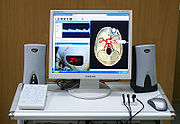
Displaying the image
Images from the sonographic scanner can be displayed, captured, and broadcast through a computer using a frame grabberFrame grabber
A frame grabber is an electronic device that captures individual, digital still frames from an analog video signal or a digital video stream. It is usually employed as a component of a computer vision system, in which video frames are captured in digital form and then displayed, stored or...
to capture and digitize the analog video signal. The captured signal can then be post-processed on the computer itself.
For computational details see also: Confocal laser scanning microscopy
Confocal laser scanning microscopy
Confocal laser scanning microscopy is a technique for obtaining high-resolution optical images with depth selectivity. The key feature of confocal microscopy is its ability to acquire in-focus images from selected depths, a process known as optical sectioning...
, Radar
Radar
Radar is an object-detection system which uses radio waves to determine the range, altitude, direction, or speed of objects. It can be used to detect aircraft, ships, spacecraft, guided missiles, motor vehicles, weather formations, and terrain. The radar dish or antenna transmits pulses of radio...
,
Sound in the body
Transducer
A transducer is a device that converts one type of energy to another. Energy types include electrical, mechanical, electromagnetic , chemical, acoustic or thermal energy. While the term transducer commonly implies the use of a sensor/detector, any device which converts energy can be considered a...
s to send pulses of sound into a material. Whenever a sound wave encounters a material with a different density (acoustical impedance), part of the sound wave is reflected back to the probe and is detected as an echo. The time it takes for the echo
Echo (phenomenon)
In audio signal processing and acoustics, an echo is a reflection of sound, arriving at the listener some time after the direct sound. Typical examples are the echo produced by the bottom of a well, by a building, or by the walls of an enclosed room and an empty room. A true echo is a single...
to travel back to the probe is measured and used to calculate the depth of the tissue interface causing the echo. The greater the difference between acoustic impedances, the larger the echo is. If the pulse hits gases or solids, the density difference is so great that most of the acoustic energy is reflected and it becomes impossible to see deeper.
The frequencies used for medical imaging are generally in the range of 1 to 18 MHz. Higher frequencies have a correspondingly smaller wavelength, and can be used to make sonograms with smaller details. However, the attenuation of the sound wave is increased at higher frequencies, so in order to have better penetration of deeper tissues, a lower frequency (3–5 MHz) is used.
Seeing deep into the body with sonography is very difficult. Some acoustic energy is lost every time an echo is formed, but most of it (approximately
) is lost from acoustic absorption.The speed of sound varies as it travels through different materials, and is dependent on the acoustical impedance
Acoustic impedance
The acoustic impedance at a particular frequency indicates how much sound pressure is generated by a given air vibration at that frequency. The acoustic impedance Z is frequency dependent and is very useful, for example, for describing the behaviour of musical wind instruments...
of the material. However, the sonographic instrument assumes that the acoustic velocity is constant at 1540 m/s. An effect of this assumption is that in a real body with non-uniform tissues, the beam becomes somewhat de-focused and image resolution is reduced.
To generate a 2D-image, the ultrasonic beam is swept. A transducer may be swept mechanically by rotating or swinging. Or a 1D phased array
Phased array
In wave theory, a phased array is an array of antennas in which the relative phases of the respective signals feeding the antennas are varied in such a way that the effective radiation pattern of the array is reinforced in a desired direction and suppressed in undesired directions.An antenna array...
transducer may be use to sweep the beam electronically. The received data is processed and used to construct the image. The image is then a 2D representation of the slice into the body.
3D
3D computer graphics
3D computer graphics are graphics that use a three-dimensional representation of geometric data that is stored in the computer for the purposes of performing calculations and rendering 2D images...
images can be generated by acquiring a series of adjacent 2D images. Commonly a specialised probe that mechanically scans a conventional 2D-image transducer is used. However, since the mechanical scanning is slow, it is difficult to make 3D images of moving tissues. Recently, 2D phased array transducers that can sweep the beam in 3D have been developed. These can image faster and can even be used to make live 3D images of a beating heart.
Doppler
Doppler effect
The Doppler effect , named after Austrian physicist Christian Doppler who proposed it in 1842 in Prague, is the change in frequency of a wave for an observer moving relative to the source of the wave. It is commonly heard when a vehicle sounding a siren or horn approaches, passes, and recedes from...
ultrasonography is used to study blood flow and muscle motion. The different detected speeds are represented in color for ease of interpretation, for example leaky heart valves: the leak shows up as a flash of unique color. Colors may alternatively be used to represent the amplitudes of the received echoes.
Modes of sonography
Several different modes of ultrasound are used in medical imaging. These are:- A-mode: A-mode is the simplest type of ultrasound. A single transducer scans a line through the body with the echoes plotted on screen as a function of depth. Therapeutic ultrasound aimed at a specific tumor or calculus is also A-mode, to allow for pinpoint accurate focus of the destructive wave energy.
- B-mode: In B-mode ultrasound, a linear array of transducers simultaneously scans a plane through the body that can be viewed as a two-dimensional image on screen.
- C-mode: A C-mode image is formed in a plane normal to a B-mode image. A gate that selects data from a specific depth from an A-mode line is used; then the transducer is moved in the 2D plane to sample the entire region at this fixed depth. When the transducer traverses the area in a spiral, an area of 100 cm2 can be scanned in around 10 seconds.
- M-mode: M stands for motion. Ultrasound pulses are emitted in quick succession – each time, either an A-mode or B-mode image is taken. Over time, this is analogous to recording a videoVideoVideo is the technology of electronically capturing, recording, processing, storing, transmitting, and reconstructing a sequence of still images representing scenes in motion.- History :...
in ultrasound. As the organ boundaries that produce reflections move relative to the probe, this can be used to determine the velocity of specific organ structures. - Doppler mode: This mode makes use of the Doppler effect in measuring and visualizing blood flow
- Color Doppler: Velocity information is presented as a color coded overlay on top of a B-mode image
- Continuous Doppler: Doppler information is sampled along a line through the body, and all velocities detected at each time point is presented (on a time line)
- Pulsed wave (PW) Doppler: Doppler information is sampled from only a small sample volume (defined in 2D image), and presented on a timeline
- Duplex: a common name for the simultaneous presentation of 2D and (usually) PW Doppler information. (Using modern ultrasound machines color Doppler is almost always also used, hence the alternative name Triplex.)
- Pulse inversion mode: In this mode two successive pulses with opposite sign are emitted and then subtracted from each other. This implies that any linearly responding constituent will disappear while gases with non-linear compressibility stands out.
- Harmonic mode: In this mode a deep penetrating fundamental frequency is emitted into the body and a harmonic overtone is detected. In this way depth penetration can be gained with improved lateral resolution.
Expansions
An additional expansion or additional technique of ultrasound is biplanar ultrasound, in which the probe has two 2D planes that are perpendicular to each other, providing more efficient localization and detection. Furthermore, an omniplane probe is one that can rotate 180° to obtain multiple images. In 3D ultrasound3D ultrasound
3D ultrasound is a medical ultrasound technique, often used in obstetric ultrasonography , providing three dimensional images of the fetus....
, many 2D planes are digitally added together to create a 3-dimensional image of the object. In contrast-enhanced ultrasound, microbubble contrast agents enhance the ultrasound waves, resulting in increased contrast.
Doppler sonography
Doppler effect
The Doppler effect , named after Austrian physicist Christian Doppler who proposed it in 1842 in Prague, is the change in frequency of a wave for an observer moving relative to the source of the wave. It is commonly heard when a vehicle sounding a siren or horn approaches, passes, and recedes from...
to assess whether structures (usually blood) are moving towards or away from the probe, and its relative velocity. By calculating the frequency shift of a particular sample volume, for example flow in an artery or a jet of blood flow over a heart valve, its speed and direction can be determined and visualised. This is particularly useful in cardiovascular studies (sonography of the vascular system and heart) and essential in many areas such as determining reverse blood flow in the liver vasculature in portal hypertension
Portal hypertension
In medicine, portal hypertension is hypertension in the portal vein and its tributaries.It is often defined as a portal pressure gradient of 10 mmHg or greater.-Causes:Causes can be divided into prehepatic, intrahepatic, and posthepatic...
. The Doppler information is displayed graphically using spectral Doppler, or as an image using color Doppler (directional Doppler) or power Doppler (non directional Doppler). This Doppler shift falls in the audible range and is often presented audibly using stereo speakers: this produces a very distinctive, although synthetic, pulsating sound.
Most modern sonographic machines use pulsed Doppler to measure velocity. Pulsed wave machines transmit and receive series of pulses. The frequency shift of each pulse is ignored, however the relative phase changes of the pulses are used to obtain the frequency shift (since frequency is the rate of change of phase). The major advantages of pulsed Doppler over continuous wave is that distance information is obtained (the time between the transmitted and received pulses can be converted into a distance with knowledge of the speed of sound) and gain correction is applied. The disadvantage of pulsed Doppler is that the measurements can suffer from aliasing
Aliasing
In signal processing and related disciplines, aliasing refers to an effect that causes different signals to become indistinguishable when sampled...
. The terminology "Doppler ultrasound" or "Doppler sonography", has been accepted to apply to both pulsed and continuous Doppler systems despite the different mechanisms by which the velocity is measured.
It should be noted here that there are no standards for the display of color Doppler. Some laboratories show arteries as red and veins as blue, as medical illustrators usually show them, even though some vessels may have portions flowing towards and portions flowing away from the transducer. This results in the illogical appearance of a vessel being partly a vein and partly an artery. Other laboratories use red to indicate flow toward the transducer and blue away from the transducer. Still other laboratories prefer to display the sonographic Doppler color map more in accord with the prior published physics with the red shift
Red shift
-Science:* Redshift, the increase of wavelength of detected electromagnetic radiation with respect to the original wavelength of the emission* Red shift, an informal term for a bathochromic shift...
representing longer waves of echoes (scattered) from blood flowing away from the transducer; and with blue representing the shorter waves of echoes reflecting from blood flowing toward the transducer. Because of this confusion and lack of standards in the various laboratories, the sonographer must understand the underlying acoustic physics of color Doppler and the physiology of normal and abnormal blood flow in the human body (see Red shift
Red shift
-Science:* Redshift, the increase of wavelength of detected electromagnetic radiation with respect to the original wavelength of the emission* Red shift, an informal term for a bathochromic shift...
.
Contrast media
The use of microbubble contrast media in medical sonography to improve ultrasound signal backscatterBackscatter
In physics, backscatter is the reflection of waves, particles, or signals back to the direction they came from. It is a diffuse reflection due to scattering, as opposed to specular reflection like a mirror...
is known as contrast-enhanced ultrasound. This technique is currently used in echocardiography
Echocardiography
An echocardiogram, often referred to in the medical community as a cardiac ECHO or simply an ECHO, is a sonogram of the heart . Also known as a cardiac ultrasound, it uses standard ultrasound techniques to image two-dimensional slices of the heart...
, and may have future applications in molecular imaging and drug delivery.
Compression ultrasonography
Compression ultrasonography is a technique used for diagnosing deep vein thrombosisDeep vein thrombosis
Deep vein thrombosis is the formation of a blood clot in a deep vein. Deep vein thrombosis commonly affects the leg veins or the deep veins of the pelvis. Occasionally the veins of the arm are affected...
and combines ultrasonography of the deep veins with venous compression. The technique can be used on deep veins of the upper and lower extremities, with some laboratories limiting the examination to the common femoral vein and the popliteal vein
Popliteal vein
The popliteal vein course runs alongside the popliteal artery but carries the blood from the knee joint and muscles in the thigh and calf back to the heart.Its origin is defined by the junction of the posterior tibial vein and anterior tibial vein....
, whereas other laboratories examine the deep veins from the inguinal region to the calf, including the calf veins.
Compression ultrasonography in B-mode has both high sensitivity and specificity
Sensitivity and specificity
Sensitivity and specificity are statistical measures of the performance of a binary classification test, also known in statistics as classification function. Sensitivity measures the proportion of actual positives which are correctly identified as such Sensitivity and specificity are statistical...
for detecting proximal deep vein thrombosis in symptomatic patients. The sensitivity lies somewhere between 90 to 100% for the diagnosis of symptomatic deep vein thrombosis, and the specificity ranges between 95% and 100%.
Attributes
As with all imaging modalities, ultrasonography has its list of positive and negative attributes.Strengths
- It images muscleMuscleMuscle is a contractile tissue of animals and is derived from the mesodermal layer of embryonic germ cells. Muscle cells contain contractile filaments that move past each other and change the size of the cell. They are classified as skeletal, cardiac, or smooth muscles. Their function is to...
, soft tissueSoft tissueIn anatomy, the term soft tissue refers to tissues that connect, support, or surround other structures and organs of the body, not being bone. Soft tissue includes tendons, ligaments, fascia, skin, fibrous tissues, fat, and synovial membranes , and muscles, nerves and blood vessels .It is sometimes...
, and bone surfaces very well and is particularly useful for delineating the interfaces between solid and fluid-filled spaces. - It renders "live" images, where the operator can dynamically select the most useful section for diagnosing and documenting changes, often enabling rapid diagnoses. Live images also allow for ultrasound-guided biopsies or injections, which can be cumbersome with other imaging modalities.
- It shows the structure of organs.
- It has no known long-term side effects and rarely causes any discomfort to the patient.
- Equipment is widely available and comparatively flexible.
- Small, easily carried scanners are available; examinations can be performed at the bedside.
- Relatively inexpensive compared to other modes of investigation, such as computed X-ray tomography, DEXADual energy X-ray absorptiometryDual-emission X-ray absorptiometry is a means of measuring bone mineral density . Two X-ray beams with differing energy levels are aimed at the patient's bones. When soft tissue absorption is subtracted out, the BMD can be determined from the absorption of each beam by bone...
or magnetic resonance imagingMagnetic resonance imagingMagnetic resonance imaging , nuclear magnetic resonance imaging , or magnetic resonance tomography is a medical imaging technique used in radiology to visualize detailed internal structures...
. - Spatial resolution is better in high frequency ultrasound transducers than it is in most other imaging modalities.
- Through the use of an Ultrasound research interfaceUltrasound Research InterfaceAn ultrasound research interface is a software tool loaded onto a diagnostic clinical ultrasound device which provides functionality beyond typical clinical modes of operation. Before an ultrasound image can be displayed to the user, it must undergo a series of transformations, typically referred...
, an ultrasound device can offer a relatively inexpensive, real-time, and flexible method for capturing data required for special research purposes for tissue characterization and development of new image processing techniques
Weaknesses
- Sonographic devices have trouble penetrating boneBoneBones are rigid organs that constitute part of the endoskeleton of vertebrates. They support, and protect the various organs of the body, produce red and white blood cells and store minerals. Bone tissue is a type of dense connective tissue...
. For example, sonography of the adult brain is very limited though improvements are being made in transcranial ultrasonography. - Sonography performs very poorly when there is a gas between the transducer and the organ of interest, due to the extreme differences in acoustic impedanceAcoustic impedanceThe acoustic impedance at a particular frequency indicates how much sound pressure is generated by a given air vibration at that frequency. The acoustic impedance Z is frequency dependent and is very useful, for example, for describing the behaviour of musical wind instruments...
. For example, overlying gas in the gastrointestinal tract often makes ultrasound scanning of the pancreasPancreasThe pancreas is a gland organ in the digestive and endocrine system of vertebrates. It is both an endocrine gland producing several important hormones, including insulin, glucagon, and somatostatin, as well as a digestive organ, secreting pancreatic juice containing digestive enzymes that assist...
difficult, and lung imaging is not possible (apart from demarcating pleural effusions). - Even in the absence of bone or air, the depth penetration of ultrasound may be limited depending on the frequency of imaging. Consequently, there might be difficulties imaging structures deep in the body, especially in obese patients.
- Body habitus has a large influence on image quality, image quality and accuracy of diagnosis is limited with obese patients, overlying subcutaneous fat attenuates the sound beam and a lower frequency tranducer is required (with lower resolution)
- The method is operator-dependent. A high level of skill and experience is needed to acquire good-quality images and make accurate diagnoses.
- There is no scout image as there is with CT and MRI. Once an image has been acquired there is no exact way to tell which part of the body was imaged.
Risks and side-effects
Ultrasonography is generally considered a safe imaging modality.Diagnostic ultrasound studies of the fetus are generally considered to be safe during pregnancy. This diagnostic procedure should be performed only when there is a valid medical indication, and the lowest possible ultrasonic exposure setting should be used to gain the necessary diagnostic information under the "as low as reasonably achievable" or ALARA
Alara
Alara may refer to:* Alara , a popular Turkish name* Alara , an insect genus in the family Derbidae* ALARA, an acronym for "As Low As Reasonably Achievable"* Alara of Nubia, the unifier of Kush...
principle.
World Health Organizations technical report series 875 (1998). supports that ultrasound is harmless:
"Diagnostic ultrasound is recognized as a safe, effective, and highly flexible imaging modality capable of providing clinically relevant information about most parts of the body in a rapid and cost-effective fashion". Although there is no evidence ultrasound could be harmful for the fetus, US Food and Drug Administration views promotion, selling, or leasing of ultrasound equipment for making "keepsake fetal videos" to be an unapproved use of a medical device.
Studies on the safety of ultrasound
- A meta-analysis of several ultrasonography studies published in 2000 found no statistically significant harmful effects from ultrasonography, but mentioned that there was a lack of data on long-term substantive outcomes such as neurodevelopment.
- A study at the Yale School of MedicineYale School of MedicineThe Yale School of Medicine at Yale University is a private medical school located in New Haven, Connecticut, U.S. It was founded in 1810 as The Medical Institution of Yale College, and formally opened its doors in 1813....
published in 2006 found a small but significant correlation between prolonged and frequent use of ultrasound and abnormal neuronal migration in mice.
Potential adverse ultrasound-related biological effects were recently reviewed and discussed on that journal's blog.
Regulation
Diagnostic and therapeutic ultrasound equipment is regulated in the USA by the FDA, and worldwide by other national regulatory agencies. The FDA limits acoustic output using several metrics. Generally other regulatory agencies around the world accept the FDA-established guidelines.Currently New Mexico is the only state in the USA which regulates diagnostic medical sonographers. Certification examinations for sonographers are available in the US from three organizations: The American Registry of Diagnostic Medical Sonography,Cardiovascular Credentialing International and the American Registry of Radiological Technologists.
The primary regulated metrics are MI (Mechanical Index) a metric associated with the cavitation bio-effect, and TI (Thermal Index) a metric associated with the tissue heating bio-effect. The FDA requires that the machine not exceed limits that they have established. This requires self-regulation on the part of the manufacturer in terms of the calibration of the machine. The established limits are reasonably conservative so as to maintain diagnostic ultrasound as a safe imaging modality.
In India, lack of social security and consequent preference for a male child has popularized the use of ultrasound technology to identify and abort female fetuses. India's Antenatal (US: Prenatal) Diagnostic Techniques act makes use of ultrasound for sex selection illegal, but unscrupulous Indian doctors and would-be parents continue to discriminate against the girl child.
United States
Ultrasonic energy was first applied to the human body for medical purposes by Dr. George LudwigGeorge Ludwig
George Döring Ludwig, M.D. was an American professor of medicine and medical researcher noted for developing the first application of ultrasound to the human body for medical purposes, at the Naval Medical Research Institute, Bethesda, Maryland, in the late 1940's. Dr, Ludwig's research was...
at the Naval Medical Research Institute, Bethesda, Maryland
Bethesda, Maryland
Bethesda is a census designated place in southern Montgomery County, Maryland, United States, just northwest of Washington, D.C. It takes its name from a local church, the Bethesda Meeting House , which in turn took its name from Jerusalem's Pool of Bethesda...
in the late 1940s. English born and educated John Wild
John J. Wild
John Julian Cuttance Wild was an English-born American physician who was part of the first group to use ultrasound for body imaging, most notably for diagnosing cancer. Modern ultrasonic diagnostic medical scans are descendants of the equipment Wild and his colleagues developed in the 1950s...
(1914–2009) first used ultrasound to assess the thickness of bowel tissue as early as 1949: for his early work he has been described as the "father of medical ultrasound".
In 1962, after about two years of work, Joseph Holmes, William Wright, and Ralph Meyerdirk developed the first compound contact B-mode scanner. Their work had been supported by U.S. Public Health Services
United States Public Health Service
The Public Health Service Act of 1944 structured the United States Public Health Service as the primary division of the Department of Health, Education and Welfare , which later became the United States Department of Health and Human Services. The PHS comprises all Agency Divisions of Health and...
and the University of Colorado
University of Colorado Denver
The University of Colorado Denver, shortened as CU Denver, UC Denver, or UCD, is a public university in the United States state of Colorado. It is one of three schools of the University of Colorado system. The university has two campuses — one in downtown Denver at the Auraria Campus, and the other...
. Wright and Meyerdirk left the University to form Physionic Engineering Inc., which launched the first commercial hand-held articulated arm compound contact B-mode scanner in 1963. This was the start of the most popular design in the history of ultrasound scanners.
The first demonstration of color Doppler was by Geoff Stevenson, who was involved in the early developments and medical use of Doppler shifted ultrasonic energy.
Sweden
Medical ultrasonography was used in 1953 at Lund UniversityLund University
Lund University , located in the city of Lund in the province of Scania, Sweden, is one of northern Europe's most prestigious universities and one of Scandinavia's largest institutions for education and research, frequently ranked among the world's top 100 universities...
by cardiologist
Cardiology
Cardiology is a medical specialty dealing with disorders of the heart . The field includes diagnosis and treatment of congenital heart defects, coronary artery disease, heart failure, valvular heart disease and electrophysiology...
Inge Edler and Carl Hellmuth Hertz
Carl Hellmuth Hertz
For the Stage Magician, see Carl Hertz.Carl Hellmuth Hertz was the son of Gustav Ludwig Hertz.During the World War II he served as a soldier for Nazi Germany. He was captured by USA troops and brought overseas...
, the son of Gustav Ludwig Hertz
Gustav Ludwig Hertz
Gustav Ludwig Hertz was a German experimental physicist and Nobel Prize winner, and a nephew of Heinrich Rudolf Hertz.-Biography:...
, who was a graduate student at the department of nuclear physics
Nuclear physics
Nuclear physics is the field of physics that studies the building blocks and interactions of atomic nuclei. The most commonly known applications of nuclear physics are nuclear power generation and nuclear weapons technology, but the research has provided application in many fields, including those...
.
Edler had asked Hertz if it was possible to use radar
Radar
Radar is an object-detection system which uses radio waves to determine the range, altitude, direction, or speed of objects. It can be used to detect aircraft, ships, spacecraft, guided missiles, motor vehicles, weather formations, and terrain. The radar dish or antenna transmits pulses of radio...
to look into the body, but Hertz said this was impossible. However, he said, it might be possible to use ultrasonography. Hertz was familiar with using ultrasonic
reflectoscopes for nondestructive materials testing
Nondestructive testing
Nondestructive testing or Non-destructive testing is a wide group of analysis techniques used in science and industry to evaluate the properties of a material, component or system without causing damage....
, and together they developed the idea of using this method in medicine.
The first successful measurement of heart activity was made on October 29, 1953 using a device borrowed from the ship construction company Kockums
Kockums
Kockums AB is a shipyard in Malmö, Sweden owned by the German shipyard Howaldtswerke-Deutsche Werft in Kiel. HDW itself is a subsidiary of the German ThyssenKrupp Marine Systems....
in Malmö
Malmö
Malmö , in the southernmost province of Scania, is the third most populous city in Sweden, after Stockholm and Gothenburg.Malmö is the seat of Malmö Municipality and the capital of Skåne County...
. On December 16 the same year, the method was used to generate an echo-encephalogram (ultrasonic probe of the brain
Brain
The brain is the center of the nervous system in all vertebrate and most invertebrate animals—only a few primitive invertebrates such as sponges, jellyfish, sea squirts and starfishes do not have one. It is located in the head, usually close to primary sensory apparatus such as vision, hearing,...
). Edler and Hertz published their findings in 1954.
Scotland
Parallel developments in GlasgowGlasgow
Glasgow is the largest city in Scotland and third most populous in the United Kingdom. The city is situated on the River Clyde in the country's west central lowlands...
, Scotland
Scotland
Scotland is a country that is part of the United Kingdom. Occupying the northern third of the island of Great Britain, it shares a border with England to the south and is bounded by the North Sea to the east, the Atlantic Ocean to the north and west, and the North Channel and Irish Sea to the...
by Professor Ian Donald
Ian Donald
Ian Donald was a Scottish physician who pioneered the use of diagnostic ultrasound in medicine. His article Investigation of Abdominal Masses by Pulsed Ultrasound, published June 7, 1958 in the medical journal The Lancet, was one of the defining publications in the field...
and colleagues at the Glasgow Royal Maternity Hospital
Glasgow Royal Maternity Hospital
Glasgow Royal Maternity Hospital in Glasgow, Scotland, was founded as the Glasgow Lying-in Hospital and Dispensary in 1834 in Greyfriars Wynd. It moved to St Andrews Square in 1841 then to Rottenrow in 1860...
(GRMH) led to the first diagnostic applications of the technique. Donald was an obstetrician
Obstetrics
Obstetrics is the medical specialty dealing with the care of all women's reproductive tracts and their children during pregnancy , childbirth and the postnatal period...
with a self-confessed "childish interest in machines, electronic and otherwise", who, having treated the wife of one of the company's directors, was invited to visit the Research Department of boilermakers Babcock & Wilcox
Doosan Babcock
Doosan Babcock, is part of Doosan Power Systems Ltd a UK-based subsidiary of Doosan Heavy Industries & Construction. DPS is a power sector utility boiler OEM and after market services company, offering specialist services and technologies to clients in the nuclear power generation, fossil-fired...
at Renfrew
Renfrew
-Local government:The town of Renfrew gave its name to a number of local government areas used at various times:*Renfrew a town to the west of Glasgow*Renfrewshire, the present unitary local council area in which Renfrew is situatated....
, where he used their industrial ultrasound equipment to conduct experiments on various morbid anatomical specimens and assess their ultrasonic characteristics. Together with the medical physicist Tom Brown and fellow obstetrician Dr John MacVicar, Donald refined the equipment to enable differentiation of pathology in live volunteer patients. These findings were reported in The Lancet
The Lancet
The Lancet is a weekly peer-reviewed general medical journal. It is one of the world's best known, oldest, and most respected general medical journals...
on 7 June 1958 as "Investigation of Abdominal Masses by Pulsed Ultrasound" – possibly one of the most important papers ever published in the field of diagnostic medical imaging
Medical imaging
Medical imaging is the technique and process used to create images of the human body for clinical purposes or medical science...
.
At GRMH, Professor Donald and Dr James Willocks then refined their techniques to obstetric applications including fetal head measurement to assess the size and growth of the fetus. With the opening of the new Queen Mother's Hospital in Yorkhill
Yorkhill
Yorkhill is a district in the Scottish city of Glasgow. It is situated north of the River Clyde in the West End of the city. It is known for its famous hospitals; the Royal Hospital for Sick Children, Yorkhill and the Queen Mother's Maternity Hospital....
in 1964, it became possible to improve these methods even further. Dr Stuart Campbell
Stuart Campbell (Obstetrician and Gynaecologist)
Professor Stuart Campbell was born in Glasgow, Scotland, and graduated from the medical school of Glasgow University. During his training he worked with Ian Donald, who had published some of the first papers on the use of ultrasound in obstetrics....
's pioneering work on
fetal cephalometry led to it acquiring long-term status as the definitive method of study of foetal growth. As the technical quality of the scans was further developed, it soon became possible to study pregnancy from start to finish and diagnose its many complications such as multiple pregnancy, fetal abnormality and placenta praevia
Placenta praevia
Placenta praevia is an obstetric complication in which the placenta is attached to the uterine wall close to or covering the cervix. It can sometimes occur in the later part of the first trimester, but usually during the second or third. It is a leading cause of antepartum haemorrhage...
. Diagnostic ultrasound has since been imported into practically every other area of medicine.
See also
- Emergency ultrasoundEmergency ultrasoundEmergency ultrasound is the specialized application of point of care ultrasound by emergency physicians in an emergency department.- Emergency department ultrasound versus radiology ultrasound :...
- 3D ultrasound3D ultrasound3D ultrasound is a medical ultrasound technique, often used in obstetric ultrasonography , providing three dimensional images of the fetus....
- Duplex ultrasonography
- Doppler fetal monitorDoppler fetal monitorInvented in 1958 by Dr. Edward H. Hon a Doppler fetal monitor or Doppler fetal heart rate monitor is a hand-held ultrasound transducer used to detect the heart beat of a fetus for prenatal care. It uses the Doppler effect to provide an audible simulation of the heart beat. Some models also...
- Global Library of Women's Medicine
- PolybiographyPolybiographyA Polybiography is the use of archived internet email services such as ListServs and WWWeb to publish historical information by those involved in the history. The historical web site and ListServ provide for formal historical articles, images, photographs, commentary, and discussion of those items...
- Prenatal nutrition and birth weightPrenatal nutrition and birth weightNutrition and weight management before and during pregnancy has a profound effect on the development of infants. This is a rather critical time for healthy fetal development as infants rely heavily on maternal stores and nutrient for optimal growth and health outcome later in life. Prenatal...
External links
- American Institute of Ultrasound in Medicine Professional Association
- About the discovery of medical ultrasonography
- History of medical sonography (ultrasound)
- Procedures in Ultrasound (Sonography) for patients, from RadiologyInfo.org
- The Global Library of Women's Medicine Imaging in Obstetrics and Gynecology Link. Non-profit offering freely downloadable expert material for healthcare professionals.
- Careers in the vascular ultrasound field
- Sonography of the female pelvic floor:clinical indications and techniques Illustrate the clinical utility of this non-invasive diagnostic technique.
- How to Become an Ultrasound Technician – a wiki article on becoming an ultrasound technician.