

Transcranial doppler
Encyclopedia
Brain
The brain is the center of the nervous system in all vertebrate and most invertebrate animals—only a few primitive invertebrates such as sponges, jellyfish, sea squirts and starfishes do not have one. It is located in the head, usually close to primary sensory apparatus such as vision, hearing,...
's blood vessels. Used to help in the diagnosis of emboli, stenosis
Stenosis
A stenosis is an abnormal narrowing in a blood vessel or other tubular organ or structure.It is also sometimes called a stricture ....
, vasospasm
Vasospasm
Vasospasm refers to a condition in which blood vessels spasm, leading to vasoconstriction. This can lead to tissue ischemia and death . Cerebral vasospasm may arise in the context of subarachnoid hemorrhage. Symptomatic vasospasm or delayed cerebral ischemia is a major contributor to...
from a subarachnoid hemorrhage (bleeding from a ruptured aneurysm
Aneurysm
An aneurysm or aneurism is a localized, blood-filled balloon-like bulge in the wall of a blood vessel. Aneurysms can commonly occur in arteries at the base of the brain and an aortic aneurysm occurs in the main artery carrying blood from the left ventricle of the heart...
), and other problems, this relatively quick and inexpensive test is growing in popularity in the United States. The equipment used for these tests is becoming increasingly portable, making it possible for a clinician to travel to a hospital, doctor's office or nursing home for both inpatient and outpatient studies. It is often used in conjunction with other tests such as MRI
Magnetic resonance imaging
Magnetic resonance imaging , nuclear magnetic resonance imaging , or magnetic resonance tomography is a medical imaging technique used in radiology to visualize detailed internal structures...
, MRA
Magnetic Resonance Angiography
Magnetic resonance angiography is a group of techniques based on Magnetic Resonance Imaging to image blood vessels. Magnetic resonance angiography is used to generate images of the arteries in order to evaluate them for stenosis , occlusion or aneurysms...
, carotid duplex ultrasound
Medical ultrasonography
Diagnostic sonography is an ultrasound-based diagnostic imaging technique used for visualizing subcutaneous body structures including tendons, muscles, joints, vessels and internal organs for possible pathology or lesions...
and CT scans.
Methods
Two methods of recording may be used for this procedure. The first uses "B-mode" imaging, which displays a 2-dimensional image as seen by the ultrasoundMedical ultrasonography
Diagnostic sonography is an ultrasound-based diagnostic imaging technique used for visualizing subcutaneous body structures including tendons, muscles, joints, vessels and internal organs for possible pathology or lesions...
probe. Once the desired blood vessel
Blood vessel
The blood vessels are the part of the circulatory system that transports blood throughout the body. There are three major types of blood vessels: the arteries, which carry the blood away from the heart; the capillaries, which enable the actual exchange of water and chemicals between the blood and...
is found, blood flow velocities may be measured with a pulsed Doppler effect
Doppler effect
The Doppler effect , named after Austrian physicist Christian Doppler who proposed it in 1842 in Prague, is the change in frequency of a wave for an observer moving relative to the source of the wave. It is commonly heard when a vehicle sounding a siren or horn approaches, passes, and recedes from...
probe, which graphs velocities over time. Together, these make a duplex test. The second method of recording uses only the second probe function, relying instead on the training and experience of the clinician in finding the correct vessels.
Applications of TCD
Clinical routine transcranialCranial cavity
The cranial cavity, or intracranial space, is the space formed inside the skull. The brain occupies the cranial cavity, which is lined by the meninges and which contains cerebrospinal fluid to cushion blows....
Doppler (TCD) ultrasound examination of the intracranial arteries was demonstrated to be possible in 1982 by Aaslid and colleagues. The value obtained for a particular artery
Artery
Arteries are blood vessels that carry blood away from the heart. This blood is normally oxygenated, exceptions made for the pulmonary and umbilical arteries....
is the velocity of blood flowing through the vessel, and unless the diameter of that vessel is established by some other means it is not possible to determine the actual blood flow. Thus TCD is primarily a technique for measuring relative changes in flow. The clinical utility of the technique is now well established for a number of different disease processes. The technology assessment report of the American Academy of Neurology
American Academy of Neurology
The American Academy of Neurology is a professional society for neurologists and neuroscientists. As a medical specialty society it was established in 1949 by A.B. Baker of the University of Minnesota to advance the art and science of neurology, and thereby promote the best possible care for...
published in 1990 stated that TCD has established value in the assessment of patients with intracranial stenosis
Stenosis
A stenosis is an abnormal narrowing in a blood vessel or other tubular organ or structure.It is also sometimes called a stricture ....
, collaterals
Circulatory anastomosis
A circulatory anastomosis is a connection between two blood vessels, such as between arteries , between veins or between an artery and a vein . Anastomoses between arteries and between veins result in a multitude of arteries and veins, respectively, serving the same volume of tissue...
, subarachnoid hemorrhage
Subarachnoid hemorrhage
A subarachnoid hemorrhage , or subarachnoid haemorrhage in British English, is bleeding into the subarachnoid space—the area between the arachnoid membrane and the pia mater surrounding the brain...
, and brain death
Brain death
Brain death is the irreversible end of all brain activity due to total necrosis of the cerebral neurons following loss of brain oxygenation. It should not be confused with a persistent vegetative state...
..
How it works
Blood flow velocity is recorded by emitting a high-pitched sound wave from the ultrasound probe, which then bounces off of various materials to be measured by the same probe. A specific frequency is used (usually a multiple of 2 MHz), and the speed of the blood in relation to the probe causes a phase shift, wherein the frequencyFrequency
Frequency is the number of occurrences of a repeating event per unit time. It is also referred to as temporal frequency.The period is the duration of one cycle in a repeating event, so the period is the reciprocal of the frequency...
is increased or decreased. This frequency change directly correlates with the speed of the blood, which is then recorded electronically for later analysis. Normally a range of depths and angles must be measured to ascertain the correct velocities, as recording from an angle to the blood vessel yields an artificially low velocity.
Because the bones of the skull
Human skull
The human skull is a bony structure, skeleton, that is in the human head and which supports the structures of the face and forms a cavity for the brain.In humans, the adult skull is normally made up of 22 bones...
block the transmission of ultrasound, regions with thinner walls – insonation windows – must be used for analyzing. For this reason, recording is performed in the temporal region above the cheekbone/zygomatic arch
Zygomatic arch
The zygomatic arch or cheek bone is formed by the zygomatic process of temporal bone and the temporal process of the zygomatic bone , the two being united by an oblique suture; the tendon of the Temporalis passes medial to the arch to gain insertion into the coronoid process...
, through the eyes, below the jaw, and from the back of the head. Patient age, gender, race and other factors affect bone thickness, making some examinations more difficult or even impossible. Most can still be performed to obtain acceptable responses, sometimes requiring using alternate sites from which to view the vessels.
Implantable Transcranial Doppler
Sometimes a patient’s history and clinical signs suggest a very high risk of stroke. Occlusive stroke causes permanent tissue damage over the following three hours (maybe even 4.5 hours), but not instantly. Various drugs (e.g. aspirin, streptokinase, and tissue plasminogen activator (TPA) in ascending order of effectiveness and cost) can reverse the stroke process. The problem is how to know immediately that a stroke is happening. One possible way is the use of an implantable transcranial Doppler device “operatively connected to a drug delivery system”. Battery-powered, it would use an RF link to a portable computer running a spectral analysis routine together with input from an oximeter (monitoring the degree of blood oxygenation, which a stroke might impair) to make the automatic decision to administer the drug.Functional Transcranial Doppler (fTCD)
Functional transcranial Doppler sonography (fTCD) is a neuroimaging tool for measuring cerebral blood flow velocity changes due to neural activation during cognitive tasks. Functional TCD utilizes pulse-wave Doppler technology to record blood flow velocities in the anterior, middle, and posterior cerebral arteries. Similar to other neuroimaging techniques such as functional magnetic resonance imaging or positron emission tomography, fTCD is based on a close coupling between regional cerebral blood flow changes and neural activation. Due to a continuous monitoring of blood flow velocity, TCD offers an excellent temporal resolution in comparison to other neuroimaging techniques. The technique is noninvasive and easy to apply. Blood flow velocity measurements are robust against movement artifacts. Since its introduction the technique has contributed substantially to the elucidation of the hemispheric organization of cognitive, motor, and sensory functions in adults and children. fTCD has been particularly useful for the study of cerebral lateralization of major brain functions such as language, facial processing, color processing, intelligence processing and gender-related differences. Moreover, most established neuroanatomical substrates for brain function are perfused by the major cerebral arteries that could be directly insonated.Functional Transcranial Doppler Spectroscopy (fTCDS)
representing pulsatile energy from reflection sites at various harmonics, which are multiples of the fundamental frequency. McDonald in 1974 showed that the first five harmonics usually contain 90% of the entire pulsatile energy within the system of pressure/flow oscillations in the peripheral circulation. It could be presumed that each arm of the vascular system represents a single viscoelastic tube terminated by impedance, creating a single reflection site . Psychophysiologic stimulation induced vasomotor activity at each terminal site sets up a standing sinusoidal wave oscillation, comprising a summation of waves due to effects of incident, reflected, and re-reflected waves from distal to proximal point of measurement. fTCDS studies are performed with the participant placed in a supine posture with their head up at about 30 degrees. The probe holder headgear (e.g. LAM-RAK, DWL, Sipplingen, Germany) are used with a base support on two earplugs and on the nasal ridge. Two 2-MHz probes are affixed in the probe holder and insonation performed to determine the optimal position for continuous monitoring of both MCA main stems at 50 mm depth from the surface of the probe. A serial recording of MFV for each stimulus is acquired and latter used for Fourier analysis. Fourier transform
Fourier transform
In mathematics, Fourier analysis is a subject area which grew from the study of Fourier series. The subject began with the study of the way general functions may be represented by sums of simpler trigonometric functions...
algorithm uses standard software (for example, Time series and forecasting module, STATISTICA
STATISTICA
STATISTICA is a statistics and analytics software package developed by StatSoft. STATISTICA provides data analysis, data management, data mining, and data visualization procedures...
, StatSoft, Inc.
StatSoft
StatSoft is a global provider of enterprise and desktop software for data analysis, data management, data visualization, data mining , and quality control.-Company History:...
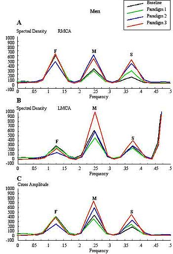
). The most efficient standard Fourier algorithm requires that the length of the input series is equal to a power of 2. If this is not the case, additional computations have to be performed. To derive the required time series, the data were averaged in 10-second segments for 1-minute duration or each stimulus, yielding 6 data points for each participant and a total of 48 data points for all eight men and women, respectively. Smoothing the periodogram values was accomplished using a weighted moving average transformation. Hamming window was applied as a smoother. The spectral density estimates, derived from single series Fourier analysis, were plotted, and the frequency regions with the highest estimates were marked as peaks. The origins of the peaks are of interest in order to determine the reliability of the present technique. The fundamental (F), cortical (C) or memory (M), and subcortical (S) peaks occurred at regular frequency intervals of 0.125, 0.25, and 0.375, respectively. These frequencies could be converted to Hz, assuming that the fundamental frequency of cardiac oscillation was the mean heart rate. The fundamental frequency (F) of the first harmonic could be determined from the mean heart rate per second. For example, a heart rate of 74 bpm, suggests 74 cycles/60 or 1.23 Hz. In other words, the F-, C-, and S-peaks occurred at multiples of the first harmonic, at second and third harmonics, respectively. The distance of the reflection site for F-peak could be presumed to emanate from a site at D1 = wavelength/4 = cf/4 = 6.15 (m/s)/(4×1.23 Hz) = 125 cm, where c is the assumed wave propagation velocity of the peripheral arterial tree according to McDonald, 1974.
Given the vascular tortuosity, the estimated distance approximates that from the measurement site in the MCA main stem, to an imaginary site of summed reflections from the upper extremities, close to the finger tips when stretched
sideways. The C-peak occurred at the second
harmonic, such that the estimated arterial length (using common carotid
c = 5.5 m/s) was given by D2 = wavelength/8 = cf2/8 = 28 cm, and a frequency f of 2.46 Hz. The distance approximates the visible arterial length from the main stem of the MCA, through vascular tortuosity and around the cerebral convexity, to the end vessels at distal cortical sites such as the occipito-temporal junction on
carotid angiograms of adults. The S-peak occurred at the third harmonic, and may have arisen from an estimated site at D3= wavelength/16 = cf3/16 = 9.3 cm and a frequency f3 of 3.69 Hz. The latter approximates the visible arterial length of the lenticulostriate vessels from the main stem of the MCA on carotid angiograms . Although not displayed, the fourth harmonic would be expected to arise from the MCA bifurcation in closest proximity to
the measurement site in the main stem of the MCA. The pre-bifurcation
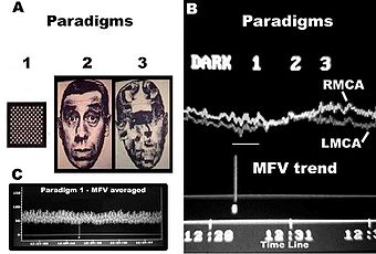
length from the measurement point would be given by D4 = wavelength/32 = cf4/32 = 3.5 cm and a frequency f4 of 4.92 Hz. The calculated distance approximates that of the segment of MCA main stem just after the carotid bifurcation, where probably the ultrasound sample volume was placed, to the MCA bifurcation. Thus, these estimates approximate actual lengths. However, it has been suggested that the estimated distances may not correlate exactly with known morphometric dimensions of the arterial tree according to Campbell et al., 1989. The method was first described by Philip Njemanze in 2007, and was referred to as functional transcranial Doppler spectroscopy (fTCDS). fTCDS examines spectral density estimates of periodic processes induced during mental tasks, and hence offers a much more comprehensive picture of changes related to effects of a given mental stimulus. The spectral density estimates would be least affected by artefacts that lack periodicity, and filtering would reduce the effect of noise . The changes at the C-peak may show cortical long-term potential (CLTP) or cortical long-term depression (CLTD), which has been proposed to be suggest equivalents of cortical activity during learning and cognitive processes. The flow velocity tracings are monitored during paradigm 1 comprising a checkerboard square as object perception are compared to whole face (paradigm 2) and facial element sorting task (paradigm 3). Fast Fourier transform calculations are used to obtain the spectral density and cross amplitude plots in the left and right middle cerebral arteries. The C-peak also called memory (M-peak) cortical peak could be seen arising during paradigm 3, a facial element sorting task requiring iterative memory recall as a subject constantly spatially fits the puzzle by matching each facial element in paradigm 3 to that stored in memory (Paradigm 2) before proceeding to form the picture of the whole face.