

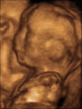
3D ultrasound
Encyclopedia
Obstetric ultrasonography
Obstetric sonography is the application of medical ultrasonography to obstetrics, in which sonography is used to visualize the embryo or foetus in its mother's uterus...
(during pregnancy
Pregnancy
Pregnancy refers to the fertilization and development of one or more offspring, known as a fetus or embryo, in a woman's uterus. In a pregnancy, there can be multiple gestations, as in the case of twins or triplets...
), providing three dimensional
Three-dimensional space
Three-dimensional space is a geometric 3-parameters model of the physical universe in which we live. These three dimensions are commonly called length, width, and depth , although any three directions can be chosen, provided that they do not lie in the same plane.In physics and mathematics, a...
images of the fetus
Fetus
A fetus is a developing mammal or other viviparous vertebrate after the embryonic stage and before birth.In humans, the fetal stage of prenatal development starts at the beginning of the 11th week in gestational age, which is the 9th week after fertilization.-Etymology and spelling variations:The...
.
There are several different scanning modes in medical and obstetric ultrasound. The standard common obstetric diagnostic mode is 2D scanning. In 3D fetal scanning, however, instead of the sound waves being sent straight down and reflected back, they are sent at different angles. The returning echoes are processed by a sophisticated computer program resulting in a reconstructed three dimensional volume image of fetus's surface or internal organs, in much the same way as a CT scan machine constructs a CT scan image from multiple x-rays. 3D ultrasounds allow one to see width, height and depth of images in much the same way as 3D movies
3-D film
A 3-D film or S3D film is a motion picture that enhances the illusion of depth perception...
but no movement is shown.
3D ultrasound was first developed by Olaf von Ramm and Stephen Smith at Duke University in 1987.
Clinical use of this technology is an area of intense research activity especially in fetal anomaly scanning but there are also popular uses that have been shown to improve fetal-maternal bonding. 4D fetal ultrasounds are similar to 3D scans, with the difference associated with time: 4D allows a 3-dimensional picture in real time, rather than delayed, due to the lag associated with the computer constructed image, as in classic 3-dimensional ultrasound.
If the system is used only in the Obstetrics Application, the ultrasound energy is limited by the manufacturer below FDA limits for obstetrical ultrasound, whether scanning 2, 3 or 4 dimensionally. (The FDA limit for obstetrical ultrasound is 94 mW/cm2.) While there has been no conclusive evidence for harmful effects of 3D ultrasounds on a developing fetus, there still remains controversy over its use in non-medical situations, and generally, the AIUM recommends that 3D ultrasounds should be undertaken with the understanding that a risk may exist.
Elective 3D Ultrasound
Although 3D ultrasound technology may be used on any part of the body, elective 3D ultrasound conventionally refers to 3D ultrasounds performed on pregnant women for the sole purpose of the woman to see her unborn baby, what the baby looks like, or to see whether the baby will be a boy or a girl. In the medical literature, these elective 3D ultrasounds are also referred to as keepsake ultrasounds, although this term is rarely used in the lay population and only sometimes used in the media.Benefits of elective 3D ultrasounds
Although there is no direct medical benefit of receiving an elective 3D ultrasound, there may be many indirect benefits, as listed below. It is important to note that there has been no conclusive evidence in the medical literature to support these benefits, and in fact, the medical literature shows conflicting studies on these benefits.A large randomized trial of 15,151 pregnant women, conducted by the RADIUS Study Group, found that in low-risk cases, high-risk subgroups and even in cases of multiple gestations or major anomalies, the use of ultrasound did not result in improved outcome in the pregnancies.
According to the World Health Organization and U.S. Department of Health and Human Services report, "It is not clear at this time whether ultrasound fetal monitoring is beneficial to the mother or fetus in terms of pregnancy outcome...If there is no generally acknowledged benefit to the monitoring, there is no reason to expose patients to increased cost and risk. The question of benefit has not yet been resolved...and the potential for delayed effects has been virtually ignored." The American Medical Association recommends against unnecessary exposure to 3D ultrasound.
Pro-life non-profit organizations have employed 3D ultrasounds for young pregnant women in order to influence their decisions regarding abortion. Charitable donations help pay for the 3D ultrasound machine.
Risks of 3D ultrasounds
According to the 1982 World Health Organization and U.S. Department of Health and Human Services report, "Effects of Ultrasound on Biological Systems," stated that "…animal studies suggest that neurological, behavioral, developmental, immunological, haematological changes and reduced fetal weight can result from exposure to ultrasound." Though generally, the risks of 3D ultrasounds mirror those of 2D ultrasounds, as it uses the same ultrasound waves at the same intensity. Unlike the comparison of CT scans to x-rays, 3D ultrasounds do not employ multiple snapshots of 2D ultrasounds but uses the 2D ultrasound images taken at various angles to construct an image. So the potential risk of 3D ultrasounds would depend on the duration of the ultrasound session rather than whether it is 2D or 3D.The risk of ultrasounds, theoretically, would depend on the following factors:
- Duration of ultrasound exposure
- Intensity of ultrasound waves
- Frequency of ultrasound sessions
Duration
Although no set standard formally exists, physicians who perform 3D/4D ultrasounds will not likely go past 30 minutes due to the increase of ultrasound waves in the extended time, the longer the ultrasound the more wave exposure the fetus is subjected to. Since doctors to do not yet know the side effects of prolonged exposure, doctors limit their sessions to be safe.Intensity
The intensity of ultrasound waves are mechanically set into the machine to not exceed FDA standards. Ultrasound machines are constructed to either shut off or give a noticeable warning if any of the built-in barriers fail to limit the ultrasound waves to below the FDA standard. Generally, a higher intensity of ultrasound waves are used to detect the baby's heartbeat, and as these waves are also directed and focused onto a single organ in the fetus, it is generally advised that use of the ultrasound machine to detect and play the baby's heartbeat be done after 12 weeks of gestation, although it may be physically detectable as early as 9 weeks of gestation.Frequency
The frequency of ultrasound sessions can also theoretically pose a risk. It is generally discouraged to perform elective 3D ultrasounds more frequently than once a month. It is important to note that women undergoing in vitro fertilization or who have possible fetal abnormalities undergo weekly 2D ultrasounds. Pregnant women in some countries undergo monthly 2D ultrasounds as routine prenatal care.Physical effects
One challenge that ultrasound operators face is keeping the transducer positioned over the part of the fetus the operator is trying to visualize. When fetuses move away from the stream of high-frequency sound waves, they may be feeling vibrations, heat or both. The FDA warned in 2004, "ultrasound is a form of energy, and even at low levels, laboratory studies have shown it can produce physical effect in tissue, such as jarring vibrations and a rise in temperature." FDA Cautions against 3D ultrasound This is consistent with research conducted in 2001 in which an ultrasound transducer aimed directly at a miniature hydrophone placed in a woman's uterus recorded sound "as loud as a subway train coming into the station." Fetuses can hear ultrasound examinationsMedical effects
Early studies showed that subtle effects of neurological damage linked to ultrasound were implicated by an increased incidence in left-handedness in boys (a marker for brain problems when not hereditary) and speech delays. Then in August 2006, Pasko Rakic, chair of Yale School of Medicine's Department of Neurobiology, announced the results of a study in which pregnant mice underwent various durations of ultrasound. The brains of the offspring showed damage consistent with that found in the brains of people with autism. The research, funded by the National Institute of Neurological Disorders and Stroke, also implicated ultrasound in neurodevelopmental problems in children, such as dyslexia, epilepsy, mental retardation and schizophrenia, and showed that damage to brain cells increased with longer exposures. .Other risks
Other unintended harm caused by 3D ultrasounds may include the finding of false positives, which can be a significant source of distress to the mother.Cysts found on the fetus that would have been harmless and may not have been found otherwise may be detected on 3D ultrasound and lead to further more invasive exams as well as cause needless anxiety to the mother. Artifacts can be seen on 3D ultrasound, particularly by those with less experience, such as duplications of body parts, holes, clefts, and missing body parts. Usually, these artifacts go away by moving the ultrasound probe slightly, but when performed by an inexperienced technician, may produce needless anxiety to the mother.
Seeing the fetus too early, under 17 weeks, may cause some harm to the mother as it may actually decrease bonding and give a feeling to the mother that the baby is alien or "not really a baby yet." It is generally advised to perform 3D ultrasounds at or after 17 weeks of gestation for this reason (Kurjak et al., How useful is 3D and 4D ultrasound in perinatal medicine? J. Perinatal Medicine, 2007).
Other incidental harms to elective 3D ultrasounds include falls or back pain when getting on and off a bed not conducive to pregnant women in 3D ultrasound centers; aggressive manipulation of the belly by an inexperienced ultrasound technician; encouragement of the pregnant mother to drink caffeine-containing beverages to stimulate the baby for a "good photo shot." Caffeine, even in small amounts, have been implicated with some controversy to result in possible harm to the fetus. However most health care professionals state that up to 200mg of caffeine a day is safe.
Substitution of prenatal care
By far, the biggest potential risk to the pregnant woman in getting a 3D ultrasound is a false sense of reassurance. Pregnant women in the United States who can not otherwise afford health care and do not have health insurance may see the payment of an elective 3D ultrasound, which may cost around $100–$200, as a cheaper alternative to the artificially raised prices of prenatal care from a clinic or hospital, where lab work alone may exceed $1000. Women who receive a 3D ultrasound may be falsely reassured that "everything is okay" and may discontinue prenatal care or taking prenatal vitaminsPrenatal vitamins
Prenatal vitamins are vitamin supplements intended to be taken before and during pregnancy and during postnatal lactation. Although not intended to replace a healthy diet, prenatal vitamins provide women of child bearing age with nutrients recognized by the various health organizations including...
. This potential risk may be averted, at least partially, by repeated instructions that the 3D ultrasound does not take place of routine prenatal care. Many 3D ultrasound centers require signatures on statements of understanding to this fact or may require proof of prenatal care. Some 3D ultrasound centers go as far as to require a note or a prescription from a physician to perform the 3D ultrasounds.
Risk reduction of 3D ultrasounds
The following can help reduce the risks of elective 3D ultrasounds:- Have a qualified medical director for 3D ultrasound centers.
Some states require that every 3D ultrasound center must have a medical director
- Employ only ARDMS-certified ultrasound technologists
Some ultrasound centers do not employ ultrasound technologists, and currently, there is no law that mandates that 3D ultrasounds must be performed by certified ultrasound technologists
- Provide adequate training to ultrasound technologists
- Have adequate communication with the ob/gyn's of the customers, and have policies put in place in regards to accidental discoveries of abnormalities
- Safety inspections of the clinic
- Regular inspections and maintenance of the ultrasound machine
- Limit ultrasound sessions to last no longer than 30 minutes
- Limit ultrasound sessions to no more than once a month
- Require proof of prenatal care prior to the 3D ultrasound session
Sex determination
Although medical textbooks state that the sex of the fetus can be seen on ultrasound as early as 12 weeks, practically speaking, most medical facilities and hospitals do not attempt to determine the sex of the fetus until the 20-week ob/gyn visit. 3D/4D ultrasound centers bridge the gap and fulfill the niche market of pregnant women who can not wait to find out the sex of the fetus, women who did not find out the sex of the fetus at their medical visit and the insurance does not pay for any more ultrasounds, and women who simply want to confirm the sex of the fetus.Some ultrasound centers attempt to determine the sex of the fetus as early as 15 weeks and advertise as such. The customer is required to pay at the first visit, but most of the time, the customer has to return when the fetus is more mature. At 15 weeks, the success rate for being able to see the sex is around 50% at best, and the accuracy at that gestational age is questionable.
At 16 weeks, the ability to find the sex and accuracy rises to 99%. Most private ultrasound centers offer sex scans from 16 weeks. The hospital do not offer it before your 20 week anatomy scan unless there is a medical reason that it is required for.
Visualization of the fetus
3D ultrasounds are best done at 24–32 weeks, and ideally between 26 and 30 weeks. Some centers advise customers to come in between 26 and 28 weeks to get the best images.After 32 weeks, there is an increased risk that the fetus has already engaged (descended into the pelvis) and at that time, getting 3D images of the fetus is nearly impossible. As the baby is bigger there is less room so it is even more difficult to get a nice image.
Methods
Unlike the medical ultrasound visit, customers of 3D ultrasound centers are not advised to hold their urine or have a full bladder before their 3D ultrasound appointment.In order to get good images, ultrasound centers advise customers to drink adequate amounts of water (32 ounces/day) for the one to two weeks prior to their appointment. This is in order to ensure there is an adequate amount of amniotic fluid around the fetus and that the fluid is clear. Drinking large amounts of water immediately before the 3D ultrasound appointment does not help to have clearer 3D ultrasound images.
Future developments
3D ultrasounds may soon become a part of routine care. Indeed, many hospitals and clinics already provide 3D ultrasounds to pregnant women as a courtesy.3D ultrasounds may soon be covered by FSA (flexible spending account
Flexible spending account
A flexible spending account , also known as a flexible spending arrangement, is one of a number of tax-advantaged financial accounts that can be set up through a cafeteria plan of an employer in the United States...
s) and may eventually be accepted by some insurance companies, as medical studies begin to show benefits of elective 3D ultrasounds.
State laws are developing to require oversight and restrictions on 3D ultrasound centers, including the requirement of a medical director, requiring verification of prenatal care, and/or requiring certified ultrasound technicians.
3D ultrasounds are already being used to detect fetal anomalies of the heart. 3D ultrasounds may be used in the near future for actual neurological and behavioral testing of the fetus to help diagnose or rule out cerebral palsy.
A novel technology enabling the use of 3D ultrasound in remote areas has been recently developed. This new technology leverages the ubiquitous mobile phone available even in the most remote corners of the world. By collecting the raw data at the patient location and
sending it for processing and expert evaluation to the central processing station, this technology reduces equipment costs and reduces the hand-eye coordination skills normally required from the technician performing the procedure.
Conversion of the 3D image files into standard CAD/CAM file formats allows the reconstruction of fetal and other images in a variety of materials including a 3d laser etched images in a crystal glass block or a solid cameo effect using a 3D printer.
Opposing views
In 1999, the American In Medicine (AIUM) released the following statement:The AIUM strongly discourages the non-medical use of ultrasound for psychosocial or entertainment purposes. The use of either two-dimensional (2D) or three-dimensional (3D) ultrasound to only view the fetus, obtain a picture of the fetus, or determine the fetal sex without a medical indication is inappropriate and contrary to responsible medical practice.
In February 2004, the American Food and Drug Administration (FDA) issued the following statement:
Persons who promote, sell or lease ultrasound equipment for making “keepsake” fetal videos should know that FDA views this as an unapproved use of a medical device. In addition, those who subject individuals to ultrasound exposure using a diagnostic ultrasound device (a prescription device) without a physician’s order may be in violation of state or local laws or regulations regarding use of a prescription medical device.
Regional Anesthesia
Real-time three dimensional ultrasound is used during peripheral nerve blockade procedures to identify relevant anatomy and monitor the spread of local anesthetic around the nerve. Peripheral nerve blockades prevent the transmission of pain signals from the site of injury to the brain without deep sedation, which makes them particularly useful for outpatient orthopedic procedures. Real-time 3D ultrasound allows muscles, nerves and vessels to be clearly identified while a needle or catheter is advanced under the skin. 3D ultrasound is able to view the needle regardless of the plane of the image, which is a substantial improvement over 2D ultrasound. Additionally, the image can be rotated or cropped in real time to reveal anatomical structures within a volume of tissue. Physicians at the Mayo Clinic in Jacksonville have been developing techniques using real time 3D ultrasound to guide peripheral nerve blocks for shoulder, knee, and ankle surgery.Further reading
- Where to get 3d/4d training
- Article: 3D and 4D Ultrasound Scanning
- Article: 3D and 4D Ultrasound Scanning
- Article: 4D: What the Medical Literature Says
- The History of Ultrasounds (including 3D ultrasounds)
- Article: What is a Elective 3D Ultrasound
- Article: Why are Ultrasound Scans Used In Pregnancy
External links
- The Endowment for Human Development provides numerous 4D ultrasounds that can be viewed online.
- Scans uncover secrets of the womb BBC News
- About the discovery of medical ultrasonography
- RadiologyTube - 3D and 4D Ultrasound Videos
- History of medical sonography (ultrasound)
- Safety Concerns Details a number of health concerns over this unregulated practice