
Craniosynostosis
Encyclopedia
Craniosynostosis is a condition in which one or more of the fibrous sutures in an infant skull prematurely fuses by ossification, thereby changing the growth pattern of the skull. Because the skull cannot expand perpendicular to the fused suture, it compensates by growing more in the direction parallel to the closed sutures. Sometimes the resulting growth pattern provides the necessary space for the growing brain, but results in an abnormal head shape and abnormal facial features. In cases in which the compensation does not effectively provide enough space for the growing brain, craniosynostosis results in increased intracranial pressure
leading possibly to visual impairment, sleeping impairment, eating difficulties, or an impairment of mental development combined with a significant reduction in IQ.
Craniosynostosis occurs one in 2000 births.
Craniosynostosis is part of a syndrome
in 15 to 40% of the patients, but it usually occurs as an isolated condition.
It is important that families seek out the opinion of a Pediatric Craniofacial Physician who has experience with craniosynostosis for proper diagnosis, surgical care, and followup.
above the meninges
undergoes intramembranous ossification
forming the neurocranium. The neurocranium consists of several bones, which are united and at the same time separated by fibrous sutures. This allows movement of the separate bones in relation to one another; the infant skull is still malleable. The fibrous sutures specifically allow the deformation of the skull during birth and absorb mechanical forces during childhood They also allow the necessary expansion during brain growth.
In the first years of life the sutures serve as the most important centers of growth in the skull.
The growth of the brain and the patency of the sutures depend on each other. Brain growth pushes the two sides of the patent sutures away from each other, thereby enabling growth of the neurocranium. This means that the neurocranium can only grow if the sutures remain open.
The neurocranium will not grow when the forces induced by brain growth are not there.
This will occur for example when the intracranial pressure drops; the sutures do not experience stretch anymore causing them to fuse.
The infant's skull consists of the metopic suture, coronal sutures, sagittal suture
, and lambdoid suture
s. The metopic suture is supposed to close between three to nine months of age. The lambdoid, sagittal and coronal sutures are supposed to close between 22 to 39 years of age.
, representing 40 to 55% of nonsyndromic cases. The second most common type is the coronal synostosis representing 20 to 25%. The metopic synostosis comes third with 5 to 15% and the lambdoid synostosis is only seen in 0 to 5% of nonsyndromic cases.
In about 5 to 15% more than one suture is affected, which referred to as complex craniosynostosis. This is generally part of a syndrome.
and genetics
, as well as the use of animal models have been of great importance in expanding our knowledge of suture fusion. Research in animal models has led to the idea that the dura mater
plays an important role in determining closure or patency of the suture. In contrast to the dura mater it appears that the periosteum
is not essential in causing closure or patency.
Instead of describing the abnormalities in structure and form, research focuses nowadays at decoding the molecular interactions that underlie them.
Despite the progress that has been made, many things are still not understood about the suture biology and the exact causative pathways remain yet to be completely understood.
Multiple potential causes of premature suture closure have been identified, such as the several genetic mutations that are associated with syndromic craniosynostosis.
The cause of nonsyndromic craniosynostosis however, is still greatly unknown.
Most likely, a role is played by biomechanical factors, as well as environmental, hormonal and genetical factors.
. These last two are both important factors influencing bone development.
On the other hand, a recent evaluation of valproic acid
(an anti-epilepticum), which has been implicated as a causative agent, has shown no association with craniosynostosis.
.
Fibroblast growth factor and fibroblast growth factor receptors regulate fetal bone growth and are expressed in cranial sutures during pregnancy.
The transcription factor gene TWIST is thought to decrease the function of FGFR, thus also indirectly regulating fetal bone growth. A relation between the mutations in these genes and craniosynostosis is therefore possible.
Moloney et al. observed a FGFR3 mutation in as many as 31% of the cases with nonsyndromic coronal synostosis, thus showing that FGFR abnormalities play an important role in nonsyndromic craniosynostosis.
In terms of syndromic craniosynostosis not only do FGFR3 and TWIST genes feature, but also FGFR1 and in particular FGFR2, which has been reported in 90% of the syndromic craniosynostoses such as Apert, Crouzon, Peiffer and Jackson-Weiss The mutations can be divided into mutations that lead to gain of function (in FGFR genes) and mutations that lead to loss of function (in TWIST genes). Craniosynostosis is therefore likely the result of a disturbance in the fine balance that regulates the multiplication and maturation of the precursor bone cells in the cranial sutures.
In addition, the following syndromes have been identified:
. The features of the phenotype are determined by which particular suture is closed. The fusion of this suture causes a certain change in the shape of the skull; a deformity of the skull.
Virchow’s law dictates that, when premature suture closure occurs, growth of the skull is typically restricted perpendicular to the fused suture and enhanced in a plane parallel to it, thus trying to provide space for the fast growing brain. Using this law, the pattern of skull deformity in craniosynostosis can often be predicted.
is a result from the premature closure of the metopic suture. Using Virchow’s law again to predict the resulting deformity, this fusion will result in a narrow forehead, which is even further emphasized by ridging of the suture. Compensatory growth occurs at both the coronal sutures, thereby pushing the forehead forwards.
The resulting shape can best be assessed from a top view again, which will reveal a somewhat triangular form of the head. Trigonocephaly is also a Greek derived word, which can be translated as triangular shaped head. A facial feature of metopic synostosis is hypotelorism
; in the frontal view, it can be seen that the width between the eyes is smaller than usual.
The sagittal suture ‘divides’ the coronal suture in two halves; unilateral meaning that either the right side or the left side to the sagittal suture is fused. This fact immediately raises an important point. Unlike closure of the sagittal or the metopic suture, right and left are not the same in unilateral coronal synostosis. This asymmetry shows in the skull deformity, as well as in the facial deformity and the complications.
This time, the skull deformity can only partly be predicted using Virchow’s law. Growth is arrested in the plane perpendicular to the fused suture and the forehead is flattened, but only at the ipsilateral side of the head. Ipsilateral indicates the same side of the head as where the suture is closed.
Compensatory growth occurs in a parallel plane, as well as in a perpendicular plane. An increase in growth at the metopic and the sagittal suture accounts for the parallel plane and will result in bulging at the temporal fossa
and an increase in width of the skull. Compensatory growth in the perpendicular plane occurs on the side of the head with the patent coronal suture, the contralateral side. Half of the forehead will bulge forwards as a result.
Assessment of the skull from a top view shows asymmetry of the frontal bones, an increased width of the skull and a forward displacement of the ear at the ipsilateral side of the head. Assessment of the skull from a frontal view will show asymmetrical features of the face, including a displacement of the chin point of the jaw and a deviation of the tip of the nose. The chin point is located more to the contralateral side of the head, due to the ipsilateral forward displacement of the temporomandibular joint
together with the ear. The tip of the nose will also point towards the contralateral side. Complications based on the skull deformation include malocclusion of the jaw and in as many as 90% - a subtle form of - strabismus
, the last being caused by the asymmetrical placement of the orbit
s.
.
Remembering Virchow’s law, restriction of growth will occur at the ipsilateral side of the head; compensatory growth will occur at the contralateral side of the head. This growth pattern exerts an effect at the base of the skull, which is not even when the child is assessed from a point of view standing behind the child, as well as on the cervical spine, which shows a curvature. In addition, an asymmetry of the ears can be seen, with the ear on the ipsilateral side placed further to the back. Also, again from a point of view standing behind the child, a bulging of the mastoid can be seen.
Minimal forehead asymmetries are typically seen.
Pansynostosis can present in several ways. The appearance can be the same as that seen with primary microcephaly
: a markedly small head, but with normal proportions. However, pansynostosis can also appear as a Kleeblattschädel (cloverleaf skull), which presents with bulging of the different bones of the cranial vault.
which is sometimes the only symptom of elevated intracranial pressure shown in these children.
Other parts of the physical examination include the measurement of the head circumference, the assessment of the skull deformity and the search for deformities affecting other parts of the body. The head circumference and the growth curve of the head provide important clues into making a differentiation between craniosynostosis, primary microcephaly and hydrocephalus
. This differentiation has an important influence on the further treatment of the child.
In a recent article Cunningham et al. described several steps in which a pediatrician should observe the patient to assess skull deformity:
The implications of the deformities that are seen are extensively discussed under ‘phenotype’.
Syndromal craniosynostosis presents with a skull deformity as well as deformities affecting other parts of the body. Clinical examination should in any case include evaluation of the neck, spine, digits and toes.
Plain radiography of the skull may be sufficient for diagnosing a single suture craniosynostosis and should therefore be performed, but the diagnostic value is outweighed by that of the CT-scan. Not only can the sutures be identified more accurately, thus objectively demonstrating a fused suture, but also evaluation of the brain for structural abnormalities and excluding other causes of asymmetric growth are possible at the same time. In addition to this, CT-scanning can visualize the extent of skull deformity, thereby enabling the surgeon to start planning surgical reconstruction.
With the tests a pediatrician should perform, as explained above, the difference is quite easy to make.
In deformational plagiocephaly the skull does not show a bulging of the mastoid and in these patients the skull base and position of the ears is level, all in contrary with plagiocephaly due to craniosynostosis. Displacement of one ear to the front is characteristic for deformational plagiocephaly.
A differentiation between these two conditions can be made with a computed tomography (CT) scan. The subarachnoid spaces are typically enlarged with primary microcephaly, whereas they are reduced or absent in true pansynostosis.
and headache.
The main risks of prolonged elevated intracranial pressure may include cognitive impairment and impaired vision through prolonged papilledema. These are the main reasons why fundoscopy should be performed during the physical examination of children with craniosynostosis.
The causes of an elevation of the intracranial pressure are best understood using the Monro-Kellie doctrine. The Monro-Kellie doctrine reduces the cranial vault to a box with rigid walls. This box contains three elements: brain, intracranial blood and liquor. The sum of volumes of these three elements is constant. An increase in one should cause a decrease in one or both of the remaining two, thereby preventing an elevation of the intracranial pressure.
A compensatory mechanism involves the movement of liquor from the cranial vault towards the spinal cord. The volume of blood in the cranial vault is auto-regulated by the brain, and will therefore not decrease that easily.
Intracranial pressure will rise as a result of continued brain growth within the rigid skull. It appears that in children with craniosynostosis, the expected decrease of intracranial liquor is probably not occurring as it should according to the Monro-Kellie hypothesis. This is shown when the brain expands in the fixed skull, which gives a faster rise in intracranial pressure than would be expected.
The main causative agent of OSA is the [midface hypoplasia], which also poses a risk to the eyes that can be seen bulging out of the eye sockets. Other factors, such as a micrognathism
and adenoid hypertrophy
, are likely to contribute in causing OSA.
The most common syndromic forms of craniosynostosis; i.e. Apert, Crouzon and Pfeiffer, have an increased risk of developing OSA. The children have nearly 50% chance of developing this condition.
A theory regarding the involvement of OSA as a causative agent for elevated intracranial pressure suggests an association with the auto-regulation of blood flow in the brain.
Certain cells in the brain respond specifically to an increase of CO2 in the blood. The response involves vasodilatation of the cranial vault blood vessels, increasing the volume of one of the elements in the Monro-Kellie doctrine. The increase of CO2 concentration in the blood is a consequence of impaired breathing, especially seen when the child suffering from OSA is sleeping. It is well documented that the highest spikes in intracranial pressure often occur during sleep.
. This causes an increase in the intracranial blood volume, thereby causing an increase in intracranial pressure.
This can be further complicated with a possible Arnold-Chiari malformation
, which can partially obstruct the flow of liquor from the neurocranium to the spinal cord. The Chiari malformation may be asymptomatic or present with ataxia
, spasticity
or abnormalities in breathing, swallowing or sleeping.
Due to the impaired venous outflow, which may be further complicated with a Arnold-Chiari malformation, there is often a clinical image of hydrocephalus
present. Hydrocephalus is seen in 6.5 to 8% of patients with Apert’s syndrome, 25.6% in patients with Crouzon’s syndrome and 27.8% of those with Pfeifer’s syndrome.
Ventriculomegaly
is a usual finding in children with the Apert syndrome.
It has been suggested that these problems are caused by a primary malformation of the brain, rather than being a consequence of the growth restriction of the skull and elevated intracranial pressure. Some evidence for this statement has been provided by studies using computed tomographic (CT scans) and magnetic resonance imaging (MRI) to identify differences between the structures of the brains of healthy children and those affected with craniosynostosis.
It has been found that corrective surgery of the cranial vault
alters the morphology of the brain compared with the situation before surgical intervention. However the structure was still abnormal in comparison to children without craniosynostosis.
In addition signs of compromised neurodevelopment have been seen amongst all the synostoses, although this may also be caused by primary maldevelopment of the brain and can thus not be prevented by surgical intervention.
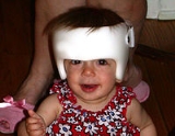
The reason why most surgeons will not intervene until after the age of six months is the greater risk that blood loss poses before this age. If possible it is preferred to wait until after three months of age when the anaesthetic risks are decreased.
Surgery is not performed in early childhood in every country. In some countries surgical intervention can take place in the late teens.
It is important that families seek out a Pediatric Craniofacial Physician who has experience with craniosynostosis for proper diagnosis, surgical care, and followup.
.
is used to asses the efficacy of the surgical intervention.
Retrospective analysis has given indication that the use of total cranial vault remodelling provides the children with a better cephalic index than does the extended strip craniectomy.
An approach that is currently evaluated is the use of springs. This intervention is likely most effective when used in the time frame between three to six months.
Metopic synostosis/trigonocephaly
The main elements of metopic suture closure are a low volume of the anterior cranial fossa
, the metopic ridging and hypotelorism
. These problems are all addressed during the surgical intervention.
The volume is increased by placing the frontal bones and the supraorbital rim further forwards. This is done by excision of the bones after which they are reshaped with greenstick fracturing
. Replacement of the bones provides a possibility for the correction of the hypotelorism at the same time. A bone graft is placed in between the two halves of the supraorbital bars, thereby increasing the width between the orbits
. The metopic ridge can be corrected with a (simple) burring.
The supraorbital bar is the rim just above the eye socket. As discussed under phenotype, the supraorbital and the frontal bone are typically recessed at the ipsilateral side of the head. The goal is to position this bar together with the frontal bone in a plane three millimetres further forwards than the vertical plane of the cornea
.
A two-dimensional sagittal image is used to pre-operatively determine the extent of movement, which can vary between seven to fifteen millimetres, depending on the severity of the deformity.
The orbital asymmetry exists typically of a narrower and taller orbit at the ipsilateral side of the head. The contralateral orbit, however, is wider than usual. The symmetry is restored by extracting a small piece of bone from the supraorbital bar at the contralateral side, thereby reducing the width. This bone fragment is introduced into the supraorbital bar on the ipsilateral side, thereby increasing the width. The height of the orbit is just altered at the ipsilateral side, by extracting a piece of bone.
The correction of the nasal tip, which points towards the contralateral side, is not performed during childhood.
are extracted and given a rounder shape by greenstick fracturing them.
, as well as the posterior cranial vault. This can be accomplished in one procedure, but is generally performed in two.
Intracranial pressure
Intracranial pressure is the pressure inside the skull and thus in the brain tissue and cerebrospinal fluid . The body has various mechanisms by which it keeps the ICP stable, with CSF pressures varying by about 1 mmHg in normal adults through shifts in production and absorption of CSF...
leading possibly to visual impairment, sleeping impairment, eating difficulties, or an impairment of mental development combined with a significant reduction in IQ.
Craniosynostosis occurs one in 2000 births.
Craniosynostosis is part of a syndrome
Syndrome
In medicine and psychology, a syndrome is the association of several clinically recognizable features, signs , symptoms , phenomena or characteristics that often occur together, so that the presence of one or more features alerts the physician to the possible presence of the others...
in 15 to 40% of the patients, but it usually occurs as an isolated condition.
It is important that families seek out the opinion of a Pediatric Craniofacial Physician who has experience with craniosynostosis for proper diagnosis, surgical care, and followup.
An overview of the cranial sutures
The mesenchymeMesenchyme
Mesenchyme, or mesenchymal connective tissue, is a type of undifferentiated loose connective tissue that is derived mostly from mesoderm, although some are derived from other germ layers; e.g. some mesenchyme is derived from neural crest cells and thus originates from the ectoderm...
above the meninges
Meninges
The meninges is the system of membranes which envelopes the central nervous system. The meninges consist of three layers: the dura mater, the arachnoid mater, and the pia mater. The primary function of the meninges and of the cerebrospinal fluid is to protect the central nervous system.-Dura...
undergoes intramembranous ossification
Intramembranous ossification
Intramembranous ossification is one of the two essential processes during fetal development of the mammalian skeletal system by which bone tissue is created. Unlike endochondral ossification, which is the other process by which bone tissue is created, cartilage is not present during intramembranous...
forming the neurocranium. The neurocranium consists of several bones, which are united and at the same time separated by fibrous sutures. This allows movement of the separate bones in relation to one another; the infant skull is still malleable. The fibrous sutures specifically allow the deformation of the skull during birth and absorb mechanical forces during childhood They also allow the necessary expansion during brain growth.
In the first years of life the sutures serve as the most important centers of growth in the skull.
The growth of the brain and the patency of the sutures depend on each other. Brain growth pushes the two sides of the patent sutures away from each other, thereby enabling growth of the neurocranium. This means that the neurocranium can only grow if the sutures remain open.
The neurocranium will not grow when the forces induced by brain growth are not there.
This will occur for example when the intracranial pressure drops; the sutures do not experience stretch anymore causing them to fuse.
The infant's skull consists of the metopic suture, coronal sutures, sagittal suture
Sagittal suture
The sagittal suture is a dense, fibrous connective tissue joint between the two parietal bones of the skull. The term is derived from the Latin word Sagitta, meaning "arrow". The derivation of this term may be demonstrated by observing how the sagittal suture is notched posteriorly, like an arrow,...
, and lambdoid suture
Lambdoid suture
The lambdoid suture is a dense, fibrous connective tissue joint on the posterior aspect of the skull that connects the parietal and temporal bones with the occipital bone.Its name comes from its lambda-like shape....
s. The metopic suture is supposed to close between three to nine months of age. The lambdoid, sagittal and coronal sutures are supposed to close between 22 to 39 years of age.
Epidemiology
It is estimated that craniosynostosis affects 1 in 2,000 to 2,500 live births worldwide. Sagittal synostosis is the most common phenotypePhenotype
A phenotype is an organism's observable characteristics or traits: such as its morphology, development, biochemical or physiological properties, behavior, and products of behavior...
, representing 40 to 55% of nonsyndromic cases. The second most common type is the coronal synostosis representing 20 to 25%. The metopic synostosis comes third with 5 to 15% and the lambdoid synostosis is only seen in 0 to 5% of nonsyndromic cases.
In about 5 to 15% more than one suture is affected, which referred to as complex craniosynostosis. This is generally part of a syndrome.
An overview of causes of premature fusion
Advances in the fields of molecular biologyMolecular biology
Molecular biology is the branch of biology that deals with the molecular basis of biological activity. This field overlaps with other areas of biology and chemistry, particularly genetics and biochemistry...
and genetics
Genetics
Genetics , a discipline of biology, is the science of genes, heredity, and variation in living organisms....
, as well as the use of animal models have been of great importance in expanding our knowledge of suture fusion. Research in animal models has led to the idea that the dura mater
Dura mater
The dura mater , or dura, is the outermost of the three layers of the meninges surrounding the brain and spinal cord. It is derived from Mesoderm. The other two meningeal layers are the pia mater and the arachnoid mater. The dura surrounds the brain and the spinal cord and is responsible for...
plays an important role in determining closure or patency of the suture. In contrast to the dura mater it appears that the periosteum
Periosteum
Periosteum is a membrane that lines the outer surface of all bones, except at the joints of long bones. Endosteum lines the inner surface of all bones....
is not essential in causing closure or patency.
Instead of describing the abnormalities in structure and form, research focuses nowadays at decoding the molecular interactions that underlie them.
Despite the progress that has been made, many things are still not understood about the suture biology and the exact causative pathways remain yet to be completely understood.
Multiple potential causes of premature suture closure have been identified, such as the several genetic mutations that are associated with syndromic craniosynostosis.
The cause of nonsyndromic craniosynostosis however, is still greatly unknown.
Most likely, a role is played by biomechanical factors, as well as environmental, hormonal and genetical factors.
Biomechanical factors
Biomechanical factors include fetal head constraint during pregnancy. It has been found by Jacob et al. that constraint inside the womb is associated with decreased expression of Indian Hedgehog protein and nogginNoggin (protein)
Noggin, also known as NOG, is a protein which in humans is encoded by the NOG gene.Noggin inhibits TGF-β signal transduction by binding to TGF-β family ligands and preventing them from binding to their corresponding receptors. Noggin plays a key role in neural induction by inhibiting BMP4, along...
. These last two are both important factors influencing bone development.
Environmental factors
Environmental factors refer for example to maternal smoking and the maternal exposure to amine-containing drugs. Several research groups have found evidence that these environmental factors are responsible for an increase in the risk of craniosynostosis, likely through effects on fibroblast growth factor receptor genes.On the other hand, a recent evaluation of valproic acid
Valproic acid
Valproic acid is a chemical compound that has found clinical use as an anticonvulsant and mood-stabilizing drug, primarily in the treatment of epilepsy, bipolar disorder, and, less commonly, major depression. It is also used to treat migraine headaches and schizophrenia...
(an anti-epilepticum), which has been implicated as a causative agent, has shown no association with craniosynostosis.
Hormonal factors
Hyperthyroid induced craniosynostosis is a hormone mediated premature closure. It is thought that the bone matures faster due to high levels of thyroid hormoneThyroid hormone
The thyroid hormones, thyroxine and triiodothyronine , are tyrosine-based hormones produced by the thyroid gland primarily responsible for regulation of metabolism. An important component in the synthesis of thyroid hormones is iodine. The major form of thyroid hormone in the blood is thyroxine ,...
.
Genetic factors
In 6 to 11% of the children born with coronal synostosis, more often involving the bilateral cases than unilateral, other members of the family have been reported that were also born with the same condition. This finding is highly suggestive of a genetic cause, which has possibly been found in the fibroblast growth factor receptor 3 (FGFR3) and TWIST genes.Fibroblast growth factor and fibroblast growth factor receptors regulate fetal bone growth and are expressed in cranial sutures during pregnancy.
The transcription factor gene TWIST is thought to decrease the function of FGFR, thus also indirectly regulating fetal bone growth. A relation between the mutations in these genes and craniosynostosis is therefore possible.
Moloney et al. observed a FGFR3 mutation in as many as 31% of the cases with nonsyndromic coronal synostosis, thus showing that FGFR abnormalities play an important role in nonsyndromic craniosynostosis.
In terms of syndromic craniosynostosis not only do FGFR3 and TWIST genes feature, but also FGFR1 and in particular FGFR2, which has been reported in 90% of the syndromic craniosynostoses such as Apert, Crouzon, Peiffer and Jackson-Weiss The mutations can be divided into mutations that lead to gain of function (in FGFR genes) and mutations that lead to loss of function (in TWIST genes). Craniosynostosis is therefore likely the result of a disturbance in the fine balance that regulates the multiplication and maturation of the precursor bone cells in the cranial sutures.
Discussion
New insights have given fuel to a debate whether there might be an intrinsic factor causing the premature fusion of the sutures. Brain structures of children with craniosynostosis were evaluated using magnetic resonance imaging. Differences were seen compared with the brain structures of normal children. The question now is whether these differences are caused by the craniosynostosis, or are the cause of craniosynostosis.An overview of various classifications of craniosynostosis
There are several ways to classify craniosynostosis.- For example, one can consider the number of closed sutures. If only one of the four sutures is prematurely closed (single suture craniosynostosis), the craniosynostosis is referred to as ‘simple’ (or ‘isolated’). Whereas when two or more sutures are no longer open, the craniosynostosis is ‘complex’.
- A second classification scheme gives a clinical description of the resulting shape of the skull. This will be further discussed under phenotype.
- A third classification involves the presence or absence of an identified craniofacial syndrome. Craniosynostosis where no extracranial deformations are present, is called non-syndromic or ‘isolated’ craniosynostosis. When there are extracranial deformations present, for instance involving the limbs, heart, central nervous system or the respiratory tract, you may speak of a syndromic form of craniosynostosis. More than 180 identified syndromes show deformations due to craniosynostosis. The following syndromes are associated with fibroblast growth factor receptorFibroblast growth factor receptorThe fibroblast growth factor receptors are, as their name implies, receptors that bind to members of the fibroblast growth factor family of proteins. Some of these receptors are involved in pathological conditions...
s:
| Name of syndrome | Other signs and symptoms (along with craniosynostosis; may not all be present) | OMIM reference | Gene |
|---|---|---|---|
| Crouzon syndrome Crouzon syndrome Crouzon syndrome is a genetic disorder known as a branchial arch syndrome. Specifically, this syndrome affects the first branchial arch, which is the precursor of the maxilla and mandible... |
wide-set, bulging eyes • beaked nose • flat face | 123500 | FGFR2, FGFR3 FGFR3 Fibroblast growth factor receptor 3 is a protein that in humans is encoded by the FGFR3 gene. FGFR3 has also been designated as CD333 .-Structure and function:-Disease linkage:... |
| Apert syndrome Apert syndrome Apert syndrome is a form of acrocephalosyndactyly, a congenital disorder characterized by malformations of the skull, face, hands and feet. It is classified as a branchial arch syndrome, affecting the first branchial arch, the precursor of the maxilla and mandible... |
fused fingers or toes • flat midface | 101200 | FGFR2 |
| Crouzonodermoskeletal syndrome Crouzonodermoskeletal syndrome Crouzonodermoskeletal syndrome is a disorder characterized by the premature joining of certain bones of the skull during development and a skin condition called acanthosis nigricans.... |
wide-set, bulging eyes • beaked nose • flat face • dark, velvety skin fold Skin fold Skin folds are areas of skin where it folds. Many skin folds are distinct, heritable anatomical features, and may be used for identification of animal species, while others are non-specific and may be produced either by individual development of an organism or by arbitrary application of force to... s • spine abnormalities • benign growths in the jaw |
134934 | FGFR3 FGFR3 Fibroblast growth factor receptor 3 is a protein that in humans is encoded by the FGFR3 gene. FGFR3 has also been designated as CD333 .-Structure and function:-Disease linkage:... |
| Jackson-Weiss syndrome Jackson-Weiss syndrome Jackson-Weiss syndrome is a genetic disorder characterized by foot abnormalities and the premature fusion of certain bones of the skull , which prevents further growth of the skull and affects the shape of the head and face... |
enlarged, bent big toes • flat midface | 123150 | FGFR1, FGFR2 |
| Muenke syndrome Muenke syndrome Muenke Syndrome, also known as FGFR3-related craniosynostosis, is a human specific condition characterized by the premature closure of certain bones of the skull during development, which affects the shape of the head and face. Muenke syndrome occurs in about 1 in 30,000 newborns... |
coronal synostosis • skeletal abnormalities of the hands or feet • hearing loss | 602849 | FGFR3 FGFR3 Fibroblast growth factor receptor 3 is a protein that in humans is encoded by the FGFR3 gene. FGFR3 has also been designated as CD333 .-Structure and function:-Disease linkage:... |
| Pfeiffer syndrome Pfeiffer syndrome Pfeiffer syndrome is a rare genetic disorder characterized by the premature fusion of certain bones of the skull , which prevents further growth of the skull and affects the shape of the head and face... |
broad, short thumbs or big toes • webbed or fused fingers or toes | 101600 | FGFR1, FGFR2 |
In addition, the following syndromes have been identified:
| Name of syndrome | Other signs and symptoms (along with craniosynostosis; may not all be present) | OMIM reference | Gene |
|---|---|---|---|
| Loeys-Dietz syndrome Loeys-Dietz syndrome Loeys-Dietz syndrome is a recently-discovered autosomal dominant genetic syndrome which has many features similar to Marfan syndrome, but which is caused by mutations in the genes encoding transforming growth factor beta receptor 1 or 2 .It was identified and characterized by American physician... |
wide-set eyes • split uvula or cleft palate • arterial tortuosity • aortic root dilatation • aneurysm Aneurysm An aneurysm or aneurism is a localized, blood-filled balloon-like bulge in the wall of a blood vessel. Aneurysms can commonly occur in arteries at the base of the brain and an aortic aneurysm occurs in the main artery carrying blood from the left ventricle of the heart... s |
609192 610168 613795 608967 610380 | TGFBR1, TGFBR2, SMAD3 |
| Saethre-Chotzen syndrome Saethre-Chotzen syndrome Saethre-Chotzen syndrome , also known as acrocephalosyndactyly type 3 and Chotzen syndrome, is a very rare autosomal dominant congenital disorder characterized by acrocephalosyndactyly, craniosynostosis... |
facial asymmetry • low frontal hairline • drooping eyelids • webbed fingers or toes • broad big toes | 101400 | TWIST1 |
| Shprintzen-Goldberg syndrome Shprintzen-Goldberg syndrome Shprintzen-Goldberg syndrome is a craniosynostosis syndrome caused, at least in some cases, by mutation in the FBN1 gene.... |
bulging eyes • flat face • hernia Hernia A hernia is the protrusion of an organ or the fascia of an organ through the wall of the cavity that normally contains it. A hiatal hernia occurs when the stomach protrudes into the mediastinum through the esophageal opening in the diaphragm.... s • long, thin fingers • developmental delay • mental retardation Mental retardation Mental retardation is a generalized disorder appearing before adulthood, characterized by significantly impaired cognitive functioning and deficits in two or more adaptive behaviors... |
182212 | FBN1 FBN1 Fibrillin-1 is a protein that in humans is encoded by the FBN1 gene.This gene encodes a member of the fibrillin family. The encoded protein is a large, extracellular matrix glycoprotein that serve as a structural component of 10-12 nm calcium-binding microfibrils... |
Phenotype
Children born with craniosynostosis have a distinct appearance, otherwise known as the phenotypePhenotype
A phenotype is an organism's observable characteristics or traits: such as its morphology, development, biochemical or physiological properties, behavior, and products of behavior...
. The features of the phenotype are determined by which particular suture is closed. The fusion of this suture causes a certain change in the shape of the skull; a deformity of the skull.
Virchow’s law dictates that, when premature suture closure occurs, growth of the skull is typically restricted perpendicular to the fused suture and enhanced in a plane parallel to it, thus trying to provide space for the fast growing brain. Using this law, the pattern of skull deformity in craniosynostosis can often be predicted.
Scaphocephaly
An illustrative example of this phenomenon is scaphocephaly; the name providing a direct hint regarding the deformity of the skull. The literal meaning of the Greek derived word ‘scaphocephaly’ is boathead. Premature sagittal suture closure restricts growth in a perpendicular plane, thus the head will not grow sideways and remain narrow. This is best seen in a view standing above the child looking downwards at the top of the head. Compensatory growth occurs forwards at the coronal suture and backwards at the lambdoid suture giving respectively a prominent forehead, called frontal bossing, and a prominent back portion of the head, called coning. When viewed from sideways the resulting shape of the head will look a bit like a boat.Trigonocephaly
TrigonocephalyTrigonocephaly
Trigonocephaly is a congenital condition of premature fusion of the metopic suture leading to a triangular shaped forehead. The merging of the two frontal bones leads to transverse growth restriction and parallel growth expansion...
is a result from the premature closure of the metopic suture. Using Virchow’s law again to predict the resulting deformity, this fusion will result in a narrow forehead, which is even further emphasized by ridging of the suture. Compensatory growth occurs at both the coronal sutures, thereby pushing the forehead forwards.
The resulting shape can best be assessed from a top view again, which will reveal a somewhat triangular form of the head. Trigonocephaly is also a Greek derived word, which can be translated as triangular shaped head. A facial feature of metopic synostosis is hypotelorism
Hypotelorism
-Causes:It is often a result of fetal alcohol syndrome caused by large alcohol intake in the first month of pregnancy.It can be associated with trisomy 8.It can also be associated with fragile X syndrome....
; in the frontal view, it can be seen that the width between the eyes is smaller than usual.
Plagiocephaly
The Greek word plagios means skew. Plagiocephaly can be subclassified in Anterior Plagiocephaly and Posterior Plagiocephaly.Anterior Plagiocephaly
Anterior plagiocephaly is a clinical description of unilateral coronal synostosis. Children born with unilateral coronal synostosis develop due to compensatory mechanisms a skew head; a plagiocephaly.The sagittal suture ‘divides’ the coronal suture in two halves; unilateral meaning that either the right side or the left side to the sagittal suture is fused. This fact immediately raises an important point. Unlike closure of the sagittal or the metopic suture, right and left are not the same in unilateral coronal synostosis. This asymmetry shows in the skull deformity, as well as in the facial deformity and the complications.
This time, the skull deformity can only partly be predicted using Virchow’s law. Growth is arrested in the plane perpendicular to the fused suture and the forehead is flattened, but only at the ipsilateral side of the head. Ipsilateral indicates the same side of the head as where the suture is closed.
Compensatory growth occurs in a parallel plane, as well as in a perpendicular plane. An increase in growth at the metopic and the sagittal suture accounts for the parallel plane and will result in bulging at the temporal fossa
Temporal fossa
The temporal fossa is a shallow depression on the side of the skull bounded by the temporal lines and terminating below the level of the zygomatic arch.-Boundaries:...
and an increase in width of the skull. Compensatory growth in the perpendicular plane occurs on the side of the head with the patent coronal suture, the contralateral side. Half of the forehead will bulge forwards as a result.
Assessment of the skull from a top view shows asymmetry of the frontal bones, an increased width of the skull and a forward displacement of the ear at the ipsilateral side of the head. Assessment of the skull from a frontal view will show asymmetrical features of the face, including a displacement of the chin point of the jaw and a deviation of the tip of the nose. The chin point is located more to the contralateral side of the head, due to the ipsilateral forward displacement of the temporomandibular joint
Temporomandibular joint
The temporomandibular joint is the joint of the jaw and is frequently referred to as TMJ. There are two TMJs, one on either side, working in unison. The name is derived from the two bones which form the joint: the upper temporal bone which is part of the cranium , and the lower jaw bone called the...
together with the ear. The tip of the nose will also point towards the contralateral side. Complications based on the skull deformation include malocclusion of the jaw and in as many as 90% - a subtle form of - strabismus
Strabismus
Strabismus is a condition in which the eyes are not properly aligned with each other. It typically involves a lack of coordination between the extraocular muscles, which prevents bringing the gaze of each eye to the same point in space and preventing proper binocular vision, which may adversely...
, the last being caused by the asymmetrical placement of the orbit
Orbit (anatomy)
In anatomy, the orbit is the cavity or socket of the skull in which the eye and its appendages are situated. "Orbit" can refer to the bony socket, or it can also be used to imply the contents...
s.
Posterior Plagiocephaly
Unilateral lambdoid synostosis is also called posterior plagiocephaly, indicating that this gives, just like unilateral coronal synostosis, a ‘skew head’. The difference is that this time, the deformity mostly shows at the occiputOcciput
The occiput is the anatomical term for the posterior portion of the head, in insects the posterior part of those head capsule.-Clinical significance:Trauma to the occiput can cause a basilar skull fracture....
.
Remembering Virchow’s law, restriction of growth will occur at the ipsilateral side of the head; compensatory growth will occur at the contralateral side of the head. This growth pattern exerts an effect at the base of the skull, which is not even when the child is assessed from a point of view standing behind the child, as well as on the cervical spine, which shows a curvature. In addition, an asymmetry of the ears can be seen, with the ear on the ipsilateral side placed further to the back. Also, again from a point of view standing behind the child, a bulging of the mastoid can be seen.
Minimal forehead asymmetries are typically seen.
Brachycephaly
Brachycephaly, or a ‘short head’, is the result of a closure of both the coronal sutures. Following Virchow’s law, this will result in a child’s head with a restriction of growth in the forward direction and in the backward direction; recessed frontal bones and a flattened occiput. Compensatory growth will occur sideways, due to the sagittal suture, and upwards, due to the lambdoid sutures.Pansynostosis
The word pansynostosis is also Greek derived and can be translated as ‘all one bone’, indicating that all of the sutures are closed. In general practice, the term is used to describe the children with three or more sutures closed.Pansynostosis can present in several ways. The appearance can be the same as that seen with primary microcephaly
Microcephaly
Microcephaly is a neurodevelopmental disorder in which the circumference of the head is more than two standard deviations smaller than average for the person's age and sex. Microcephaly may be congenital or it may develop in the first few years of life...
: a markedly small head, but with normal proportions. However, pansynostosis can also appear as a Kleeblattschädel (cloverleaf skull), which presents with bulging of the different bones of the cranial vault.
Diagnosis and preoperative assessment
The evaluation of a child suspected to have craniosynostosis is preferentially performed in a craniofacial center. The three main elements of analysis include medical history, physical examination and radiographic analysis.Medical history
Medical history should in any case include questions about risk factors during pregnancy, the familial rate and the presence of symptoms of elevated intracranial pressure (ICP).Risk factors during pregnancy
- Certain medication (like amine-containing drugs) can increase the risk of craniosynostosis when taken during pregnancy, these are so called teratogenic factors.
The familial rate
- The familial rate, which is different for nonsyndromic and syndromic cases, provides an important clue. In the nonsyndromic cases, a positive family history is found in 2% of the cases with sagittal suture closure and in 6 to 11% of the cases with coronal suture closure. In the syndromic cases, approximately 50% of the children may present with a positive family history.
Symptoms of elevated ICP
- Symptoms of increased intracranial pressure – such as headache and vomiting – should be questioned after. An elevation of ICP can be present in 4 to 20% of the children where only a single suture is affected. The incidence of ICP in children with more than one suture involved can be as high as 62%. However, even though the children are affected, symptoms are not always present.
Physical examination
Fundoscopy should always be performed in children with craniosynostosis. It is used to find papilledemaPapilledema
Papilledema is optic disc swelling that is caused by increased intracranial pressure. The swelling is usually bilateral and can occur over a period of hours to weeks. Unilateral presentation is extremely rare....
which is sometimes the only symptom of elevated intracranial pressure shown in these children.
Other parts of the physical examination include the measurement of the head circumference, the assessment of the skull deformity and the search for deformities affecting other parts of the body. The head circumference and the growth curve of the head provide important clues into making a differentiation between craniosynostosis, primary microcephaly and hydrocephalus
Hydrocephalus
Hydrocephalus , also known as "water in the brain," is a medical condition in which there is an abnormal accumulation of cerebrospinal fluid in the ventricles, or cavities, of the brain. This may cause increased intracranial pressure inside the skull and progressive enlargement of the head,...
. This differentiation has an important influence on the further treatment of the child.
In a recent article Cunningham et al. described several steps in which a pediatrician should observe the patient to assess skull deformity:
- The first is looking with a bird’s eye view at the patient while the patient preferably faces the parent while sitting on the parents lap. The goal is to assess the shape of the forehead, the skull length, the width of the skull, position of the ears and the symmetry of the frontal bones and [occiput].
- The second is looking at the patient from behind while preferably the child is in the same position as described above. It is important to look at the skull base (to determine whether it is level or not), the position of the ears and to the mastoid (to spot the possible presence of a bulge).
- The third point of view is the frontal view. The points to look at are: eye position, eye symmetry and twisting of the nasal tip.
The implications of the deformities that are seen are extensively discussed under ‘phenotype’.
Syndromal craniosynostosis presents with a skull deformity as well as deformities affecting other parts of the body. Clinical examination should in any case include evaluation of the neck, spine, digits and toes.
Radiographic analysis
Radiographic analysis by performing a computed axial tomographic scan is the gold standard for diagnosing craniosynostosis.Plain radiography of the skull may be sufficient for diagnosing a single suture craniosynostosis and should therefore be performed, but the diagnostic value is outweighed by that of the CT-scan. Not only can the sutures be identified more accurately, thus objectively demonstrating a fused suture, but also evaluation of the brain for structural abnormalities and excluding other causes of asymmetric growth are possible at the same time. In addition to this, CT-scanning can visualize the extent of skull deformity, thereby enabling the surgeon to start planning surgical reconstruction.
Deformational Plagiocephaly
The main difference between plagiocephaly based on craniosynostosis and deformational plagiocephaly is that there is no suture fusion in the latter one. The malleability of the neonatal skull allows the skull to change shape due to extrinsic forces.With the tests a pediatrician should perform, as explained above, the difference is quite easy to make.
In deformational plagiocephaly the skull does not show a bulging of the mastoid and in these patients the skull base and position of the ears is level, all in contrary with plagiocephaly due to craniosynostosis. Displacement of one ear to the front is characteristic for deformational plagiocephaly.
Primary microcephaly
Primary microcephaly shows a clinical image of craniosynostosis, but due to a different cause. The primary failure is the absence of growth of the brain, rendering the sutures of the cranial vault useless. As a consequence, the sutures close, presenting a pansynostosis like image.A differentiation between these two conditions can be made with a computed tomography (CT) scan. The subarachnoid spaces are typically enlarged with primary microcephaly, whereas they are reduced or absent in true pansynostosis.
An overview of presenting symptoms
Not all cranial abnormalities seen in children with craniosynostosis are solely a consequence of the premature fusion of a cranial suture. This is especially true in the cases with syndromic craniosynostosis. Findings include elevation of the intracranial pressure; obstructive sleep apnoea(OSA); abnormalities in the skull base and neurobehavioral impairment.Elevated intracranial pressure
When the ICP is elevated the following symptomes may occur: vomiting, visual disturbance, bulging of the anterior fontanel, altered mental status, papilledemaPapilledema
Papilledema is optic disc swelling that is caused by increased intracranial pressure. The swelling is usually bilateral and can occur over a period of hours to weeks. Unilateral presentation is extremely rare....
and headache.
The main risks of prolonged elevated intracranial pressure may include cognitive impairment and impaired vision through prolonged papilledema. These are the main reasons why fundoscopy should be performed during the physical examination of children with craniosynostosis.
The causes of an elevation of the intracranial pressure are best understood using the Monro-Kellie doctrine. The Monro-Kellie doctrine reduces the cranial vault to a box with rigid walls. This box contains three elements: brain, intracranial blood and liquor. The sum of volumes of these three elements is constant. An increase in one should cause a decrease in one or both of the remaining two, thereby preventing an elevation of the intracranial pressure.
A compensatory mechanism involves the movement of liquor from the cranial vault towards the spinal cord. The volume of blood in the cranial vault is auto-regulated by the brain, and will therefore not decrease that easily.
Intracranial pressure will rise as a result of continued brain growth within the rigid skull. It appears that in children with craniosynostosis, the expected decrease of intracranial liquor is probably not occurring as it should according to the Monro-Kellie hypothesis. This is shown when the brain expands in the fixed skull, which gives a faster rise in intracranial pressure than would be expected.
Obstructive sleep apnea
The short stops in breathing during the sleep are the mainstay of OSA. Other symptoms can be difficulty in breathing, snoring, day-time sleepiness and perspiration.The main causative agent of OSA is the [midface hypoplasia], which also poses a risk to the eyes that can be seen bulging out of the eye sockets. Other factors, such as a micrognathism
Micrognathism
Micrognathism is a condition where the jaw is undersized. It is also sometimes called "Mandibular hypoplasia". It is common in infants, but is usually self-corrected during growth, due to the jaws increasing in size. It may be a cause of abnormal tooth alignment and in severe cases can hamper...
and adenoid hypertrophy
Adenoid hypertrophy
Adenoid hypertrophy is the unusual growth of the adenoid tonsil. Firstly described and adenoidectomy performed by the Danish physician Wilhelm Meyer in Copenhagen in 1868. He described that a long term adenoid hypertrophy will cause a nasal obstruction airways...
, are likely to contribute in causing OSA.
The most common syndromic forms of craniosynostosis; i.e. Apert, Crouzon and Pfeiffer, have an increased risk of developing OSA. The children have nearly 50% chance of developing this condition.
A theory regarding the involvement of OSA as a causative agent for elevated intracranial pressure suggests an association with the auto-regulation of blood flow in the brain.
Certain cells in the brain respond specifically to an increase of CO2 in the blood. The response involves vasodilatation of the cranial vault blood vessels, increasing the volume of one of the elements in the Monro-Kellie doctrine. The increase of CO2 concentration in the blood is a consequence of impaired breathing, especially seen when the child suffering from OSA is sleeping. It is well documented that the highest spikes in intracranial pressure often occur during sleep.
Abnormalities in the skull base
Impaired venous outflow is often caused by a hypoplastic jugular foramenJugular foramen
The jugular foramen is a large aperture in the base of the skull. It is located behind the carotid canal and is formed in front by the petrous portion of the temporal, and behind by the occipital; it is generally larger on the right than on the left side....
. This causes an increase in the intracranial blood volume, thereby causing an increase in intracranial pressure.
This can be further complicated with a possible Arnold-Chiari malformation
Arnold-Chiari malformation
Arnold–Chiari malformation, or often simply Chiari malformation, is a malformation of the brain. It consists of a downward displacement of the cerebellar tonsils through the foramen magnum , sometimes causing non-communicating hydrocephalus as a result of obstruction of cerebrospinal fluid outflow...
, which can partially obstruct the flow of liquor from the neurocranium to the spinal cord. The Chiari malformation may be asymptomatic or present with ataxia
Ataxia
Ataxia is a neurological sign and symptom that consists of gross lack of coordination of muscle movements. Ataxia is a non-specific clinical manifestation implying dysfunction of the parts of the nervous system that coordinate movement, such as the cerebellum...
, spasticity
Spasticity
Spasticity is a feature of altered skeletal muscle performance in muscle tone involving hypertonia, which is also referred to as an unusual "tightness" of muscles...
or abnormalities in breathing, swallowing or sleeping.
Due to the impaired venous outflow, which may be further complicated with a Arnold-Chiari malformation, there is often a clinical image of hydrocephalus
Hydrocephalus
Hydrocephalus , also known as "water in the brain," is a medical condition in which there is an abnormal accumulation of cerebrospinal fluid in the ventricles, or cavities, of the brain. This may cause increased intracranial pressure inside the skull and progressive enlargement of the head,...
present. Hydrocephalus is seen in 6.5 to 8% of patients with Apert’s syndrome, 25.6% in patients with Crouzon’s syndrome and 27.8% of those with Pfeifer’s syndrome.
Ventriculomegaly
Ventriculomegaly
Ventriculomegaly is a brain condition that occurs when the lateral ventricles become dilated. The most common definition uses a width of the atrium of the lateral ventricle of greater than 10 mm. This occurs in around 1% of pregnancies. When this measurement is between 10 and 15 mm, the...
is a usual finding in children with the Apert syndrome.
Neurobehavioural impairment
Neurobehavioural impairment includes problems with attention and planning, processing speed, visual spatial skills, language, reading and spelling. A decreased IQ may also be part of the problems.It has been suggested that these problems are caused by a primary malformation of the brain, rather than being a consequence of the growth restriction of the skull and elevated intracranial pressure. Some evidence for this statement has been provided by studies using computed tomographic (CT scans) and magnetic resonance imaging (MRI) to identify differences between the structures of the brains of healthy children and those affected with craniosynostosis.
It has been found that corrective surgery of the cranial vault
Cranial vault
The cranial vault is the space in the skull within the neurocranium, occupied by the brain. In humans, the size and shape of the brain, may be affected by the size of the vault as shown in craniometry, but studies relating it to intelligence have been ambivalent...
alters the morphology of the brain compared with the situation before surgical intervention. However the structure was still abnormal in comparison to children without craniosynostosis.
Goal of surgery
The primary goal in surgical intervention is to allow normal cranial vault development to occur. This can be achieved by excision of the prematurely fused suture and correction of the associated skull deformities. If the synostosis goes uncorrected, the deformity will progressively worsen not only threatening the aesthetic aspect, but also the functional aspect. This is especially the case in the asymmetric conditions, such as unilateral coronal synostosis, with compromised function of the eyes and the jaw.In addition signs of compromised neurodevelopment have been seen amongst all the synostoses, although this may also be caused by primary maldevelopment of the brain and can thus not be prevented by surgical intervention.
Timing of surgery
The prevention of the complications mentioned above plays an important role in the discussion about the timing of the surgery. The general consensus is now to perform surgery in early infancy, i.e. between six to twelve months. In this time frame the efficacy of surgery is enhanced due to several reasons:- The bone is still more malleable and can be remodelled relatively ‘simple’ by greenstick fractureGreenstick fractureA greenstick, buckle or torus fracture is a fracture in a young, soft bone in which the bone bends and partially breaks. A person's bones become harder and more brittle with age. Greenstick fractures usually occur most often during infancy and childhood when bones are soft...
s of the bone. At approximately one year of age the bone has become more mineralized and brittle and needs to be fastened to the surrounding bone with sutures or an absorbable plate. - Reshaping of the cranial vault most commonly means excision of the bones and adjustment of the shape. Replacement of the bones can leave ‘gaps’ which are readily re-ossified before the age of one year, but need bony filling thereafter.
The reason why most surgeons will not intervene until after the age of six months is the greater risk that blood loss poses before this age. If possible it is preferred to wait until after three months of age when the anaesthetic risks are decreased.
Surgery is not performed in early childhood in every country. In some countries surgical intervention can take place in the late teens.
It is important that families seek out a Pediatric Craniofacial Physician who has experience with craniosynostosis for proper diagnosis, surgical care, and followup.
Basic elements of surgery
There are a few basic elements involved in the surgical intervention aimed at normalization of the cranial vaultCranial vault
The cranial vault is the space in the skull within the neurocranium, occupied by the brain. In humans, the size and shape of the brain, may be affected by the size of the vault as shown in craniometry, but studies relating it to intelligence have been ambivalent...
.
- One is minimization of blood loss, which is attempted by injection of vasoconstrictive agents (i.e. epinephrineEpinephrineEpinephrine is a hormone and a neurotransmitter. It increases heart rate, constricts blood vessels, dilates air passages and participates in the fight-or-flight response of the sympathetic nervous system. In chemical terms, adrenaline is one of a group of monoamines called the catecholamines...
) seven to ten minutes before scalp incision. In addition is the initiation of surgery delayed until blood products are physically present in the operating room.
- Another general agreement is the avoidance of the use of titanium plates in the fixation of the skull. The complication following this procedure is gradual movement of the titanium plates towards the brain, induced by resorption of the innermost bone layer of the skull and deposition of new bone on the outermost layer, thereby integrating the titanium plates. In some cases, the plates were even seen coming in direct contact with the brain. Absorbable plates are now used instead.
Sagittal craniosynostosis/scaphocephaly
There are two surgical procedures which are commonly used to treat sagittal synostosis. Which of the two is the best is still heavily debated amongst the surgeons treating this condition. It is generally agreed upon that the cephalic indexCephalic index
Cephalic index is the ratio of the maximum width of the head multiplied by 100 divided by its maximum length ....
is used to asses the efficacy of the surgical intervention.
- Firstly, the extended strip craniectomy will be discussed, which is a further developed form of the traditional craniectomy. The traditional procedure involved only the excision of the closed suture, hoping that the compensatory skull deformations would automatically be corrected by the fast growth of the brain during the first year of life.
This did not quite result in a true normalization of the cranial vault, causing the development of numerous modifications. One of those is the extended strip craniectomy. This procedure can be performed by using a endoscope and became more popular due to rapid recovery of the child and the reduced need for blood transfusionBlood transfusionBlood transfusion is the process of receiving blood products into one's circulation intravenously. Transfusions are used in a variety of medical conditions to replace lost components of the blood...
. The patient is afterwards placed in a custom made molding helmet to correct the resting deformities of the skull.
- With the total cranial vault remodelling, which is the other widely used surgical procedure, the compensatory deformities are corrected during the operation, in addition to excision of the fused suture. Most of the bones that together form the cranial vault – i.e. the frontal, the parietal and the occipital bones – are removed and reshaped to normalize the contours. The bones are then replaced and fixated to form a cranial vault with a normalized shape.
Retrospective analysis has given indication that the use of total cranial vault remodelling provides the children with a better cephalic index than does the extended strip craniectomy.
An approach that is currently evaluated is the use of springs. This intervention is likely most effective when used in the time frame between three to six months.
Metopic synostosis/trigonocephalyTrigonocephalyTrigonocephaly is a congenital condition of premature fusion of the metopic suture leading to a triangular shaped forehead. The merging of the two frontal bones leads to transverse growth restriction and parallel growth expansion...
The main elements of metopic suture closure are a low volume of the anterior cranial fossaAnterior cranial fossa
The floor of the anterior fossa is formed by the orbital plates of the frontal, the cribriform plate of the ethmoid, and the small wings and front part of the body of the sphenoid; it is limited behind by the posterior borders of the small wings of the sphenoid and by the anterior margin of the...
, the metopic ridging and hypotelorism
Hypotelorism
-Causes:It is often a result of fetal alcohol syndrome caused by large alcohol intake in the first month of pregnancy.It can be associated with trisomy 8.It can also be associated with fragile X syndrome....
. These problems are all addressed during the surgical intervention.
The volume is increased by placing the frontal bones and the supraorbital rim further forwards. This is done by excision of the bones after which they are reshaped with greenstick fracturing
Greenstick fracture
A greenstick, buckle or torus fracture is a fracture in a young, soft bone in which the bone bends and partially breaks. A person's bones become harder and more brittle with age. Greenstick fractures usually occur most often during infancy and childhood when bones are soft...
. Replacement of the bones provides a possibility for the correction of the hypotelorism at the same time. A bone graft is placed in between the two halves of the supraorbital bars, thereby increasing the width between the orbits
Orbit (anatomy)
In anatomy, the orbit is the cavity or socket of the skull in which the eye and its appendages are situated. "Orbit" can refer to the bony socket, or it can also be used to imply the contents...
. The metopic ridge can be corrected with a (simple) burring.
Unilateral coronal synostosis/anterior plagiocephaly
The treatment of unilateral coronal synostosis typically exists out of two parts: the forward advancement of the supraorbital bar and the correction of the orbital asymmetry.The supraorbital bar is the rim just above the eye socket. As discussed under phenotype, the supraorbital and the frontal bone are typically recessed at the ipsilateral side of the head. The goal is to position this bar together with the frontal bone in a plane three millimetres further forwards than the vertical plane of the cornea
Cornea
The cornea is the transparent front part of the eye that covers the iris, pupil, and anterior chamber. Together with the lens, the cornea refracts light, with the cornea accounting for approximately two-thirds of the eye's total optical power. In humans, the refractive power of the cornea is...
.
A two-dimensional sagittal image is used to pre-operatively determine the extent of movement, which can vary between seven to fifteen millimetres, depending on the severity of the deformity.
The orbital asymmetry exists typically of a narrower and taller orbit at the ipsilateral side of the head. The contralateral orbit, however, is wider than usual. The symmetry is restored by extracting a small piece of bone from the supraorbital bar at the contralateral side, thereby reducing the width. This bone fragment is introduced into the supraorbital bar on the ipsilateral side, thereby increasing the width. The height of the orbit is just altered at the ipsilateral side, by extracting a piece of bone.
The correction of the nasal tip, which points towards the contralateral side, is not performed during childhood.
Unilateral lambdoid synostosis/posterior plagiocephaly
An excision of the flattened occipital bone with release of the fused suture tends to correct the cranial vault deformity.Bilateral coronal synostosis/brachycephaly
The treatment of bilateral coronal synostosis shows great overlap with the treatment of unilateral coronal synostosis; in both surgical interventions is the forward advancement of the supraorbital rim together with the frontal bones an important part of the procedure. Again is the plane three millimetres further forwards than the vertical plane of the [cornea] the appropriate position to place the bone. The increased height of the skull is addressed in the same procedure. Parts of the flattened occiputOcciput
The occiput is the anatomical term for the posterior portion of the head, in insects the posterior part of those head capsule.-Clinical significance:Trauma to the occiput can cause a basilar skull fracture....
are extracted and given a rounder shape by greenstick fracturing them.
Pansynostosis/kleeblattschädel
The treatment of pansynostosis comprises the expansion of the anterior cranial vaultCranial vault
The cranial vault is the space in the skull within the neurocranium, occupied by the brain. In humans, the size and shape of the brain, may be affected by the size of the vault as shown in craniometry, but studies relating it to intelligence have been ambivalent...
, as well as the posterior cranial vault. This can be accomplished in one procedure, but is generally performed in two.
External links
- Craniosynostosis support and awareness Cranio Kids
- Overview of craniosynostosis from Children's Hospital Boston
- Craniosynostosis — comprehensive overview (American Family Physician)
- GeneReview/NIH/UW entry on FGFR-Related Craniosynostosis Syndromes