

Orbit (anatomy)
Encyclopedia
In anatomy
, the orbit is the cavity or socket of the skull
in which the eye
and its appendages
are situated. "Orbit" can refer to the bony socket, or it can also be used to imply the contents. In the adult human, the volume of the orbit is 30 ml, of which the eye occupies 6.5 ml.
The base, which opens in the face, has four borders. The following bones take part in their formation:
The apex lies near the medial end of superior orbital fissure and contains the optic canal which communicates with middle cranial fossa
.
The roof (superior wall) is formed by the orbital plate frontal bone
and the lesser wing of sphenoid
. The orbital surface presents medially by trochlear fovea and laterally by lacrimal fossa
The floor (inferior wall) is formed by the orbital surface of maxilla
, the orbital surface of zygomatic bone
and the orbital process of palatine bone
. Medially near the orbital margin is located the groove for nasolacrimal duct
. Near the middle of the floor, located infraorbital groove, which leads to the infraorbital foramen. The floor is separated from the lateral wall by inferior orbital fissure
, which connects the orbit to pterygopalatine
and infratemporal fossa
.
The medial wall is formed by the frontal process of maxilla, lacrimal bone
, orbital plate of ethmoid and a small part of the body of the sphenoid.
The Lateral wall is formed by the orbital process of zygomatic and the orbital plate of greater wing of sphenoid. The bones meet at the zygomaticosphenoid suture. The lateral wall is the thickest wall of the orbit. The optic foramen, which contains the optic nerve and the large ophthalmic artery, is at the nasal side of the apex, while a larger entry, the superior orbital fissure, through which veins, motor nerves, and non-visual sensory nerves (e.g., those for pain), among other fissures.
). Alternately, the eye may make an illusion of protrusion in extreme fear, not from the contraction of smooth muscle of the orbit, but based on the widening of the eyelids and dilation of the pupil (all commanded by the sympathetic nervous system.
 Enlargement of the lacrimal gland
, located superotemporally within the orbit, produces protrusion of the eye inferiorly and medially (away from the location of the lacrimal gland). Lacrimal gland may be enlarged from inflammation
(e.g. sarcoid
) or neoplasm (e.g. lymphoma
or adenoid cystic carcinoma
).
Tumors (e.g. glioma
and meningioma
of the optic nerve
) within the cone formed by the horizontal rectus muscles produce axial protrusion (bulging forward) of the eye.
Graves disease may also cause axial protrusion of the eye, known as Graves' ophthalmopathy, due to buildup of extracellular matrix
protein
s and fibrosis
in the rectus muscles. Development of Graves' ophthalmopathy may be independent of thyroid function.
Anatomy
Anatomy is a branch of biology and medicine that is the consideration of the structure of living things. It is a general term that includes human anatomy, animal anatomy , and plant anatomy...
, the orbit is the cavity or socket of the skull
Human skull
The human skull is a bony structure, skeleton, that is in the human head and which supports the structures of the face and forms a cavity for the brain.In humans, the adult skull is normally made up of 22 bones...
in which the eye
Human eye
The human eye is an organ which reacts to light for several purposes. As a conscious sense organ, the eye allows vision. Rod and cone cells in the retina allow conscious light perception and vision including color differentiation and the perception of depth...
and its appendages
Accessory visual structures
Accessory visual structures is a term used to collectively refer to the adnexa of the eye.Included are the eyebrow, eyelids, and lacrimal apparatus.One source defines "ocular adnexa" as the orbit, conjunctiva, and eyelids....
are situated. "Orbit" can refer to the bony socket, or it can also be used to imply the contents. In the adult human, the volume of the orbit is 30 ml, of which the eye occupies 6.5 ml.
Definition
The orbits are conical or four-sided pyramidal cavities, which open into the midline of the face and point back into the head. Each consists of a base, an apex and four walls. They protect the eye from mechanical injury.The base, which opens in the face, has four borders. The following bones take part in their formation:
- Superior margin: frontal bone
- Inferior margin: maxilla and zygomatic
- Medial margin: frontal, lacrimal and maxilla
- Lateral margin: zygomatic and frontal
The apex lies near the medial end of superior orbital fissure and contains the optic canal which communicates with middle cranial fossa
Middle cranial fossa
The middle fossa, deeper than the anterior cranial fossa, is narrow medially and widens laterally to the sides of the skull. It is separated from the posterior fossa by the clivus and the petrous crest....
.
The roof (superior wall) is formed by the orbital plate frontal bone
Frontal bone
The frontal bone is a bone in the human skull that resembles a cockleshell in form, and consists of two portions:* a vertical portion, the squama frontalis, corresponding with the region of the forehead....
and the lesser wing of sphenoid
Sphenoid bone
The sphenoid bone is an unpaired bone situated at the base of the skull in front of the temporal bone and basilar part of the occipital bone.The sphenoid bone is one of the seven bones that articulate to form the orbit...
. The orbital surface presents medially by trochlear fovea and laterally by lacrimal fossa
The floor (inferior wall) is formed by the orbital surface of maxilla
Maxilla
The maxilla is a fusion of two bones along the palatal fissure that form the upper jaw. This is similar to the mandible , which is also a fusion of two halves at the mental symphysis. Sometimes The maxilla (plural: maxillae) is a fusion of two bones along the palatal fissure that form the upper...
, the orbital surface of zygomatic bone
Zygomatic bone
The zygomatic bone is a paired bone of the human skull. It articulates with the maxilla, the temporal bone, the sphenoid bone and the frontal bone. The zygomatic is homologous to the jugal bone of other tetrapods...
and the orbital process of palatine bone
Palatine bone
The palatine bone is a bone in many species of the animal kingdom, commonly termed the palatum .-Human anatomy:...
. Medially near the orbital margin is located the groove for nasolacrimal duct
Nasolacrimal duct
The nasolacrimal duct carries tears from the lacrimal sac into the nasal cavity. Excess tears flow through nasolacrimal duct which drains into the inferior nasal meatus...
. Near the middle of the floor, located infraorbital groove, which leads to the infraorbital foramen. The floor is separated from the lateral wall by inferior orbital fissure
Inferior orbital fissure
Not to be confused with the infraorbital groove, infraorbital canal, and infraorbital foramen.The lateral wall and the floor of the orbit are separated posteriorly by the inferior orbital fissure which transmits the maxillary nerve and its zygomatic branch, and the ascending branches from the...
, which connects the orbit to pterygopalatine
Pterygopalatine fossa
The pterygopalatine fossa is a fossa in the skull. It is the indented area medial to the pterygomaxillary fissure leading into the sphenopalatine foramen.-Boundaries:It has the following boundaries:...
and infratemporal fossa
Infratemporal fossa
The infratemporal fossa is an irregularly shaped cavity, situated below and medial to the zygomatic arch.* anteriorly, by the infratemporal surface of the maxilla and the ridge which descends from its zygomatic process...
.
The medial wall is formed by the frontal process of maxilla, lacrimal bone
Lacrimal bone
The lacrimal bone, the smallest and most fragile bone of the face, is situated at the front part of the medial wall of the orbit. It has two surfaces and four borders.-Lateral or orbital surface:...
, orbital plate of ethmoid and a small part of the body of the sphenoid.
The Lateral wall is formed by the orbital process of zygomatic and the orbital plate of greater wing of sphenoid. The bones meet at the zygomaticosphenoid suture. The lateral wall is the thickest wall of the orbit. The optic foramen, which contains the optic nerve and the large ophthalmic artery, is at the nasal side of the apex, while a larger entry, the superior orbital fissure, through which veins, motor nerves, and non-visual sensory nerves (e.g., those for pain), among other fissures.
Protrusion
In the orbit, fat tissue, which surrounds the eyeball and its muscles, keeps the rotation smooth with respect to the center of the eye. If excess liquid is collected in the fat cushion tissue, the eye may protrude (known also as exophthalmosExophthalmos
Exophthalmos is a bulging of the eye anteriorly out of the orbit. Exophthalmos can be either bilateral or unilateral . Measurement of the degree of exophthalmos is performed using an exophthalmometer...
). Alternately, the eye may make an illusion of protrusion in extreme fear, not from the contraction of smooth muscle of the orbit, but based on the widening of the eyelids and dilation of the pupil (all commanded by the sympathetic nervous system.
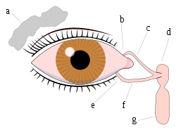
Lacrimal gland
The lacrimal glands are paired almond-shaped glands, one for each eye, that secrete the aqueous layer of the tear film. They are situated in the upper, outer portion of each orbit, in the lacrimal fossa of the orbit formed by the frontal bone. Inflammation of the lacrimal glands is called...
, located superotemporally within the orbit, produces protrusion of the eye inferiorly and medially (away from the location of the lacrimal gland). Lacrimal gland may be enlarged from inflammation
Inflammation
Inflammation is part of the complex biological response of vascular tissues to harmful stimuli, such as pathogens, damaged cells, or irritants. Inflammation is a protective attempt by the organism to remove the injurious stimuli and to initiate the healing process...
(e.g. sarcoid
Sarcoidosis
Sarcoidosis , also called sarcoid, Besnier-Boeck disease or Besnier-Boeck-Schaumann disease, is a disease in which abnormal collections of chronic inflammatory cells form as nodules in multiple organs. The cause of sarcoidosis is unknown...
) or neoplasm (e.g. lymphoma
Lymphoma
Lymphoma is a cancer in the lymphatic cells of the immune system. Typically, lymphomas present as a solid tumor of lymphoid cells. Treatment might involve chemotherapy and in some cases radiotherapy and/or bone marrow transplantation, and can be curable depending on the histology, type, and stage...
or adenoid cystic carcinoma
Adenoid cystic carcinoma
Adenoid cystic cancer is a rare type of cancer that can exist in many different body sites. It most often occurs in the areas of the head and neck, in particular the salivary glands; but has also been reported in the breast, lacrimal gland of the eye, lung, brain, bartholin gland, trachea, and...
).
Tumors (e.g. glioma
Glioma
A glioma is a type of tumor that starts in the brain or spine. It is called a glioma because it arises from glial cells. The most common site of gliomas is the brain.-By type of cell:...
and meningioma
Meningioma
The word meningioma was first used by Harvey Cushing in 1922 to describe a tumor originating from the meninges, the membranous layers surrounding the CNS ....
of the optic nerve
Optic nerve
The optic nerve, also called cranial nerve 2, transmits visual information from the retina to the brain. Derived from the embryonic retinal ganglion cell, a diverticulum located in the diencephalon, the optic nerve doesn't regenerate after transection.-Anatomy:The optic nerve is the second of...
) within the cone formed by the horizontal rectus muscles produce axial protrusion (bulging forward) of the eye.
Graves disease may also cause axial protrusion of the eye, known as Graves' ophthalmopathy, due to buildup of extracellular matrix
Extracellular matrix
In biology, the extracellular matrix is the extracellular part of animal tissue that usually provides structural support to the animal cells in addition to performing various other important functions. The extracellular matrix is the defining feature of connective tissue in animals.Extracellular...
protein
Protein
Proteins are biochemical compounds consisting of one or more polypeptides typically folded into a globular or fibrous form, facilitating a biological function. A polypeptide is a single linear polymer chain of amino acids bonded together by peptide bonds between the carboxyl and amino groups of...
s and fibrosis
Fibrosis
Fibrosis is the formation of excess fibrous connective tissue in an organ or tissue in a reparative or reactive process. This is as opposed to formation of fibrous tissue as a normal constituent of an organ or tissue...
in the rectus muscles. Development of Graves' ophthalmopathy may be independent of thyroid function.
Contents
- Eyeball
- FasciaFasciaA fascia is a layer of fibrous tissue that permeates the human body. A fascia is a connective tissue that surrounds muscles, groups of muscles, blood vessels, and nerves, binding those structures together in much the same manner as plastic wrap can be used to hold the contents of sandwiches...
s: Orbital, Bulbar - Extraocular musclesMuscles of orbitThe muscles of the orbit are a group of six muscles that control movement of the eye. Four of the muscles control the movement of the eye in the four cardinal directions: up, down, left and right...
(Levator Palpebrae Superioris, Superior, Inferior, Lateral and Medial Rectus muscles, Superior and Inferior Oblique Muscles) - NerveNerveA peripheral nerve, or simply nerve, is an enclosed, cable-like bundle of peripheral axons . A nerve provides a common pathway for the electrochemical nerve impulses that are transmitted along each of the axons. Nerves are found only in the peripheral nervous system...
s: cranial nerves IIOptic nerveThe optic nerve, also called cranial nerve 2, transmits visual information from the retina to the brain. Derived from the embryonic retinal ganglion cell, a diverticulum located in the diencephalon, the optic nerve doesn't regenerate after transection.-Anatomy:The optic nerve is the second of...
, IIIOculomotor nerveThe oculomotor nerve is the 3rd of 12 paired cranial nerves. It enters the orbit via the superior orbital fissure and controls most of the eye's movements, including constriction of the pupil and maintaining an open eyelid by innervating the Levator palpebrae superiors muscle. The optic nerve is...
, IVTrochlear nerveThe trochlear nerve is a motor nerve that innervates a single muscle: the superior oblique muscle of the eye....
, VTrigeminal nerveThe trigeminal nerve contains both sensory and motor fibres. It is responsible for sensation in the face and certain motor functions such as biting, chewing, and swallowing. Sensory information from the face and body is processed by parallel pathways in the central nervous system...
, and VI - Blood vesselBlood vesselThe blood vessels are the part of the circulatory system that transports blood throughout the body. There are three major types of blood vessels: the arteries, which carry the blood away from the heart; the capillaries, which enable the actual exchange of water and chemicals between the blood and...
s - Extraocular FatFatFats consist of a wide group of compounds that are generally soluble in organic solvents and generally insoluble in water. Chemically, fats are triglycerides, triesters of glycerol and any of several fatty acids. Fats may be either solid or liquid at room temperature, depending on their structure...
- Lacrimal glandLacrimal glandThe lacrimal glands are paired almond-shaped glands, one for each eye, that secrete the aqueous layer of the tear film. They are situated in the upper, outer portion of each orbit, in the lacrimal fossa of the orbit formed by the frontal bone. Inflammation of the lacrimal glands is called...
, Lacrimal sacLacrimal sacThe lacrimal sac is the upper dilated end of the nasolacrimal duct, and is lodged in a deep groove formed by the lacrimal bone and frontal process of the maxilla...
, Nasolacrimal ductNasolacrimal ductThe nasolacrimal duct carries tears from the lacrimal sac into the nasal cavity. Excess tears flow through nasolacrimal duct which drains into the inferior nasal meatus... - Eyelids
- Medial palpebral ligamentMedial palpebral ligamentThe medial palpebral ligament , about 4 mm. in length and 2 mm. in breadth, is attached to the frontal process of the maxilla in front of the lacrimal groove....
and Lateral palpebral ligament - Medial and Lateral Check ligaments
- Suspensory ligamentZonule of ZinnThe zonule of Zinn is a ring of fibrous strands connecting the ciliary body with the crystalline lens of the eye....
of the eyeball - ConjunctivaConjunctivaThe conjunctiva covers the sclera and lines the inside of the eyelids. It is composed of rare stratified columnar epithelium.-Function:...
- Trochlea of superior obliqueTrochlea of superior obliqueThe Trochlea of superior oblique is a pulley structure in the eye. The tendon of the superior oblique muscle passes through it. Situated on the superior nasal aspect of the frontal bone, it is the only cartilage found in the normal orbit....
- Orbital septumOrbital septumThe orbital septum is a membranous sheet that acts as the anterior boundary of the orbit. It extends from the orbital rims to the eyelids...
- Ciliary ganglionCiliary ganglionThe ciliary ganglion is a parasympathetic ganglion located in the posterior orbit. It measures 1–2 millimeters in diameter and contains approximately 2,500 neurons. Preganglionic axons from the Edinger-Westphal nucleus travel along the oculomotor nerve and form synapses with these cells...
and short ciliary nerves
Bones
In humans, seven bones make up the bony orbit:- Frontal boneFrontal boneThe frontal bone is a bone in the human skull that resembles a cockleshell in form, and consists of two portions:* a vertical portion, the squama frontalis, corresponding with the region of the forehead....
(Pars orbitalisPars orbitalisThe orbital or horizontal part of the frontal bone consists of two thin triangular plates, the orbital plates, which form the vaults of the orbits, and are separated from one another by a median gap, the ethmoidal notch.- Surfaces :...
) - Lacrimal boneLacrimal boneThe lacrimal bone, the smallest and most fragile bone of the face, is situated at the front part of the medial wall of the orbit. It has two surfaces and four borders.-Lateral or orbital surface:...
- Ethmoid boneEthmoid boneThe ethmoid bone is a bone in the skull that separates the nasal cavity from the brain. As such, it is located at the roof of the nose, between the two orbits. The cubical bone is lightweight due to a spongy construction. The ethmoid bone is one of the bones that makes up the orbit of the eye...
(Lamina papyracea) - Zygomatic boneZygomatic boneThe zygomatic bone is a paired bone of the human skull. It articulates with the maxilla, the temporal bone, the sphenoid bone and the frontal bone. The zygomatic is homologous to the jugal bone of other tetrapods...
(Orbital process of the zygomatic boneOrbital process of the zygomatic boneThe orbital process of the zygomatic bone is a thick, strong plate, projecting backward and medialward from the orbital margin.Its antero-medial surface forms, by its junction with the orbital surface of the maxilla and with the great wing of the sphenoid, part of the floor and lateral wall of the...
) - Maxillary bone (Orbital surface of the body of the maxillaOrbital surface of the body of the maxillaThe orbital surface is smooth and triangular, and forms the greater part of the floor of the orbit.It is bounded medially by an irregular margin which in front presents a notch, the lacrimal notch; behind this notch the margin articulates with the lacrimal, the lamina papyracea of the ethmoid and...
) - Palatine bonePalatine boneThe palatine bone is a bone in many species of the animal kingdom, commonly termed the palatum .-Human anatomy:...
(Orbital process of palatine boneOrbital process of palatine boneThe orbital process of the palatine bone is placed on a higher level than the sphenoidal, and is directed upward and lateralward from the front of the vertical part, to which it is connected by a constricted neck. It presents five surfaces, which enclose an air cell...
) - Sphenoid boneSphenoid boneThe sphenoid bone is an unpaired bone situated at the base of the skull in front of the temporal bone and basilar part of the occipital bone.The sphenoid bone is one of the seven bones that articulate to form the orbit...
(Greater and lesser wings)
Foramina and openings
- Optic canal
- Superior orbital fissure
- Inferior orbital fissureInferior orbital fissureNot to be confused with the infraorbital groove, infraorbital canal, and infraorbital foramen.The lateral wall and the floor of the orbit are separated posteriorly by the inferior orbital fissure which transmits the maxillary nerve and its zygomatic branch, and the ascending branches from the...
- Anterior ethmoidal foramenAnterior ethmoidal foramenLateral to either olfactory groove are the internal openings of the anterior and posterior ethmoidal foramina .The anterior ethmoidal foramen, situated about the middle of the lateral margin of the olfactory groove, transmits the anterior ethmoidal vessels and the anterior ethmoidal nerve; the...
- Posterior ethmoidal foramenPosterior ethmoidal foramenLateral to either olfactory groove are the internal openings of the anterior and posterior ethmoidal foramina .The posterior ethmoidal foramen opens at the back part of this margin under cover of the projecting lamina of the sphenoid, and transmits the posterior ethmoidal vessels and nerve....
- Infraorbital foramenInfraorbital foramenAbove the canine fossa is the infraorbital foramen, the end of the infraorbital canal; it transmits the infraorbital artery, vein, and infraorbital nerve.-External links: *...
- Supraorbital foramenSupraorbital foramenThe supraorbital foramen is a bony elongated path located above the orbit and under the forehead. The supraorbital foramen lies directly under the eyebrow....
- Naso-lacrimal canal opening
- Zygomatic orbital foramen