

Western blot
Encyclopedia
Analytical technique
An analytical technique is a method that is used to determine the concentration of a chemical compound or chemical element. There are a wide variety of techniques used for analysis, from simple weighing to titrations to very advanced techniques using highly specialized instrumentation...
used to detect specific proteins in the given sample of tissue homogenate or extract. It uses gel electrophoresis
Gel electrophoresis
Gel electrophoresis is a method used in clinical chemistry to separate proteins by charge and or size and in biochemistry and molecular biology to separate a mixed population of DNA and RNA fragments by length, to estimate the size of DNA and RNA fragments or to separate proteins by charge...
to separate native proteins by 3-D structure or denatured proteins by the length of the polypeptide. The proteins are then transferred to a membrane (typically nitrocellulose
Nitrocellulose
Nitrocellulose is a highly flammable compound formed by nitrating cellulose through exposure to nitric acid or another powerful nitrating agent. When used as a propellant or low-order explosive, it is also known as guncotton...
or PVDF), where they are probed (detected) using antibodies
Antibody
An antibody, also known as an immunoglobulin, is a large Y-shaped protein used by the immune system to identify and neutralize foreign objects such as bacteria and viruses. The antibody recognizes a unique part of the foreign target, termed an antigen...
specific to the target protein.
There are now many reagent companies that specialize in providing antibodies (both monoclonal
Monoclonal antibodies
Monoclonal antibodies are monospecific antibodies that are the same because they are made by identical immune cells that are all clones of a unique parent cell....
and polyclonal antibodies) against tens of thousands of different proteins. Commercial antibodies can be expensive, although the unbound antibody can be reused between experiments. This method is used in the fields of molecular biology
Molecular biology
Molecular biology is the branch of biology that deals with the molecular basis of biological activity. This field overlaps with other areas of biology and chemistry, particularly genetics and biochemistry...
, biochemistry
Biochemistry
Biochemistry, sometimes called biological chemistry, is the study of chemical processes in living organisms, including, but not limited to, living matter. Biochemistry governs all living organisms and living processes...
, immunogenetics
Immunogenetics
Immunogenetics is the branch of medical research that explores the relationship between the immune system and genetics.Autoimmune diseases, such as type 1 diabetes, are complex genetic traits which result from defects in the immune system. Identification of genes defining the immune defects may...
and other molecular biology disciplines.
Other related techniques include using antibodies to detect proteins in tissues and cells by immunostaining
Immunostaining
Immunostaining is a general term in biochemistry that applies to any use of an antibody-based method to detect a specific protein in a sample. The term immunostaining was originally used to refer to the immunohistochemical staining of tissue sections, as first described by Albert Coons in 1941...
and enzyme-linked immunosorbent assay (ELISA
ELISA
Enzyme-linked immunosorbent assay , is a popular format of a "wet-lab" type analytic biochemistry assay that uses one sub-type of heterogeneous, solid-phase enzyme immunoassay to detect the presence of a substance in a liquid sample."Wet lab" analytic biochemistry assays involves detection of an...
).
The method originated in the laboratory of George Stark at Stanford
Stanford University
The Leland Stanford Junior University, commonly referred to as Stanford University or Stanford, is a private research university on an campus located near Palo Alto, California. It is situated in the northwestern Santa Clara Valley on the San Francisco Peninsula, approximately northwest of San...
. The name Western blot was given to the technique by W. Neal Burnette and Sushant Bhat and is a play on the name Southern blot
Southern blot
A Southern blot is a method routinely used in molecular biology for detection of a specific DNA sequence in DNA samples. Southern blotting combines transfer of electrophoresis-separated DNA fragments to a filter membrane and subsequent fragment detection by probe hybridization. The method is named...
, a technique for DNA
DNA
Deoxyribonucleic acid is a nucleic acid that contains the genetic instructions used in the development and functioning of all known living organisms . The DNA segments that carry this genetic information are called genes, but other DNA sequences have structural purposes, or are involved in...
detection developed earlier by Edwin Southern
Edwin Southern
Sir Edwin Mellor Southern, FRS is an English 2005 Lasker Award-winning molecular biologist. His award was for the invention of the Southern blot, now a common laboratory procedure, when he was working at the University of Edinburgh....
. Detection of RNA is termed Northern blot
Northern blot
The northern blot is a technique used in molecular biology research to study gene expression by detection of RNA in a sample. With northern blotting it is possible to observe cellular control over structure and function by determining the particular gene expression levels during differentiation,...
.
Tissue preparation
Samples can not be taken from whole tissue or from cell culture. solid tissues are first broken down mechanically using a blenderBlender
A blender is a kitchen appliance for chopping or liquefying food.Blender may also refer to:Media:* Blender , a music-themed magazine* Blender , a free and open-source software program for 3D modeling, animation, and rendering...
(for larger sample volumes), using a homogenizer
Homogenizer
A homogenizer is a piece of laboratory equipment used for the homogenization of various types of material, such as tissue, plant, food, soil, and many others. Many different models have been developed using various physical technologies for disruption. The 'mortar and pestle', already used for...
(smaller volumes), or by sonication
Sonication
thumb|right|A sonicator at the [[Weizmann Institute of Science]] during sonicationSonication is the act of applying sound energy to agitate particles in a sample, for various purposes. In the laboratory, it is usually applied using an ultrasonic bath or an ultrasonic probe, colloquially known as...
. Cells may also be broken open by one of the above mechanical methods. However, virus or environmental samples can be the source of protein and thus western blotting is not restricted to cellular studies only.
Assorted detergent
Detergent
A detergent is a surfactant or a mixture of surfactants with "cleaning properties in dilute solutions." In common usage, "detergent" refers to alkylbenzenesulfonates, a family of compounds that are similar to soap but are less affected by hard water...
s, salts, and buffer
Buffer solution
A buffer solution is an aqueous solution consisting of a mixture of a weak acid and its conjugate base or a weak base and its conjugate acid. It has the property that the pH of the solution changes very little when a small amount of strong acid or base is added to it. Buffer solutions are used as a...
s may be employed to encourage lysis
Lysis
Lysis refers to the breaking down of a cell, often by viral, enzymic, or osmotic mechanisms that compromise its integrity. A fluid containing the contents of lysed cells is called a "lysate"....
of cells and to solubilize proteins. Protease
Protease
A protease is any enzyme that conducts proteolysis, that is, begins protein catabolism by hydrolysis of the peptide bonds that link amino acids together in the polypeptide chain forming the protein....
and phosphatase
Alkaline phosphatase
Alkaline phosphatase is a hydrolase enzyme responsible for removing phosphate groups from many types of molecules, including nucleotides, proteins, and alkaloids. The process of removing the phosphate group is called dephosphorylation...
inhibitors are often added to prevent the digestion of the sample by its own enzymes. Tissue preparation is often done at cold temperatures to avoid protein denaturing
Denaturation (biochemistry)
Denaturation is a process in which proteins or nucleic acids lose their tertiary structure and secondary structure by application of some external stress or compound, such as a strong acid or base, a concentrated inorganic salt, an organic solvent , or heat...
and degradation.
A combination of biochemical and mechanical techniques – comprising various types of filtration and centrifugation
Centrifuge
A centrifuge is a piece of equipment, generally driven by an electric motor , that puts an object in rotation around a fixed axis, applying a force perpendicular to the axis...
– can be used to separate different cell compartments and organelle
Organelle
In cell biology, an organelle is a specialized subunit within a cell that has a specific function, and is usually separately enclosed within its own lipid bilayer....
s.
Gel electrophoresis
The proteins of the sample are separated using gel electrophoresisGel electrophoresis
Gel electrophoresis is a method used in clinical chemistry to separate proteins by charge and or size and in biochemistry and molecular biology to separate a mixed population of DNA and RNA fragments by length, to estimate the size of DNA and RNA fragments or to separate proteins by charge...
. Separation of proteins may be by isoelectric point
Isoelectric point
The isoelectric point , sometimes abbreviated to IEP, is the pH at which a particular molecule or surface carries no net electrical charge....
(pI), molecular weight, electric charge, or a combination of these factors. The nature of the separation depends on the treatment of the sample and the nature of the gel. This is a very useful way to identify a protein.
By far the most common type of gel electrophoresis employs polyacrylamide
Polyacrylamide
Polyacrylamide is a polymer formed from acrylamide subunits. It can be synthesized as a simple linear-chain structure or cross-linked, typically using N,N-methylenebisacrylamide. Polyacrylamide is not toxic...
gels and buffers loaded with sodium dodecyl sulfate
Sodium dodecyl sulfate
Sodium dodecyl sulfate , sodium laurilsulfate or sodium lauryl sulfate is an organic compound with the formula CH311OSO3Na). It is an anionic surfactant used in many cleaning and hygiene products...
(SDS). SDS-PAGE
SDS-PAGE
SDS-PAGE, sodium dodecyl sulfate polyacrylamide gel electrophoresis, describes a collection of related techniques widely used in biochemistry, forensics, genetics and molecular biology to separate proteins according to their electrophoretic mobility...
(SDS polyacrylamide gel electrophoresis) maintains polypeptides in a denatured state once they have been treated with strong reducing agents to remove secondary and tertiary structure (e.g. disulfide bonds [S-S] to sulfhydryl groups [SH and SH]) and thus allows separation of proteins by their molecular weight. Sampled proteins become covered in the negatively charged SDS and move to the positively charged electrode through the acrylamide
Acrylamide
Acrylamide is a chemical compound with the chemical formula C3H5NO. Its IUPAC name is prop-2-enamide. It is a white odourless crystalline solid, soluble in water, ethanol, ether, and chloroform. Acrylamide is incompatible with acids, bases, oxidizing agents, iron, and iron salts...
mesh of the gel. Smaller proteins migrate faster through this mesh and the proteins are thus separated according to size (usually measured in kilodaltons, kDa
KDA
KDA may refer to:* Karachi Development Authority* Kongsberg Defence & Aerospace* Kotelawala Defence Academy* Kramer Design Associates* Lithium diisopropylamide, KDA is the potassium analogue of lithium diisopropylamideOr kDa may refer to:...
). The concentration of acrylamide determines the resolution of the gel - the greater the acrylamide concentration the better the resolution of lower molecular weight proteins. The lower the acrylamide concentration the better the resolution of higher molecular weight proteins. Proteins travel only in one dimension along the gel for most blots.
Samples are loaded into wells in the gel. One lane is usually reserved for a marker or ladder, a commercially available mixture of proteins having defined molecular weights, typically stained so as to form visible, coloured bands. When voltage
Voltage
Voltage, otherwise known as electrical potential difference or electric tension is the difference in electric potential between two points — or the difference in electric potential energy per unit charge between two points...
is applied along the gel, proteins migrate into it at different speeds. These different rates of advancement (different electrophoretic mobilities) separate into bands within each lane.
It is also possible to use a two-dimensional (2-D) gel which spreads the proteins from a single sample out in two dimensions. Proteins are separated according to isoelectric point (pH at which they have neutral net charge) in the first dimension, and according to their molecular weight in the second dimension.
Transfer
In order to make the proteins accessible to antibody detection, they are moved from within the gel onto a membrane made of nitrocelluloseNitrocellulose
Nitrocellulose is a highly flammable compound formed by nitrating cellulose through exposure to nitric acid or another powerful nitrating agent. When used as a propellant or low-order explosive, it is also known as guncotton...
or polyvinylidene difluoride (PVDF). The membrane is placed on top of the gel, and a stack of filter papers placed on top of that. The entire stack is placed in a buffer solution which moves up the paper by capillary action
Capillary action
Capillary action, or capilarity, is the ability of a liquid to flow against gravity where liquid spontanously rise in a narrow space such as between the hair of a paint-brush, in a thin tube, or in porous material such as paper or in some non-porous material such as liquified carbon fiber, or in a...
, bringing the proteins with it. In practice this method is not used as it takes to much time; another method is used. The other method for transferring the proteins is called electroblotting
Electroblotting
Electroblotting is a method in molecular biology/biochemistry/immunogenetics to transfer proteins or nucleic acids onto a membrane by using PVDF or nitrocellulose, after gel electrophoresis. The protein or nucleic acid can then be further analyzed using probes such as specific antibodies, ligands...
and uses an electric current to pull proteins from the gel into the PVDF or nitrocellulose membrane. The proteins move from within the gel onto the membrane while maintaining the organization they had within the gel. As a result of this "blotting" process, the proteins are exposed on a thin surface layer for detection (see below). Both varieties of membrane are chosen for their non-specific protein binding properties (i.e. binds all proteins equally well). Protein binding is based upon hydrophobic interactions, as well as charged interactions between the membrane and protein. Nitrocellulose membranes are cheaper than PVDF, but are far more fragile and do not stand up well to repeated probings.
The uniformity and overall effectiveness of transfer of protein from the gel to the membrane can be checked by staining the membrane with Coomassie Brilliant Blue or Ponceau S
Ponceau S
Ponceau S, Acid Red 112, or C.I. 27195 is a sodium salt of a diazo dye that may be used to prepare a stain for rapid reversible detection of protein bands on nitrocellulose or PVDF membranes , as well as on cellulose acetate membranes...
dyes. Ponceau S is the more common of the two, due to its higher sensitivity and water solubility, the latter making it easier to subsequently destain and probe the membrane, as described below.
Blocking
Since the membrane has been chosen for its ability to bind protein and as both antibodies and the target are proteins, steps must be taken to prevent interactions between the membrane and the antibody used for detection of the target protein. Blocking of non-specific binding is achieved by placing the membrane in a dilute solution of protein - typically 3-5% Bovine serum albuminBovine serum albumin
Bovine serum albumin is a serum albumin protein derived from cows. It is often used as a protein concentration standard....
(BSA) or non-fat dry milk
Powdered milk
Powdered milk is a manufactured dairy product made by evaporating milk to dryness. One purpose of drying milk is to preserve it; milk powder has a far longer shelf life than liquid milk and does not need to be refrigerated, due to its low moisture content. Another purpose is to reduce its bulk for...
(both are inexpensive) in Tris-Buffered Saline
Tris-Buffered Saline
Tris-buffered saline is a buffer used in some biochemical techniques to maintain the pH within a relatively narrow range. Tris has a slightly alkaline buffering capacity in the 7–9.2 range....
(TBS), with a minute percentage of detergent such as Tween 20
Tween 20
Polysorbate 20 is a polysorbate surfactant whose stability and relative non-toxicity allows it to be used as a detergent and emulsifier in a number of domestic, scientific, and pharmacological applications...
or Triton X-100. The protein in the dilute solution attaches to the membrane in all places where the target proteins have not attached. Thus, when the antibody is added, there is no room on the membrane for it to attach other than on the binding sites of the specific target protein. This reduces "noise" in the final product of the western blot, leading to clearer results, and eliminates false positives.
Detection
During the detection process the membrane is "probed" for the protein of interest with a modified antibody which is linked to a reporter enzyme; when exposed to an appropriate substrate this enzyme drives a colourimetric reaction and produces a colour. For a variety of reasons, this traditionally takes place in a two-step process, although there are now one-step detection methods available for certain applications.Two steps
- Primary antibody
Primary antibodies are generated when a host species or immune cell culture is exposed to the protein of interest (or a part thereof). Normally, this is part of the immune response, whereas here they are harvested and used as sensitive and specific detection tools that bind the protein directly.
After blocking, a dilute solution of primary antibody (generally between 0.5 and 5 micrograms/mL) is incubated with the membrane under gentle agitation. Typically, the solution is composed of buffered saline solution with a small percentage of detergent, and sometimes with powdered milk or BSA. The antibody solution and the membrane can be sealed and incubated together for anywhere from 30 minutes to overnight. It can also be incubated at different temperatures, with warmer temperatures being associated with more binding, both specific (to the target protein, the "signal") and non-specific ("noise").
- Secondary antibody
After rinsing the membrane to remove unbound primary antibody, the membrane is exposed to another antibody, directed at a species-specific portion of the primary antibody. Antibodies come from animal sources (or animal sourced hybridoma
Hybridoma
Hybridoma technology is a technology of forming hybrid cell lines by fusing a specific antibody-producing B cell with a myeloma cell that is selected for its ability to grow in tissue culture and for an absence of antibody chain synthesis...
cultures); an anti-mouse secondary will bind to almost any mouse-sourced primary antibody, which allows some cost savings by allowing an entire lab to share a single source of mass-produced antibody, and provides far more consistent results. This is known as a secondary antibody, and due to its targeting properties, tends to be referred to as "anti-mouse," "anti-goat," etc. The secondary antibody is usually linked to biotin
Biotin
Biotin, also known as Vitamin H or Coenzyme R, is a water-soluble B-complex vitamin discovered by Bateman in 1916. It is composed of a ureido ring fused with a tetrahydrothiophene ring. A valeric acid substituent is attached to one of the carbon atoms of the tetrahydrothiophene ring...
or to a reporter enzyme
Enzyme
Enzymes are proteins that catalyze chemical reactions. In enzymatic reactions, the molecules at the beginning of the process, called substrates, are converted into different molecules, called products. Almost all chemical reactions in a biological cell need enzymes in order to occur at rates...
such as alkaline phosphatase
Alkaline phosphatase
Alkaline phosphatase is a hydrolase enzyme responsible for removing phosphate groups from many types of molecules, including nucleotides, proteins, and alkaloids. The process of removing the phosphate group is called dephosphorylation...
or horseradish peroxidase
Horseradish peroxidase
The enzyme horseradish peroxidase , found in horseradish, is used extensively in biochemistry applications primarily for its ability to amplify a weak signal and increase detectability of a target molecule.-Applications:...
. This means that several secondary antibodies will bind to one primary antibody and enhance the signal.
Most commonly, a horseradish peroxidase
Horseradish peroxidase
The enzyme horseradish peroxidase , found in horseradish, is used extensively in biochemistry applications primarily for its ability to amplify a weak signal and increase detectability of a target molecule.-Applications:...
-linked secondary is used to cleave a chemiluminescent agent, and the reaction product produces luminescence
Luminescence
Luminescence is emission of light by a substance not resulting from heat; it is thus a form of cold body radiation. It can be caused by chemical reactions, electrical energy, subatomic motions, or stress on a crystal. This distinguishes luminescence from incandescence, which is light emitted by a...
in proportion to the amount of protein. A sensitive sheet of photographic film is placed against the membrane, and exposure to the light from the reaction creates an image of the antibodies bound to the blot. A cheaper but less sensitive approach utilizes a 4-chloronaphthol stain with 1% hydrogen peroxide
Hydrogen peroxide
Hydrogen peroxide is the simplest peroxide and an oxidizer. Hydrogen peroxide is a clear liquid, slightly more viscous than water. In dilute solution, it appears colorless. With its oxidizing properties, hydrogen peroxide is often used as a bleach or cleaning agent...
; reaction of peroxide radicals with 4-chloronaphthol produces a dark purple stain that can be photographed without using specialized photographic film.
As with the ELISPOT
ELISPOT
The Enzyme-linked immunosorbent spot assay is a common method for monitoring immune responses in humans and animals. It was developed by Cecil Czerkinsky in 1983....
and ELISA
ELISA
Enzyme-linked immunosorbent assay , is a popular format of a "wet-lab" type analytic biochemistry assay that uses one sub-type of heterogeneous, solid-phase enzyme immunoassay to detect the presence of a substance in a liquid sample."Wet lab" analytic biochemistry assays involves detection of an...
procedures, the enzyme can be provided with a substrate molecule that will be converted by the enzyme to a colored reaction product that will be visible on the membrane (see the figure below with blue bands).
Another method of secondary antibody detection utilizes a near-infrared (NIR) fluorophore-linked antibody. Light produced from the excitation of a fluorescent dye is static, making fluorescent detection a more precise and accurate measure of the difference in signal produced by labeled antibodies bound to proteins on a western blot. Proteins can be accurately quantified because the signal generated by the different amounts of proteins on the membranes is measured in a static state, as compared to chemiluminescence, in which light is measured in a dynamic state.
A third alternative is to use a radioactive label rather than an enzyme coupled to the secondary antibody, such as labeling an antibody-binding protein like Staphylococcus
Staphylococcus
Staphylococcus is a genus of Gram-positive bacteria. Under the microscope they appear round , and form in grape-like clusters....
Protein A or Streptavidin with a radioactive isotope of iodine. Since other methods are safer, quicker, and cheaper, this method is now rarely used; however, an advantage of this approach is the sensitivity of auto-radiography based imaging, which enables highly accurate protein quantification when combined with optical software (e.g. Optiquant).
One step
Historically, the probing process was performed in two steps because of the relative ease of producing primary and secondary antibodies in separate processes. This gives researchers and corporations huge advantages in terms of flexibility, and adds an amplification step to the detection process. Given the advent of high-throughput protein analysis and lower limits of detection, however, there has been interest in developing one-step probing systems that would allow the process to occur faster and with less consumables. This requires a probe antibody which both recognizes the protein of interest and contains a detectable label, probes which are often available for known protein tags. The primary probe is incubated with the membrane in a manner similar to that for the primary antibody in a two-step process, and then is ready for direct detection after a series of wash steps.
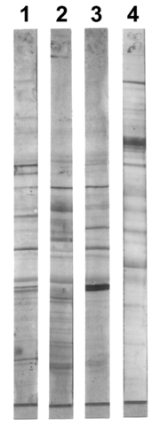
Analysis
After the unbound probes are washed away, the western blot is ready for detection of the probes that are labeled and bound to the protein of interest. In practical terms, not all westerns reveal protein only at one band in a membrane. Size approximations are taken by comparing the stained bands to that of the marker or ladder loaded during electrophoresis. The process is repeated for a structural protein, such as actin or tubulin, that should not change between samples. The amount of target protein is normalizedNormalization (statistics)
In one usage in statistics, normalization is the process of isolating statistical error in repeated measured data. A normalization is sometimes based on a property...
to the structural protein to control between groups. This practice ensures correction for the amount of total protein on the membrane in case of errors or incomplete transfers.
Colorimetric detection
The colorimetric detection method depends on incubation of the western blot with a substrate that reacts with the reporter enzyme (such as peroxidasePeroxidase
Peroxidases are a large family of enzymes that typically catalyze a reaction of the form:For many of these enzymes the optimal substrate is hydrogen peroxide, but others are more active with organic hydroperoxides such as lipid peroxides...
) that is bound to the secondary antibody. This converts the soluble dye into an insoluble form of a different color that precipitates next to the enzyme and thereby stains the membrane. Development of the blot is then stopped by washing away the soluble dye. Protein levels are evaluated through densitometry
Densitometry
Densitometry is the quantitative measurement of optical density in light-sensitive materials, such as photographic paper or film, due to exposure to light...
(how intense the stain is) or spectrophotometry
Spectrophotometry
In chemistry, spectrophotometry is the quantitative measurement of the reflection or transmission properties of a material as a function of wavelength...
.
Chemiluminescent detection
Chemiluminescent detection methods depend on incubation of the western blot with a substrate that will luminesce when exposed to the reporter on the secondary antibody. The light is then detected by photographic film, and more recently by CCD cameras which capture a digital image of the western blot. The image is analysed by densitometry, which evaluates the relative amount of protein staining and quantifies the results in terms of optical density. Newer software allows further data analysis such as molecular weight analysis if appropriate standards are used.Radioactive detection
Radioactive labels do not require enzyme substrates, but rather allow the placement of medical X-ray film directly against the western blot which develops as it is exposed to the label and creates dark regions which correspond to the protein bands of interest (see image to the right). The importance of radioactive detections methods is declining due to its hazardous radiation , because it is very expensive, health and safety risks are high, and ECL (enhanced chemiluminescence) provides a useful alternative.Fluorescent detection
The fluorescently labeled probe is excited by light and the emission of the excitation is then detected by a photosensor such as CCD camera equipped with appropriate emission filters which captures a digital image of the western blot and allows further data analysis such as molecular weight analysis and a quantitative western blot analysis. Fluorescence is considered to be one of the best methods for quantification, but is less sensitive than chemiluminescence.Secondary probing
One major difference between nitrocellulose and PVDF membranes relates to the ability of each to support "stripping" antibodies off and reusing the membrane for subsequent antibody probes. While there are well-established protocols available for stripping nitrocellulose membranes, the sturdier PVDF allows for easier stripping, and for more reuse before background noise limits experiments. Another difference is that, unlike nitrocellulose, PVDF must be soaked in 95% ethanol, isopropanol or methanol before use. PVDF membranes also tend to be thicker and more resistant to damage during use.2-D gel electrophoresis
2-dimensional SDS-PAGE uses the principles and techniques outlined above. 2-D SDS-PAGE, as the name suggests, involves the migration of polypeptides in 2 dimensions. For example, in the first dimension polypeptides are separated according to isoelectric pointIsoelectric point
The isoelectric point , sometimes abbreviated to IEP, is the pH at which a particular molecule or surface carries no net electrical charge....
, while in the second dimension polypeptides are separated according to their molecular weight. The isoelectric point of a given protein is determined by the relative number of positively (e.g. lysine and arginine) and negatively (e.g. glutamate and aspartate) charged amino acids, with negatively charged amino acids contributing to a high isoelectric point and positively charged amino acids contributing to a low isoelectric point. Samples could also be separated first under nonreducing conditions using SDS-PAGE and under reducing conditions in the second dimension, which breaks apart disulfide bonds that hold subunits together. SDS-PAGE might also be coupled with urea-PAGE for a 2-dimensional gel.
In principle, this method allows for the separation of all cellular proteins on a single large gel. A major advantage of this method is that it often distinguishes between different isoforms of a particular protein - e.g. a protein that has been phosphorylated (by addition of a negatively charged group). Proteins that have been separated can be cut out of the gel and then analysed by mass spectrometry
Mass spectrometry
Mass spectrometry is an analytical technique that measures the mass-to-charge ratio of charged particles.It is used for determining masses of particles, for determining the elemental composition of a sample or molecule, and for elucidating the chemical structures of molecules, such as peptides and...
, which identifies the protein.
Please refer to reference articles for examples of the application of 2-D SDS PAGE.
Medical diagnostic applications
- The confirmatory HIV testHIV testHIV tests are used to detect the presence of the human immunodeficiency virus , the virus that causes acquired immunodeficiency syndrome , in serum, saliva, or urine. Such tests may detect antibodies, antigens, or RNA.- Terminology :...
employs a western blot to detect anti-HIV antibody in a human serumBlood plasmaBlood plasma is the straw-colored liquid component of blood in which the blood cells in whole blood are normally suspended. It makes up about 55% of the total blood volume. It is the intravascular fluid part of extracellular fluid...
sample. Proteins from known HIVHIVHuman immunodeficiency virus is a lentivirus that causes acquired immunodeficiency syndrome , a condition in humans in which progressive failure of the immune system allows life-threatening opportunistic infections and cancers to thrive...
-infected cells are separated and blotted on a membrane as above. Then, the serum to be tested is applied in the primary antibody incubation step; free antibody is washed away, and a secondary anti-human antibody linked to an enzyme signal is added. The stained bands then indicate the proteins to which the patient's serum contains antibody. - A western blot is also used as the definitive test for Bovine spongiform encephalopathyBovine spongiform encephalopathyBovine spongiform encephalopathy , commonly known as mad-cow disease, is a fatal neurodegenerative disease in cattle that causes a spongy degeneration in the brain and spinal cord. BSE has a long incubation period, about 30 months to 8 years, usually affecting adult cattle at a peak age onset of...
(BSE, commonly referred to as 'mad cow disease'). - Some forms of Lyme diseaseLyme diseaseLyme disease, or Lyme borreliosis, is an emerging infectious disease caused by at least three species of bacteria belonging to the genus Borrelia. Borrelia burgdorferi sensu stricto is the main cause of Lyme disease in the United States, whereas Borrelia afzelii and Borrelia garinii cause most...
testing employ western blotting. - Western blot can also be used as a confirmatory test for Hepatitis B infection.
- In veterinary medicine, western blot is sometimes used to confirm FIV+ status in cats.
Protocols
- Western Blotting Protocol from Protocolmonkey
- Modified western blotting protocols from Biotechniques
- Dr. Mark Barton Frank Lab protocol
- Kamps's Western Blotting Protocol
- http://www.natureprotocols.com/2006/09/15/western_blot_analysis_of_subce.php
- http://www.prosci-inc.com/Western-Blot-Protocol.html
- CSH Protocols collection: Immunoblotting
- western blot protocols, troubleshooting, scientific resources and research articles