

Ventricle (heart)
Encyclopedia
In the heart
, a ventricle is one of two large chambers that collect and expel blood
received from an atrium towards the peripheral beds within the body and lungs. The Atria (an adjacent/upper heart chamber that is smaller than a ventricle) primes the Pump
. Interventricular means between two or more ventricles (for example the interventricular septum
), while intraventricular means within one ventricle (for example an intraventricular block
).
In a four-chambered heart, such as that in human
s, there are two ventricles: the right ventricle
pumps blood into the pulmonary circulation
to/for the lung
s, and the left ventricle
pumps blood into the systemic circulation
through the aorta
(systemic circulation). (See Double circulatory system
for details.)
Ventricles have thicker walls than atria and must allow and withstand higher incoming and outgoing blood pressure
s. The physiologic load on the ventricles requiring pumping of blood throughout the body and lungs is much greater than the pressure generated by the atria to fill the ventricles. Further, the left ventricle has thicker walls than the right because it needs to pump blood to most of the body while the right ventricle fills only the lungs.
The mass of the left ventricle, as estimated by magnetic resonance imaging
, averages 143 g ± 38.4 g, with a range of 87 - 224 g.
, the ventricles contract, pumping blood through the body. During diastole
, the ventricles relax and fill with blood again.
, the performance of the ventricles are measured with several volumetric parameters, including end-diastolic volume
(EDV), end-systolic volume
(ESV), stroke volume
(SV)and ejection fraction
(Ef).
(mostly used for animal model research). Optimally, it is specified with which plane the distance is measured in, e.g. the dimension of the longitudinal plane.
Fractional shortening (FS) is the fraction
of any diastolic dimension that is lost in systole. When referring to endocardial luminal
distances, it is EDD minus ESD divided by EDD (times 100 when measured in percentage). Normal values may differ somewhat dependent on which anatomical plane is used to measure the distances, but a range from 30 to 42% is considered normal with 26 to 30% representing a mild decrease in function. Midwall fractional shortening may also be used to measure diastolic/systolic changes for inter-ventricular septal dimensions and posterior wall dimensions. However, both endocardial and midwall fractional shortening are dependent on myocardial wall thickness, and thereby dependent on long-axis function. By comparison, a measure of short-axis function termed epicardial volume change (EVC) is independent of myocardial wall thickness and represents isolated short-axis function.
Heart
The heart is a myogenic muscular organ found in all animals with a circulatory system , that is responsible for pumping blood throughout the blood vessels by repeated, rhythmic contractions...
, a ventricle is one of two large chambers that collect and expel blood
Blood
Blood is a specialized bodily fluid in animals that delivers necessary substances such as nutrients and oxygen to the cells and transports metabolic waste products away from those same cells....
received from an atrium towards the peripheral beds within the body and lungs. The Atria (an adjacent/upper heart chamber that is smaller than a ventricle) primes the Pump
Pump
A pump is a device used to move fluids, such as liquids, gases or slurries.A pump displaces a volume by physical or mechanical action. Pumps fall into three major groups: direct lift, displacement, and gravity pumps...
. Interventricular means between two or more ventricles (for example the interventricular septum
Interventricular septum
Interventricular septum , abbreviated IVS, is the stout wall separating the lower chambers of the heart from one another....
), while intraventricular means within one ventricle (for example an intraventricular block
Intraventricular block
A intraventricular block is a heart block of the ventricles of the heart.An example is a right bundle branch block....
).
In a four-chambered heart, such as that in human
Human
Humans are the only living species in the Homo genus...
s, there are two ventricles: the right ventricle
Right ventricle
The right ventricle is one of four chambers in the human heart. It receives deoxygenated blood from the right atrium via the tricuspid valve, and pumps it into the pulmonary artery via the pulmonary valve and pulmonary trunk....
pumps blood into the pulmonary circulation
Pulmonary circulation
Pulmonary circulation is the half portion of the cardiovascular system which carries Oxygen-depleted Blood away from the heart, to the Lungs, and returns oxygenated blood back to the heart. Encyclopedic description and discovery of the pulmonary circulation is widely attributed to Doctor Ibn...
to/for the lung
Lung
The lung is the essential respiration organ in many air-breathing animals, including most tetrapods, a few fish and a few snails. In mammals and the more complex life forms, the two lungs are located near the backbone on either side of the heart...
s, and the left ventricle
Left ventricle
The left ventricle is one of four chambers in the human heart. It receives oxygenated blood from the left atrium via the mitral valve, and pumps it into the aorta via the aortic valve.-Shape:...
pumps blood into the systemic circulation
Systemic circulation
Systemic circulation is the part of the cardiovascular system which carries oxygenated blood away from the heart to the body, and returns deoxygenated blood back to the heart. This physiologic theory of circulation was first described by William Harvey...
through the aorta
Aorta
The aorta is the largest artery in the body, originating from the left ventricle of the heart and extending down to the abdomen, where it branches off into two smaller arteries...
(systemic circulation). (See Double circulatory system
Double circulatory system
In a first order circulatory circuit, blood is pumped to the lungs, thus acquiring oxygen while simultaneously releasing carbon dioxide. Fully oxygenated blood then enters the second order circuit, going to the brain and body...
for details.)
Ventricles have thicker walls than atria and must allow and withstand higher incoming and outgoing blood pressure
Blood pressure
Blood pressure is the pressure exerted by circulating blood upon the walls of blood vessels, and is one of the principal vital signs. When used without further specification, "blood pressure" usually refers to the arterial pressure of the systemic circulation. During each heartbeat, BP varies...
s. The physiologic load on the ventricles requiring pumping of blood throughout the body and lungs is much greater than the pressure generated by the atria to fill the ventricles. Further, the left ventricle has thicker walls than the right because it needs to pump blood to most of the body while the right ventricle fills only the lungs.
The mass of the left ventricle, as estimated by magnetic resonance imaging
Magnetic resonance imaging
Magnetic resonance imaging , nuclear magnetic resonance imaging , or magnetic resonance tomography is a medical imaging technique used in radiology to visualize detailed internal structures...
, averages 143 g ± 38.4 g, with a range of 87 - 224 g.
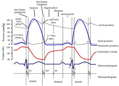
In systole and diastole
During systoleSystole (medicine)
Systole is the contraction of the heart. Used alone, it usually means the contraction of the left ventricle.In all mammals, the heart has 4 chambers. The left and right ventricles pump together. The atria and ventricles pump in sequence...
, the ventricles contract, pumping blood through the body. During diastole
Diastole
Diastole is the period of time when the heart fills with blood after systole . Ventricular diastole is the period during which the ventricles are relaxing, while atrial diastole is the period during which the atria are relaxing...
, the ventricles relax and fill with blood again.
Volumes
In cardiologyCardiology
Cardiology is a medical specialty dealing with disorders of the heart . The field includes diagnosis and treatment of congenital heart defects, coronary artery disease, heart failure, valvular heart disease and electrophysiology...
, the performance of the ventricles are measured with several volumetric parameters, including end-diastolic volume
End-diastolic volume
In cardiovascular physiology, end-diastolic volume is the volume of blood in the right and/or left ventricle at end Load or filling in . Because greater EDVs cause greater distention of the ventricle, EDV is often used synonymously with preload, which refers to the length of the sarcomeres in...
(EDV), end-systolic volume
End-systolic volume
End-systolic volume is the volume of blood in a ventricle at the end of contraction, or systole, and the beginning of filling, or diastole.ESV is the lowest volume of blood in the ventricle at any point in the cardiac cycle....
(ESV), stroke volume
Stroke volume
In cardiovascular physiology, stroke volume is the volume of blood pumped from one ventricle of the heart with each beat. SV is calculated using measurements of ventricle volumes from an echocardiogram and subtracting the volume of the blood in the ventricle at the end of a beat from the volume...
(SV)and ejection fraction
Ejection fraction
In cardiovascular physiology, ejection fraction is the fraction of Blood pumped out of the Right Ventricle of the heart to the Pulmonary Circulation and Left Ventricle of the heart to the Systemic Circulation with each Heart beat or Cardiac cycle...
(Ef).
Dimensions
The heart and its performance are also commonly measured in terms of dimensions, which in this case means one-dimensional distances, usually measured in millimeters. This is not as informative as volumes, but may be much easier to estimate with e.g. M-Mode echocardiography or with sonomicrometrySonomicrometry
Sonomicrometry is a technique of measuring the distance between piezoelectric crystals based on the speed of acoustic signals through the medium they are embedded in. Typically, the crystals will be coated with an epoxy 'lens' and placed into the material facing each other...
(mostly used for animal model research). Optimally, it is specified with which plane the distance is measured in, e.g. the dimension of the longitudinal plane.
| Dimension | Abbreviation | Definition | | Normally |
|---|---|---|---|
| End-diastolic dimension | EDD | The diameter across a ventricle at the end of diastole Diastole Diastole is the period of time when the heart fills with blood after systole . Ventricular diastole is the period during which the ventricles are relaxing, while atrial diastole is the period during which the atria are relaxing... , if not else specified then usually referring to the transverse (left-to-right) internal (luminal Lumen (anatomy) A lumen in biology is the inside space of a tubular structure, such as an artery or intestine... ) distance, excluding thickness of walls, although it can also be measured as the external distance. |
|
|
LVEDD or sometimes LVDD | The end-diastolic dimension of the left ventricle. | 48 mm, Range 36 – 56 mm |
|
|
RVEDD or sometimes RVDD | The end-diastolic dimension of the right ventricle. | Range 10 – 26 mm |
| End-systolic dimension | ESD | ESD is similar to the end-diastolic dimension, but is measured at the end of systole Systole (medicine) Systole is the contraction of the heart. Used alone, it usually means the contraction of the left ventricle.In all mammals, the heart has 4 chambers. The left and right ventricles pump together. The atria and ventricles pump in sequence... (after the ventricles have pumped out blood) rather than at the end of diastole Diastole Diastole is the period of time when the heart fills with blood after systole . Ventricular diastole is the period during which the ventricles are relaxing, while atrial diastole is the period during which the atria are relaxing... . |
|
|
|
LVESD or sometimes LVSD | The end-systolic dimension of the left ventricle. | Range 20 – 40 mm |
|
|
RVESD or sometimes RVSD | The end-systolic dimension of the right ventricle. | Range 10 – 26 mm |
| Interventricular septal end diastolic dimension | IVSd | The thickness of the interventricular septum Interventricular septum Interventricular septum , abbreviated IVS, is the stout wall separating the lower chambers of the heart from one another.... . |
8.3 mm, Range 7 – 11 mm |
| Left ventricular end diastolic posterior wall dimension | LVPWd | The thickness of the posterior left ventricular wall. | 8.3 mm, Range 7 – 11 mm |
| Left atrial dimension | LA | Range 24 – 40 mm |
Fractional shortening (FS) is the fraction
Fraction (mathematics)
A fraction represents a part of a whole or, more generally, any number of equal parts. When spoken in everyday English, we specify how many parts of a certain size there are, for example, one-half, five-eighths and three-quarters.A common or "vulgar" fraction, such as 1/2, 5/8, 3/4, etc., consists...
of any diastolic dimension that is lost in systole. When referring to endocardial luminal
Lumen (anatomy)
A lumen in biology is the inside space of a tubular structure, such as an artery or intestine...
distances, it is EDD minus ESD divided by EDD (times 100 when measured in percentage). Normal values may differ somewhat dependent on which anatomical plane is used to measure the distances, but a range from 30 to 42% is considered normal with 26 to 30% representing a mild decrease in function. Midwall fractional shortening may also be used to measure diastolic/systolic changes for inter-ventricular septal dimensions and posterior wall dimensions. However, both endocardial and midwall fractional shortening are dependent on myocardial wall thickness, and thereby dependent on long-axis function. By comparison, a measure of short-axis function termed epicardial volume change (EVC) is independent of myocardial wall thickness and represents isolated short-axis function.