

Systole (medicine)
Encyclopedia

Left ventricle
The left ventricle is one of four chambers in the human heart. It receives oxygenated blood from the left atrium via the mitral valve, and pumps it into the aorta via the aortic valve.-Shape:...
.
In all mammals, the heart has 4 chambers. The left and right ventricles pump together. The atria and ventricles pump in sequence. The atria feed blood into the ventricles.
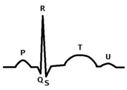
When the smaller, upper atria chambers contract in the first phase of systole, they send blood down to the larger, lower ventricle
Ventricle (heart)
In the heart, a ventricle is one of two large chambers that collect and expel blood received from an atrium towards the peripheral beds within the body and lungs. The Atria primes the Pump...
chambers. When the lower chambers are filled and the valves to the atria are closed, the ventricles contract in the second phase, sending blood from the left ventricle
Left ventricle
The left ventricle is one of four chambers in the human heart. It receives oxygenated blood from the left atrium via the mitral valve, and pumps it into the aorta via the aortic valve.-Shape:...
to the aorta
Aorta
The aorta is the largest artery in the body, originating from the left ventricle of the heart and extending down to the abdomen, where it branches off into two smaller arteries...
and body, and from the right ventricle
Right ventricle
The right ventricle is one of four chambers in the human heart. It receives deoxygenated blood from the right atrium via the tricuspid valve, and pumps it into the pulmonary artery via the pulmonary valve and pulmonary trunk....
to the lungs.
Cardiac systole is the contraction of specialized muscle in response to an electrochemical stimulus to the heart cells (cardiomyocytes).
The cardiac output
Cardiac output
Cardiac output is the volume of blood being pumped by the heart, in particular by a left or right ventricle in the time interval of one minute. CO may be measured in many ways, for example dm3/min...
(CO) is the volume of blood pumped by the left ventricle in 1 minute. The ejection fraction
Ejection fraction
In cardiovascular physiology, ejection fraction is the fraction of Blood pumped out of the Right Ventricle of the heart to the Pulmonary Circulation and Left Ventricle of the heart to the Systemic Circulation with each Heart beat or Cardiac cycle...
(EF) is the fraction of blood pumped divided by the total amount of blood in the left ventricle.
Electrical systole
Electrical systole is staged and first derived from sympatheticSympathetic nervous system
The sympathetic nervous system is one of the three parts of the autonomic nervous system, along with the enteric and parasympathetic systems. Its general action is to mobilize the body's nervous system fight-or-flight response...
charge from the sinoatrial node
Sinoatrial node
The sinoatrial node is the impulse-generating tissue located in the right atrium of the heart, and thus the generator of normal sinus rhythm. It is a group of cells positioned on the wall of the right atrium, near the entrance of the superior vena cava...
. The subsequent physiologic discharge from the SA node then finds it way through the atrial mass, eventually meeting at the atrioventicular node to be gated through the available channels from the atria to the ventricles to allow ventricular systole or [LVEF] + [RVEF].
Mechanical systole
Electrical systole opens sodium, potassium and calcium voltage gated channels triggering the essential binding of actin and myosin in the presence of ATP (see Physiological mechanism below). The contraction of myocardium induces conformational change of the muscle mass enabling expedient ejection of blood mass or mechanical systole.Mechanical systole is the origin of the pulse
Pulse
In medicine, one's pulse represents the tactile arterial palpation of the heartbeat by trained fingertips. The pulse may be palpated in any place that allows an artery to be compressed against a bone, such as at the neck , at the wrist , behind the knee , on the inside of the elbow , and near the...
. The pulse is readily palpated at many points on the body and represents a universally accepted tactile (and sometimes visual) method of observing peak or systolic blood pressure
Blood pressure
Blood pressure is the pressure exerted by circulating blood upon the walls of blood vessels, and is one of the principal vital signs. When used without further specification, "blood pressure" usually refers to the arterial pressure of the systemic circulation. During each heartbeat, BP varies...
.
Mechanical forces enabled by electrical systole further allow movement of the muscle mass around long and short axes. It is well understood that the mass rotates clockwise through systole on the long axis, a process understood as "wringing" of the ventricles.
Atrial systole
Atrial systole represents the contraction of myocardium of the left and right atriaAtria
Atria may refer to:*Atrium , an anatomical structure of the heart*Atrium , a large open space within a building*Atria or Alpha Trianguli Australis, a star in the constellation Triangulum Australe...
. Atrial systole occurs late in ventricular diastole
Diastole
Diastole is the period of time when the heart fills with blood after systole . Ventricular diastole is the period during which the ventricles are relaxing, while atrial diastole is the period during which the atria are relaxing...
. One force driving blood from the atria to the ventricles is the decrease in ventricular pressure that occurs during ventricular diastole.
The drop in ventricular pressure that occurs during ventricular diastole allows the atrioventricular valves to open, emptying the contents of the atria into the ventricles. Contraction of the atrium confers a relatively minor, additive effect toward ventricular filling; atrial contraction becomes significant in left ventricular hypertrophy
Left ventricular hypertrophy
Left ventricular hypertrophy is the thickening of the myocardium of the left ventricle of the heart.-Causes:While ventricular hypertrophy occurs naturally as a reaction to aerobic exercise and strength training, it is most frequently referred to as a pathological reaction to cardiovascular...
, in which the ventricle does not fully relax during ventricular diastole. Loss of normal electrical conduction in the heart, as seen during atrial fibrillation
Atrial fibrillation
Atrial fibrillation is the most common cardiac arrhythmia . It is a common cause of irregular heart beat, identified clinically by taking a pulse. Chaotic electrical activity in the two upper chambers of the heart result in the muscle fibrillating , instead of achieving coordinated contraction...
, atrial flutter
Atrial flutter
Atrial flutter is an abnormal heart rhythm that occurs in the atria of the heart. When it first occurs, it is usually associated with a fast heart rate or tachycardia , and falls into the category of supra-ventricular tachycardias. While this rhythm occurs most often in individuals with...
, and complete heart block
Third degree heart block
-Presentation:Third-degree AV block, also known as complete heart block, is a medical condition in which the impulse generated in the SA node in the atrium does not propagate to the ventricles....
, may abolish atrial systole. The aortic valve and pulmonary valve remain closed, while the atrioventricular mitral and tricuspid valves remain open because the pressure gradient between the atrium and ventricle is preserved during late ventricular diastole.
Atrial fibrillation
Atrial fibrillation
Atrial fibrillation is the most common cardiac arrhythmia . It is a common cause of irregular heart beat, identified clinically by taking a pulse. Chaotic electrical activity in the two upper chambers of the heart result in the muscle fibrillating , instead of achieving coordinated contraction...
represents a common electrical malady apparent during the time interval of atrial systole. Theory suggests that an ectopic focus, usually within the pulmonary trunks, competes with the sinoatrial node for electrical control of the atrial chambers to the detriment of atrial myocardial performance. Ordered sinoatrial control of atrial electrical activity is lost, as a result coordinated pressure generation does not occur in the upper cardiac chambers. Atrial fibrillation represents an electrically disordered but well Blood
Blood
Blood is a specialized bodily fluid in animals that delivers necessary substances such as nutrients and oxygen to the cells and transports metabolic waste products away from those same cells....
perfused atrial Mass
Mass
Mass can be defined as a quantitive measure of the resistance an object has to change in its velocity.In physics, mass commonly refers to any of the following three properties of matter, which have been shown experimentally to be equivalent:...
working in an uncoordinated fashion with an electrically (comparatively) healthy ventricle.
The ventricles are histologically and electrically isolated from the atria by the unique and electrically impermeable Collagen
Collagen
Collagen is a group of naturally occurring proteins found in animals, especially in the flesh and connective tissues of mammals. It is the main component of connective tissue, and is the most abundant protein in mammals, making up about 25% to 35% of the whole-body protein content...
layers of connective tissue known as the Cardiac Skeleton
Cardiac skeleton
The cardiac skeleton, sometimes called the fibrous skeleton of the heart, is the structure of dense connective tissue in the heart that separates the atria from the ventricles...
. The bulwarks of this entity stem from the central body
Central body
In astrodynamics a central body is a body that is being orbited by an secondary body, or satellite .The central body is properly referred to as the primary body.Under standard assumptions in astrodynamics:...
to form the four valve rings. Collagen extensions from the valve rings seal and limit atrial electrical influence from ventricular electrical influence to the SA/AV/Purkinje pathways. Exceptions such as accessory pathways may occur in this firewall between atrial and ventricular electrical influence but are rare.
The compromised load of atrial fibrillation detracts from overall performance but the ventricles continue to work as a physiologically effective pump. Given this pathology, Ejection Fraction
Ejection fraction
In cardiovascular physiology, ejection fraction is the fraction of Blood pumped out of the Right Ventricle of the heart to the Pulmonary Circulation and Left Ventricle of the heart to the Systemic Circulation with each Heart beat or Cardiac cycle...
may deteriorate by ten to thirty percent. Uncorrected atrial fibrillation can lead to heart rates approaching 200 beats per minute. If one can slow this rate down to a normal range of approximately 80 beats per minute, the filling time of the heart cycle is longer and confers additional benefit to the pumping ability of the heart. Breathless individuals with uncontrolled atrial fibrillation can be rapidly returned to normal breathing when conversion with medication or electrical Cardioversion
Cardioversion
Cardioversion is a medical procedure by which an abnormally fast heart rate or cardiac arrhythmia is converted to a normal rhythm, using electricity or drugs. Synchronized electrical cardioversion uses a therapeutic dose of electric current to the heart, at a specific moment in the cardiac cycle...
is attempted.
Pharmacological manipulation of rate control, for example, by beta blocker|beta adrenoceptor antagonists, non-dyhydropyridine calcium channel blockers and digoxin are important historical interventions in this condition. Individuals prone to a hypercoagulable state are at a decided risk of Thromboembolism, thus requiring therapy with warfarin
Warfarin
Warfarin is an anticoagulant. It is most likely to be the drug popularly referred to as a "blood thinner," yet this is a misnomer, since it does not affect the thickness or viscosity of blood...
for life if the defined pathology cannot be corrected.
Right atrial systole
Right atrial systole coincides with right ventricular diastole, driving the blood through the tricuspid valveTricuspid valve
The tricuspid valve, or right atrioventricular valve, is on the right dorsal side of the mammalian heart, between the right atrium and the right ventricle. The normal tricuspid valve usually has three leaflets and three papillary muscles. They are connected to the papillary muscles by the chordae...
into the right ventricle. The Time
Time
Time is a part of the measuring system used to sequence events, to compare the durations of events and the intervals between them, and to quantify rates of change such as the motions of objects....
variable of right atrial systole is tricuspid valve (TV) open to (TV) close.
Left atrial systole
Left atrial systole coincides with left ventricular diastole, driving blood through the mitral valveMitral valve
The mitral valve is a dual-flap valve in the heart that lies between the left atrium and the left ventricle...
(MV) into the left ventricle. The Time
Time
Time is a part of the measuring system used to sequence events, to compare the durations of events and the intervals between them, and to quantify rates of change such as the motions of objects....
variable of left atrial systole is mitral valve (MV) open to (MV) close.
The atria contains two valves named as Bicuspid and Tricuspid valves which open during late stages of Diastole.
Athma
Ventricular systole
Ventricle (heart)
In the heart, a ventricle is one of two large chambers that collect and expel blood received from an atrium towards the peripheral beds within the body and lungs. The Atria primes the Pump...
. Ventricular systole induces increased pressure in the left and right ventricles. Pressure in the ventricles rises to a level above that of the atria, thus closing the tricuspid and mitral valves, which are prevented from inverting by chordae tendineae
Chordae tendineae
The chordae tendineae, or heart strings, are cord-like tendons that connect the papillary muscles to the tricuspid valve and the mitral valve in the heart....
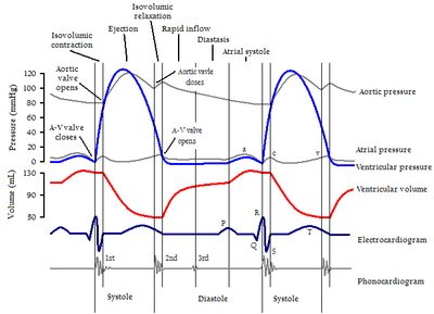
and associated papillary muscles. Ventricular pressure continues to rise in isovolumetric contraction
Isovolumetric contraction
Isovolumetric contraction is a term used in cardiac physiology to refer to an event occurring in early systole, during which the ventricles contract with no corresponding volume change.-Description:...
with maximal pressure generation (max dP/dt) occurring during this phase, until the pulmonary and aortic valves open in the ejection phase
Cardiac cycle
The cardiac cycle is a term referring to all or any of the events related to the flow or blood pressure that occurs from the beginning of one heartbeat to the beginning of the next. The frequency of the cardiac cycle is described by the heart rate. Each beat of the heart involves five major stages...
. In the ejection phase, blood flows down its pressure gradient through the aorta and pulmonary artery from left and right ventricles respectively. It is important to note that cardiac muscle perfusion through coronary vessels does not occur during ventricular systole, but occurs during ventricular diastole.
Ventricular systole is the origin of the pulse
Pulse
In medicine, one's pulse represents the tactile arterial palpation of the heartbeat by trained fingertips. The pulse may be palpated in any place that allows an artery to be compressed against a bone, such as at the neck , at the wrist , behind the knee , on the inside of the elbow , and near the...
.
Right ventricular systole
Right ventricular systole drives blood through the pulmonary valvePulmonary valve
The pulmonary valve is the semilunar valve of the heart that lies between the right ventricle and the pulmonary artery and has three cusps. Similar to the aortic valve, the pulmonary valve opens in ventricular systole, when the pressure in the right ventricle rises above the pressure in the...
(PV) into the lung
Lung
The lung is the essential respiration organ in many air-breathing animals, including most tetrapods, a few fish and a few snails. In mammals and the more complex life forms, the two lungs are located near the backbone on either side of the heart...
s. Right heart systole is volumetrically defined as right ventricular ejection fraction
Ejection fraction
In cardiovascular physiology, ejection fraction is the fraction of Blood pumped out of the Right Ventricle of the heart to the Pulmonary Circulation and Left Ventricle of the heart to the Systemic Circulation with each Heart beat or Cardiac cycle...
(RVEF). The Time
Time
Time is a part of the measuring system used to sequence events, to compare the durations of events and the intervals between them, and to quantify rates of change such as the motions of objects....
variable of right ventricular systole is PV open to PV close. Increased RVEF is indicative of Pulmonary Hypertension
Pulmonary hypertension
In medicine, pulmonary hypertension is an increase in blood pressure in the pulmonary artery, pulmonary vein, or pulmonary capillaries, together known as the lung vasculature, leading to shortness of breath, dizziness, fainting, and other symptoms, all of which are exacerbated by exertion...
.
Left ventricular Systole
Left Ventricular Systole drives blood through the aortic valveAortic valve
The aortic valve is one of the valves of the heart. It is normally tricuspid , although in 1% of the population it is found to be congenitally bicuspid . It lies between the left ventricle and the aorta....
(AoV) to the body and representative end organs excluding the lungs Pulmonary System. Left ventricular systole is volumetrically defined as left ventricular ejection fraction (LVEF). The Time
Time
Time is a part of the measuring system used to sequence events, to compare the durations of events and the intervals between them, and to quantify rates of change such as the motions of objects....
variable of left ventricular systole is AoV open to AoV close.
Physiological mechanism
Systole of the heart is initiated by the electrically excitable cells of the sinoatrial nodeSinoatrial node
The sinoatrial node is the impulse-generating tissue located in the right atrium of the heart, and thus the generator of normal sinus rhythm. It is a group of cells positioned on the wall of the right atrium, near the entrance of the superior vena cava...
. These cells are activated spontaneously by depolarization
Depolarization
In biology, depolarization is a change in a cell's membrane potential, making it more positive, or less negative. In neurons and some other cells, a large enough depolarization may result in an action potential...
of their membranes beyond a given threshold for excitation. At this point, voltage-gated calcium channels on the cell membrane open and allow calcium ions to pass through, into the sarcoplasm
Sarcoplasm
The Sarcoplasm of a muscle fiber is comparable to the cytoplasm of other cells, but it houses unusually large amounts of glycosomes and significant amounts of myoglobin, an oxygen binding protein...
of muscle cell. Calcium ions bind to ryanodine receptor
Ryanodine receptor
Ryanodine receptors form a class of intracellular calcium channels in various forms of excitable animal tissue like muscles and neurons...
s on the sarcoplasmic reticulum causing a flux of calcium ions to the sarcoplasm.
Calcium ions bind to troponin C
Troponin C
Troponin C is a part of the troponin complex. It contains four calcium-binding EF hands. It is a component of thin filaments . It contains an N lobe and a C lobe. The C lobe serves a structural purpose and binds to the N domain of TnI. The C lobe can bind either Ca2+ or Mg2+...
, causing a conformational change in the troponin-tropomyosin complex, and thus allowing myosin
Myosin
Myosins comprise a family of ATP-dependent motor proteins and are best known for their role in muscle contraction and their involvement in a wide range of other eukaryotic motility processes. They are responsible for actin-based motility. The term was originally used to describe a group of similar...
head binding sites on F-Actin
Actin
Actin is a globular, roughly 42-kDa moonlighting protein found in all eukaryotic cells where it may be present at concentrations of over 100 μM. It is also one of the most highly-conserved proteins, differing by no more than 20% in species as diverse as algae and humans...
to be exposed. This transition allows cross bridge cycling
Muscle contraction
Muscle fiber generates tension through the action of actin and myosin cross-bridge cycling. While under tension, the muscle may lengthen, shorten, or remain the same...
to occur.
The cardiac action potential
Cardiac action potential
In electrocardiography, the cardiac action potential is a specialized action potential in the heart, necessary for the electrical conduction system of the heart....
spreads distally to the small branches of the Purkinje tree via the flux of cations through gap junctions that connect the sarcoplasm of adjacent myocytes.
The electrical activity of ventricular systole is coordinated by the atrioventricular node
Atrioventricular node
The atrioventricular node is a part of the electrical control system of the heart that coordinates heart rate. It electrically connects atrial and ventricular chambers...
, this discrete collection of cells receives electrical stimulation from the atrium, but also has a slower intrinsic pacemaker activity. The cardiac action potential is propagated down the bundle of His
Bundle of His
The bundle of His, known as the AV bundle or atrioventricular bundle, is a collection of heart muscle cells specialized for electrical conduction that transmits the electrical impulses from the AV node to the point of the apex of the fascicular branches...
to Purkinje fibres which rapidly causes coordinated depolarisation, and excitation-contraction coupling from the apex of the heart
Apex of the heart
The apex of the heart is the lowest superficial part of the heart.It is directed downward, forward, and to the left, and is overlapped by the left lung and pleura.-External anatomy:...
up.