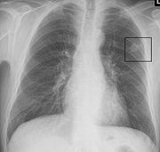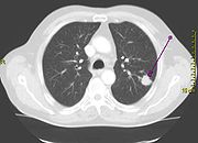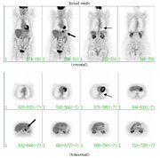
Solitary pulmonary nodule
Encyclopedia
In radiology
, a solitary pulmonary nodule (SPN) or coin lesion is a mass
in the lung
smaller than 3 centimeters in diameter. It can be an incidental finding
found in up to 0.2% of chest X-ray
s and around 1% of CT scans.
The nodule
most commonly represents a benign
tumor such as a granuloma
or hamartoma
, but in around 20% of cases it represents a malignant
cancer
, especially in older adults and smokers
. Conversely, 10 to 20% of patients with lung cancer
are diagnosed in this way. Thus, the possibility of cancer needs to be excluded through further radiological studies and interventions, possibly including surgical resection. The prognosis
depends on the underlying condition.
(functional tissue) with a diameter less than 3 cm and without associated pneumonia
, atelectasis
(lung collapse) or lymphadenopathies
(swollen lymph nodes).
or electrocardiographic monitoring.
The most important cause to exclude is a form of lung cancer, including rare forms such as primary pulmonary lymphoma
, carcinoid tumor and a solitary metastasis
to the lung (common unrecognised primary tumor sites are melanoma
s, sarcoma
s or testicular cancer
). Benign tumors in the lung include hamartoma
s and chondroma
s.
The most common benign coin lesion is a granuloma
(inflammatory nodule
), for example due to tuberculosis
or a fungal infection. Other infectious causes include a pulmonary abscess, pneumonia
(including Pneumocystis carinii pneumonia) or rarely nocardia
l infection or worm infection (such as dirofilariasis
or dog heartworm infestation). Lung nodules can also occur in immune disorder
s such as rheumatoid arthritis
or Wegener's granulomatosis
.
An SPN can be found to be an arteriovenous malformation
, a hematoma
or an infarction
zone. It may also be caused by bronchial atresia, sequestration, an inhaled foreign body
or pleural plaque.
 Several features help to distinguish benign conditions from possible lung cancer. The first parameter is the size of the lesion: the smaller, the less risk for malignant cancer. Benign causes tend to have a well defined border, whereas lobulated lesions or those with an irregular margin extending into the neighbouring tissue tend to be malignant.
If there is a central cavity, then a thin wall points to a benign cause whereas a thick wall is associated with malignancy (especially 4mm or less versus 16mm or more). In lung cancer, cavitation can represent central tumor necrosis
(tissue death) or secondary abscess
formation. If the walls of an airway are visible (air bronchogram), bronchioloalveolar carcinoma
is a possibility.
An SPN often contains calcifications. Certain patterns of calcification are reassuring, such as the popcorn-like appearance of hamartoma. An SPN with a density below 15 Hounsfield units on computed tomography
tends to be benign, whereas malignant tumors often measure more than 20 Hounsfield units. Fatty tissue inside hamartomas will have a strongly negative value on the Hounsfield scale
.
The growth velocity of a lesion is also informative: very fast or very slow growing tumors are rarely malignant, in contrary to inflammatory or congenital conditions. It is therefore important to retrieve previous imaging studies to see if a lesion was presented and how fast its volume is increasing. This is more difficult for nodules smaller than 1 centimeter. Moreover, the predictive value of stable lesion over a period of 2 years has been found to be rather low and unreliable.
s such as asbestos
, and previously diagnosed cancer or respiratory infections. A patient with airway symptoms, especially coughing up blood (hemoptysis
), is more likely to have cancer compared to a patient with no respiratory symptoms.
 The work-up in patients with a solitary pulmonary nodule is based on an initial risk assessment. If the risk of malignancy is thought to be low, follow-up imaging (usually serial CT scans) can be planned at a later time. The frequency of further scans can be determined by the patient's risk for cancer and the size of the nodule. If the initial impression is that there is a high likelihood of cancer, than a surgical intervention is appropriate (provided that the patient is fit for surgery).
If there is an intermediate risk of malignancy, further imaging with positron emission tomography
(PET scan) is appropriate (if available). Around 95% of patients with a malignant nodule will have an abnormal PET scan, while around 78% of patients with a benign nodule will look normal on PET (this is the test sensitivity and specificity
). Thus, an abnormal PET scan will reliably pick up cancer, but several other types of nodules (inflammatory or infectious, for example) will also show up on a PET scan. If the nodule has a diameter below 1 centimeter, PET scans are often avoided because there is an increased risk of falsely normal results. Cancerous lesions usually have a high metabolism
on PET, as demonstrated by their high uptake of FDG
(a radioactive sugar). If the lesion is found on further imaging to be suspicious, it should be surgically excised (via thoracotomy
or video-assisted thoracic surgery) to confirm the diagnosis by microscopical examination
.
In selected cases, nodules can also be sampled through the airways using bronchoscopy
or through the chest wall using needle aspiration
(which can be done under CT guidance). Needle aspiration can only retrieve groups of cells for cytology
and not a tissue cylinder or biopsy
, precluding evaluation of the tissue architecture. Theoretically, this makes the diagnosis of benign conditions more difficult, although rates higher than 90% have been reported. Complications of the latter technique include hemorrhage into the lung and air leak in the pleural space between the lung and the chest wall (pneumothorax
). However, not all these cases of pneumothorax need treatment with a chest tube
.
Other imaging techniques include PET-CT
(simultaneous PET scan and CT scan with superposition of the images), magnetic resonance imaging
(MRI) or single photon emission computed tomography
(SPECT).
Radiology
Radiology is a medical specialty that employs the use of imaging to both diagnose and treat disease visualized within the human body. Radiologists use an array of imaging technologies to diagnose or treat diseases...
, a solitary pulmonary nodule (SPN) or coin lesion is a mass
Tumor
A tumor or tumour is commonly used as a synonym for a neoplasm that appears enlarged in size. Tumor is not synonymous with cancer...
in the lung
Lung
The lung is the essential respiration organ in many air-breathing animals, including most tetrapods, a few fish and a few snails. In mammals and the more complex life forms, the two lungs are located near the backbone on either side of the heart...
smaller than 3 centimeters in diameter. It can be an incidental finding
Incidentaloma
In medicine, an incidentaloma is a tumor found by coincidence without clinical symptoms or suspicion. It is a common problem: up to 7% of all patients over 60 may harbor a benign growth, often of the adrenal gland, which is detected when diagnostic imaging is used for the analysis of unrelated...
found in up to 0.2% of chest X-ray
Chest X-ray
In medicine, a chest radiograph, commonly called a chest X-ray , is a projection radiograph of the chest used to diagnose conditions affecting the chest, its contents, and nearby structures...
s and around 1% of CT scans.
The nodule
Nodule (medicine)
For use of the term nodule in dermatology, see Nodule In medicine, a nodule refers to a relatively hard, roughly spherical abnormal structure....
most commonly represents a benign
Benign
A benign tumor is a tumor that lacks the ability to metastasize. Common examples of benign tumors include moles and uterine fibroids.The term "benign" implies a mild and nonprogressive disease. Indeed, many kinds of benign tumors are harmless to human health...
tumor such as a granuloma
Granuloma
Granuloma is a medical term for a tiny collection of immune cells known as macrophages. Granulomas form when the immune system attempts to wall off substances that it perceives as foreign but is unable to eliminate. Such substances include infectious organisms such as bacteria and fungi as well as...
or hamartoma
Hamartoma
A hamartoma is a benign, focal malformation that resembles a neoplasm in the tissue of its origin. This is not a malignant tumor, and it grows at the same rate as the surrounding tissues. It is composed of tissue elements normally found at that site, but which are growing in a disorganized mass...
, but in around 20% of cases it represents a malignant
Malignant
Malignancy is the tendency of a medical condition, especially tumors, to become progressively worse and to potentially result in death. Malignancy in cancers is characterized by anaplasia, invasiveness, and metastasis...
cancer
Cancer
Cancer , known medically as a malignant neoplasm, is a large group of different diseases, all involving unregulated cell growth. In cancer, cells divide and grow uncontrollably, forming malignant tumors, and invade nearby parts of the body. The cancer may also spread to more distant parts of the...
, especially in older adults and smokers
Tobacco smoking
Tobacco smoking is the practice where tobacco is burned and the resulting smoke is inhaled. The practice may have begun as early as 5000–3000 BCE. Tobacco was introduced to Eurasia in the late 16th century where it followed common trade routes...
. Conversely, 10 to 20% of patients with lung cancer
Lung cancer
Lung cancer is a disease characterized by uncontrolled cell growth in tissues of the lung. If left untreated, this growth can spread beyond the lung in a process called metastasis into nearby tissue and, eventually, into other parts of the body. Most cancers that start in lung, known as primary...
are diagnosed in this way. Thus, the possibility of cancer needs to be excluded through further radiological studies and interventions, possibly including surgical resection. The prognosis
Prognosis
Prognosis is a medical term to describe the likely outcome of an illness.When applied to large statistical populations, prognostic estimates can be very accurate: for example the statement "45% of patients with severe septic shock will die within 28 days" can be made with some confidence, because...
depends on the underlying condition.
Definition
A solitary pulmonary nodulus needs to be separated from larger lung tumors, smaller infiltrates or masses with other accompanying characteristics. An often used formal radiological definition is the following: a single lesion in the lung completely surrounded by lung parenchymaParenchyma
Parenchyma is a term used to describe a bulk of a substance. It is used in different ways in animals and in plants.The term is New Latin, f. Greek παρέγχυμα - parenkhuma, "visceral flesh", f. παρεγχεῖν - parenkhein, "to pour in" f. para-, "beside" + en-, "in" + khein, "to pour"...
(functional tissue) with a diameter less than 3 cm and without associated pneumonia
Pneumonia
Pneumonia is an inflammatory condition of the lung—especially affecting the microscopic air sacs —associated with fever, chest symptoms, and a lack of air space on a chest X-ray. Pneumonia is typically caused by an infection but there are a number of other causes...
, atelectasis
Atelectasis
Atelectasis is defined as the collapse or closure of alveoli resulting in reduced or absent gas exchange. It may affect part or all of one lung. It is a condition where the alveoli are deflated, as distinct from pulmonary consolidation.It is a very common finding in chest x-rays and other...
(lung collapse) or lymphadenopathies
Lymphadenopathy
Lymphadenopathy is a term meaning "disease of the lymph nodes." It is, however, almost synonymously used with "swollen/enlarged lymph nodes". It could be due to infection, auto-immune disease, or malignancy....
(swollen lymph nodes).
Causes
Not every round spot on a radiological image is a coin lesion: it should not be confused with the projection of a structure of the chest wall or skin, such as a nipple, a healing rib fractureRib fracture
A rib fracture is a break or fracture in one or more of the bones making up the rib cage.The first rib is rarely fractured because of its protected position behind the clavicle . However, if it is broken serious damage can occur to the brachial plexus of nerves and the subclavian vessels...
or electrocardiographic monitoring.
The most important cause to exclude is a form of lung cancer, including rare forms such as primary pulmonary lymphoma
Lymphoma
Lymphoma is a cancer in the lymphatic cells of the immune system. Typically, lymphomas present as a solid tumor of lymphoid cells. Treatment might involve chemotherapy and in some cases radiotherapy and/or bone marrow transplantation, and can be curable depending on the histology, type, and stage...
, carcinoid tumor and a solitary metastasis
Metastasis
Metastasis, or metastatic disease , is the spread of a disease from one organ or part to another non-adjacent organ or part. It was previously thought that only malignant tumor cells and infections have the capacity to metastasize; however, this is being reconsidered due to new research...
to the lung (common unrecognised primary tumor sites are melanoma
Melanoma
Melanoma is a malignant tumor of melanocytes. Melanocytes are cells that produce the dark pigment, melanin, which is responsible for the color of skin. They predominantly occur in skin, but are also found in other parts of the body, including the bowel and the eye...
s, sarcoma
Sarcoma
A sarcoma is a cancer that arises from transformed cells in one of a number of tissues that develop from embryonic mesoderm. Thus, sarcomas include tumors of bone, cartilage, fat, muscle, vascular, and hematopoietic tissues...
s or testicular cancer
Testicular cancer
Testicular cancer is cancer that develops in the testicles, a part of the male reproductive system.In the United States, between 7,500 and 8,000 diagnoses of testicular cancer are made each year. In the UK, approximately 2,000 men are diagnosed each year. Over his lifetime, a man's risk of...
). Benign tumors in the lung include hamartoma
Hamartoma
A hamartoma is a benign, focal malformation that resembles a neoplasm in the tissue of its origin. This is not a malignant tumor, and it grows at the same rate as the surrounding tissues. It is composed of tissue elements normally found at that site, but which are growing in a disorganized mass...
s and chondroma
Chondroma
A chondroma is a benign cartilaginous tumor, which is encapsulated with a lobular growing pattern.Tumor cells resemble normal cells and produce the cartilaginous matrix ....
s.
The most common benign coin lesion is a granuloma
Granuloma
Granuloma is a medical term for a tiny collection of immune cells known as macrophages. Granulomas form when the immune system attempts to wall off substances that it perceives as foreign but is unable to eliminate. Such substances include infectious organisms such as bacteria and fungi as well as...
(inflammatory nodule
Nodule (medicine)
For use of the term nodule in dermatology, see Nodule In medicine, a nodule refers to a relatively hard, roughly spherical abnormal structure....
), for example due to tuberculosis
Tuberculosis
Tuberculosis, MTB, or TB is a common, and in many cases lethal, infectious disease caused by various strains of mycobacteria, usually Mycobacterium tuberculosis. Tuberculosis usually attacks the lungs but can also affect other parts of the body...
or a fungal infection. Other infectious causes include a pulmonary abscess, pneumonia
Pneumonia
Pneumonia is an inflammatory condition of the lung—especially affecting the microscopic air sacs —associated with fever, chest symptoms, and a lack of air space on a chest X-ray. Pneumonia is typically caused by an infection but there are a number of other causes...
(including Pneumocystis carinii pneumonia) or rarely nocardia
Nocardia
Nocardia is a genus of weakly staining Gram-positive, catalase-positive, rod-shaped bacteria. It forms partially acid-fast beaded branching filaments . It has a total of 85 species. Some species are non-pathogenic while others are responsible for nocardiosis. Nocardia are found worldwide in soil...
l infection or worm infection (such as dirofilariasis
Dirofilariasis
Dirofilariasis is an infection by parasites in the Dirofilaria genus.It can be caused by:* Dirofilaria immitis* Dirofilaria repens...
or dog heartworm infestation). Lung nodules can also occur in immune disorder
Immune disorder
An immune disorder is a dysfunction of the immune system. These disorders can be characterized in several different ways:* By the component of the immune system affected* By whether the immune system is overactive or underactive...
s such as rheumatoid arthritis
Rheumatoid arthritis
Rheumatoid arthritis is a chronic, systemic inflammatory disorder that may affect many tissues and organs, but principally attacks synovial joints. The process produces an inflammatory response of the synovium secondary to hyperplasia of synovial cells, excess synovial fluid, and the development...
or Wegener's granulomatosis
Wegener's granulomatosis
Wegener's granulomatosis , more recently granulomatosis with polyangiitis , is an incurable form of vasculitis that affects the nose, lungs, kidneys and other organs. Due to its end-organ damage, it is life-threatening and requires long-term immunosuppression...
.
An SPN can be found to be an arteriovenous malformation
Arteriovenous malformation
Arteriovenous malformation or AVM is an abnormal connection between veins and arteries, usually congenital. This pathology is widely known because of its occurrence in the central nervous system, but can appear in any location. An arteriovenous malformation is a vascular anomaly. It is a...
, a hematoma
Hematoma
A hematoma, or haematoma, is a localized collection of blood outside the blood vessels, usually in liquid form within the tissue. This distinguishes it from an ecchymosis, which is the spread of blood under the skin in a thin layer, commonly called a bruise...
or an infarction
Infarction
In medicine, infarction refers to tissue death that is caused by a local lack of oxygen due to obstruction of the tissue's blood supply. The resulting lesion is referred to as an infarct.-Causes:...
zone. It may also be caused by bronchial atresia, sequestration, an inhaled foreign body
Foreign body
A foreign body is any object originating outside the body. In machinery, it can mean any unwanted intruding object.Most references to foreign bodies involve propulsion through natural orifices into hollow organs....
or pleural plaque.
Initial evaluation
The medical history, physical examination and imaging results form the basis of the initial risk assessment and determine the further course of action. Most patients will have a CT scan.Radiological features

If there is a central cavity, then a thin wall points to a benign cause whereas a thick wall is associated with malignancy (especially 4mm or less versus 16mm or more). In lung cancer, cavitation can represent central tumor necrosis
Necrosis
Necrosis is the premature death of cells in living tissue. Necrosis is caused by factors external to the cell or tissue, such as infection, toxins, or trauma. This is in contrast to apoptosis, which is a naturally occurring cause of cellular death...
(tissue death) or secondary abscess
Abscess
An abscess is a collection of pus that has accumulated in a cavity formed by the tissue in which the pus resides due to an infectious process or other foreign materials...
formation. If the walls of an airway are visible (air bronchogram), bronchioloalveolar carcinoma
Bronchioloalveolar carcinoma
Bronchioloalveolar carcinoma is a term describing certain variants of lung cancer arising in the distal bronchioles or alveoli that initially exhibit a specific non-invasive growth pattern.-Classification:...
is a possibility.
An SPN often contains calcifications. Certain patterns of calcification are reassuring, such as the popcorn-like appearance of hamartoma. An SPN with a density below 15 Hounsfield units on computed tomography
Computed tomography
X-ray computed tomography or Computer tomography , is a medical imaging method employing tomography created by computer processing...
tends to be benign, whereas malignant tumors often measure more than 20 Hounsfield units. Fatty tissue inside hamartomas will have a strongly negative value on the Hounsfield scale
Hounsfield scale
The Hounsfield scale, named after Sir Godfrey Newbold Hounsfield, is a quantitative scale for describing radiodensity.-Definition:The Hounsfield unit scale is a linear transformation of the original linear attenuation coefficient measurement into one in which the radiodensity of distilled water at...
.
The growth velocity of a lesion is also informative: very fast or very slow growing tumors are rarely malignant, in contrary to inflammatory or congenital conditions. It is therefore important to retrieve previous imaging studies to see if a lesion was presented and how fast its volume is increasing. This is more difficult for nodules smaller than 1 centimeter. Moreover, the predictive value of stable lesion over a period of 2 years has been found to be rather low and unreliable.
Patient features
Several patient factors may influence the likelihood of a benign versus a malignant condition: these include previous exposure to smoke or other carcinogenCarcinogen
A carcinogen is any substance, radionuclide, or radiation that is an agent directly involved in causing cancer. This may be due to the ability to damage the genome or to the disruption of cellular metabolic processes...
s such as asbestos
Asbestos
Asbestos is a set of six naturally occurring silicate minerals used commercially for their desirable physical properties. They all have in common their eponymous, asbestiform habit: long, thin fibrous crystals...
, and previously diagnosed cancer or respiratory infections. A patient with airway symptoms, especially coughing up blood (hemoptysis
Hemoptysis
Hemoptysis or haemoptysis is the expectoration of blood or of blood-stained sputum from the bronchi, larynx, trachea, or lungs Hemoptysis or haemoptysis is the expectoration (coughing up) of blood or of blood-stained sputum from the bronchi, larynx, trachea, or lungs Hemoptysis or haemoptysis ...
), is more likely to have cancer compared to a patient with no respiratory symptoms.
Work-up

If there is an intermediate risk of malignancy, further imaging with positron emission tomography
Positron emission tomography
Positron emission tomography is nuclear medicine imaging technique that produces a three-dimensional image or picture of functional processes in the body. The system detects pairs of gamma rays emitted indirectly by a positron-emitting radionuclide , which is introduced into the body on a...
(PET scan) is appropriate (if available). Around 95% of patients with a malignant nodule will have an abnormal PET scan, while around 78% of patients with a benign nodule will look normal on PET (this is the test sensitivity and specificity
Sensitivity and specificity
Sensitivity and specificity are statistical measures of the performance of a binary classification test, also known in statistics as classification function. Sensitivity measures the proportion of actual positives which are correctly identified as such Sensitivity and specificity are statistical...
). Thus, an abnormal PET scan will reliably pick up cancer, but several other types of nodules (inflammatory or infectious, for example) will also show up on a PET scan. If the nodule has a diameter below 1 centimeter, PET scans are often avoided because there is an increased risk of falsely normal results. Cancerous lesions usually have a high metabolism
Metabolism
Metabolism is the set of chemical reactions that happen in the cells of living organisms to sustain life. These processes allow organisms to grow and reproduce, maintain their structures, and respond to their environments. Metabolism is usually divided into two categories...
on PET, as demonstrated by their high uptake of FDG
Fluorodeoxyglucose
Fludeoxyglucose or fluorodeoxyglucose , commonly abbreviated 18F-FDG or FDG, is a radiopharmaceutical used in the medical imaging modality positron emission tomography...
(a radioactive sugar). If the lesion is found on further imaging to be suspicious, it should be surgically excised (via thoracotomy
Thoracotomy
Thoracotomy is an incision into the pleural space of the chest. It is performed by a surgeon, and, rarely, by emergency physicians, to gain access to the thoracic organs, most commonly the heart, the lungs, the esophagus or thoracic aorta, or for access to the anterior spine such as is necessary...
or video-assisted thoracic surgery) to confirm the diagnosis by microscopical examination
Histopathology
Histopathology refers to the microscopic examination of tissue in order to study the manifestations of disease...
.
In selected cases, nodules can also be sampled through the airways using bronchoscopy
Bronchoscopy
Bronchoscopy is a technique of visualizing the inside of the airways for diagnostic and therapeutic purposes. An instrument is inserted into the airways, usually through the nose or mouth, or occasionally through a tracheostomy. This allows the practitioner to examine the patient's airways for...
or through the chest wall using needle aspiration
Needle aspiration biopsy
Needle aspiration biopsy , may refer to fine needle aspiration cytology , fine needle aspiration biopsy and fine needle aspiration , is a diagnostic procedure sometimes used to investigate superficial lumps or masses...
(which can be done under CT guidance). Needle aspiration can only retrieve groups of cells for cytology
Cytopathology
Cytopathology is a branch of pathology that studies and diagnoses diseases on the cellular level. The discipline was founded by Rudolf Virchow in 1858. A common application of cytopathology is the Pap smear, used as a screening tool, to detect precancerous cervical lesions and prevent cervical...
and not a tissue cylinder or biopsy
Biopsy
A biopsy is a medical test involving sampling of cells or tissues for examination. It is the medical removal of tissue from a living subject to determine the presence or extent of a disease. The tissue is generally examined under a microscope by a pathologist, and can also be analyzed chemically...
, precluding evaluation of the tissue architecture. Theoretically, this makes the diagnosis of benign conditions more difficult, although rates higher than 90% have been reported. Complications of the latter technique include hemorrhage into the lung and air leak in the pleural space between the lung and the chest wall (pneumothorax
Pneumothorax
Pneumothorax is a collection of air or gas in the pleural cavity of the chest between the lung and the chest wall. It may occur spontaneously in people without chronic lung conditions as well as in those with lung disease , and many pneumothoraces occur after physical trauma to the chest, blast...
). However, not all these cases of pneumothorax need treatment with a chest tube
Chest tube
A chest tube is a flexible plastic tube that is inserted through the side of the chest into the pleural space. It is used to remove air or fluid , or pus from the intrathoracic space...
.
Other imaging techniques include PET-CT
PET-CT
Positron emission tomography - computed tomography is a medical imaging device which combines in a single gantry system both a Positron Emission Tomography and an x-ray Computed Tomography, so that images acquired from both devices can be taken sequentially, in the same session from the patient...
(simultaneous PET scan and CT scan with superposition of the images), magnetic resonance imaging
Magnetic resonance imaging
Magnetic resonance imaging , nuclear magnetic resonance imaging , or magnetic resonance tomography is a medical imaging technique used in radiology to visualize detailed internal structures...
(MRI) or single photon emission computed tomography
Single photon emission computed tomography
Single-photon emission computed tomography is a nuclear medicine tomographic imaging technique using gamma rays. It is very similar to conventional nuclear medicine planar imaging using a gamma camera. However, it is able to provide true 3D information...
(SPECT).

