

Slit lamp
Encyclopedia

Anterior segment
The anterior segment is the front third of the eye that includes the structures in front of the vitreous humour: the cornea, iris, ciliary body, and lens.Within the anterior segment are two fluid-filled spaces:...
, or frontal structures and posterior segment
Posterior segment
The posterior segment is the back two-thirds of the eye that includes the anterior hyaloid membrane and all of the optical structures behind it: the vitreous humor, retina, choroid, and optic nerve....
, of the human eye
Human eye
The human eye is an organ which reacts to light for several purposes. As a conscious sense organ, the eye allows vision. Rod and cone cells in the retina allow conscious light perception and vision including color differentiation and the perception of depth...
, which includes the eyelid
Eyelid
An eyelid is a thin fold of skin that covers and protects an eye. With the exception of the prepuce and the labia minora, it has the thinnest skin of the whole body. The levator palpebrae superioris muscle retracts the eyelid to "open" the eye. This can be either voluntarily or involuntarily...
, sclera
Sclera
The sclera , also known as the white or white of the eye, is the opaque , fibrous, protective, outer layer of the eye containing collagen and elastic fiber. In the development of the embryo, the sclera is derived from the neural crest...
, conjunctiva
Conjunctiva
The conjunctiva covers the sclera and lines the inside of the eyelids. It is composed of rare stratified columnar epithelium.-Function:...
, iris
Iris (anatomy)
The iris is a thin, circular structure in the eye, responsible for controlling the diameter and size of the pupils and thus the amount of light reaching the retina. "Eye color" is the color of the iris, which can be green, blue, or brown. In some cases it can be hazel , grey, violet, or even pink...
, natural crystalline lens, and cornea
Cornea
The cornea is the transparent front part of the eye that covers the iris, pupil, and anterior chamber. Together with the lens, the cornea refracts light, with the cornea accounting for approximately two-thirds of the eye's total optical power. In humans, the refractive power of the cornea is...
. The binocular slit-lamp examination provides a stereoscopic magnified view of the eye structures in detail, enabling anatomical diagnoses to be made for a variety of eye conditions. A second, hand-held lens is used to examine the retina
Retina
The vertebrate retina is a light-sensitive tissue lining the inner surface of the eye. The optics of the eye create an image of the visual world on the retina, which serves much the same function as the film in a camera. Light striking the retina initiates a cascade of chemical and electrical...
.
History
To fully understand the development of the slit lamp one must consider that with this inventionInvention
An invention is a novel composition, device, or process. An invention may be derived from a pre-existing model or idea, or it could be independently conceived, in which case it may be a radical breakthrough. In addition, there is cultural invention, which is an innovative set of useful social...
and its improvements, it had to be accompanied by the introduction of new examination techniques. Two conflicting trends emerged in the development of the slit lamp. One trend originated from clinical research
Clinical research
Clinical research is a branch of medical science that determines the safety and effectiveness of medications, devices, diagnostic products and treatment regimens intended for human use...
and aimed at an increase in functions and the introduction and application of the increasingly complex and advanced technology of the time The second trend originated from ophthalmologic practice and aimed at technical perfection and a restriction to useful methods and the applications of the instrument. The first man credited with developments in this field was Hermann Von Helmholtz
Hermann von Helmholtz
Hermann Ludwig Ferdinand von Helmholtz was a German physician and physicist who made significant contributions to several widely varied areas of modern science...
(1850) when he invented the ophthalmoscope.
In ophthalmology
Ophthalmology
Ophthalmology is the branch of medicine that deals with the anatomy, physiology and diseases of the eye. An ophthalmologist is a specialist in medical and surgical eye problems...
and optometry
Optometry
Optometry is a health care profession concerned with eyes and related structures, as well as vision, visual systems, and vision information processing in humans. Optometrists, or Doctors of Optometry, are state licensed medical professionals trained to prescribe and fit lenses to improve vision,...
, the term “slit lamp” is the most commonly referred to term however it would be more correct to call it the “slit lamp instrument”. Today’s instrument however is a combination of two separate developments in instruments. The two developments are the corneal microscope and that of the slit lamp itself. Though the slit lamp is a combination of these two developments, the first concept of the slit lamp dates back to 1911 credited to Alvar Gullstrand and his “large reflection-free ophthalmoscope.” The instrument was manufactured by the company Zeiss and consisted of a special illuminator that was connected by a small stand base through a vertical adjustable column. The base was able to move freely on a glass plate. The illuminator employed a Nernst glower
Nernst glower
The Nernst glower is an obsolete device for providing a continuous source of infrared radiation for use in spectroscopy. Typically it was in the form of a cylindrical rod or tube composed of a mixture of certain oxides such as zirconium oxide , yttrium oxide and erbium oxide at a ratio of...
which was later converted into a slit through a simple optical system. However, the instrument never received much attention and the term “slit lamp” did not appear and any literature again until 1914.
It wasn’t until 1919 that several improvements were made to the Gullstrand slit lamp made by Vogt Henker. First, a mechanical connection was made between lamp and ophthalmoscopic lens. This illumination unit was mounted to the table column with a double articulated arm. The binocular microscope was supported on a small stand and could be moved freely across the tabletop. Later, a cross slide stage was used for this purpose. Vogt introduced Koehler illumination, and the reddish shining Nernst glower
Nernst glower
The Nernst glower is an obsolete device for providing a continuous source of infrared radiation for use in spectroscopy. Typically it was in the form of a cylindrical rod or tube composed of a mixture of certain oxides such as zirconium oxide , yttrium oxide and erbium oxide at a ratio of...
was replaced with the brighter and whiter incandescent lamp
Incandescent light bulb
The incandescent light bulb, incandescent lamp or incandescent light globe makes light by heating a metal filament wire to a high temperature until it glows. The hot filament is protected from air by a glass bulb that is filled with inert gas or evacuated. In a halogen lamp, a chemical process...
. Special mention should be paid to the experiments that followed Henker’s improvements in 1919. On his improvements the Nitra lamp was replaced with a carbon arc lamp with a liquid filter. At this time the great importance of color temperature and the luminance of the light source for slit lamp examinations were recognized and the basis created for examinations in red-free light.
In the year 1926, the slit lamp instrument was redesigned. The vertical arrangement of the slit projector (slit lamp) made it an easy to handle instrument. For the first time, the axis through the patient’s eye
Eye
Eyes are organs that detect light and convert it into electro-chemical impulses in neurons. The simplest photoreceptors in conscious vision connect light to movement...
was fixed at a common swiveling axis. This was a fundamental principle that was adopted for every slit lamp instrument developed. A limitation still with the instrument was it lacked a coordinate cross-slide stage for instrument adjustment but only a laterally adjustable chin rest for the patient. The importance of focal illumination had not yet been fully recognized.
In 1927, stereo camera
Stereo camera
A stereo camera is a type of camera with two or more lenses with a separate image sensor or film frame for each lens. This allows the camera to simulate human binocular vision, and therefore gives it the ability to capture three-dimensional images, a process known as stereo photography. Stereo...
s were developed and added to the slit lamp to further its use and application. In 1930, a man named Rudolf Theil presented the further development of the slit lamp was encouraged by a company named Goldmann. Horizontal and vertical co-ordinate adjustments were performed with three control elements on the cross-slide stage. The common swivel axis for microscope and illumination system was connected to the cross-slide stage, which allowed it to be brought to any part of the eye to be examined. A further improvement was made in 1938. A control lever or joystick
Joystick
A joystick is an input device consisting of a stick that pivots on a base and reports its angle or direction to the device it is controlling. Joysticks, also known as 'control columns', are the principal control in the cockpit of many civilian and military aircraft, either as a center stick or...
was used for the first time to allow for horizontal movement.
Following World War II
World War II
World War II, or the Second World War , was a global conflict lasting from 1939 to 1945, involving most of the world's nations—including all of the great powers—eventually forming two opposing military alliances: the Allies and the Axis...
the slit lamp was improved again. On this particular improvement the slit projector could be swiveled continuously across the front of the microscope
Microscope
A microscope is an instrument used to see objects that are too small for the naked eye. The science of investigating small objects using such an instrument is called microscopy...
. This was then improved again in 1950. In 1950, a company named Littmann redesigned the slit lamp again. The adopted the joystick control from the Goldmann instrument and the illumination path present in the Comberg instrument. Additionally Littmann added the stereo telescope system with a common objective magnification changer.
In 1965, the Model 100/16 Slit Lamp was produced based on the slit lamp by Littmann. This was soon followed by the Model 125/16 Slit Lamp in 1972. The only difference between the two models was their operating distances of 100 mm to 125 mm. With the introduction of the photo slit lamp further advancements were possible. In 1976, the development of the Model 110 Slit Lamp and the 210/211 Photo Slit Lamps were an innovation by which each were constructed from standard modules allowing for a wide range of different configurations. At the same time, halogen lamp
Halogen lamp
A halogen lamp, also known as a tungsten halogen lamp, is an incandescent lamp with a tungsten filament contained within an inert gas and a small amount of a halogen such as iodine or bromine. The chemical halogen cycle redeposits evaporated tungsten back on to the filament, extending the life of...
s replaced the old illumination systems to make them brighter and essentially daylight quality. From 1994 onwards, new slit lamps were introduced which took advantage of new technologies. The last major development was in 1996 in which included the advantages of new slit lamp optics.
Observation by Optical Section
Observation with an optical section or direct focal illumination is the most frequently applied method of examination with the slit lamp. With this method, the axes of illuminating and viewing path intersect in the area of the anterior eye media to be examined, for example, the individual corneal layersDirect diffuse illumination
If media, especially that of the cornea, are opaque, optical section images are often impossible depending on severity. In these cases, direct diffuse illumination may be used to advantage. For this, the slit is opened very wide and a diffuse, attenuated survey illumination is produced by inserting a ground glass screen or diffuser in the illuminating path. "Wide beam" illumination is the only type that has the light source set wide open. Its main purpose is to illuminate as much of the eye and its adnexaAdnexa
In anatomy, adnexa refers to the appendages of an organ. The term adnexa stems from a Latin word meaning appendages.More specifically, it can refer to:* Adnexa of eye * Adnexa of uterus...
at once for general observation.
Indirect illumination
With this method, light enters the eye through a narrow to medium slit (2 to 4 mm) to one side of the area to be examined. The axes of illuminating and viewing path do not intersect at the point of image focus, to achieve this; the illuminating prismPrism (optics)
In optics, a prism is a transparent optical element with flat, polished surfaces that refract light. The exact angles between the surfaces depend on the application. The traditional geometrical shape is that of a triangular prism with a triangular base and rectangular sides, and in colloquial use...
is decentered by rotating it about its vertical axis off the normal position. In this way, reflected, indirect light illuminates the area of the anterior chamber or cornea to be examined. The observed corneal area then lies between the incident light section through the cornea and the irradiated area of the iris. Observation is thus against a comparatively dark background.
Retro-illumination
In certain cases, illumination by optical section does not yield sufficient information or is impossible. This is the case, for example, when larger, extensive zones or spaces of the ocular media are opaque. Then the scattered light that is not very bright normally is absorbed. A similar situation arises when areas behind the crystalline lens are to be observed. In this case the observation beam must pass a number of interfaces that may reflect and attenuate the light.Scattering sclero-corneal illumination
With this type of illumination, a wide light beam is directed onto the limbal region of the cornea at an extremely low angle of incidence and with a laterally de-centered illuminating prism. Adjustment must allow the light beam to transmit through the corneal parenchymal layers according to the principle of total reflection allowing the interface with the cornea to be brightly illuminated. The magnification should be selected so that the entire cornea can be seen at a glance.Fundus observation and gonioscopy with the slit lamp
Fundus (eye)Fundus (eye)
The fundus of the eye is the interior surface of the eye, opposite the lens, and includes the retina, optic disc, macula and fovea, and posterior pole. The fundus can be viewed with an ophthalmoscope. The term may also be inclusive of Bruch's membrane and the choroid.The color of the fundus varies...
observation is known by the ophthalmic and the use of fundus camera
Fundus camera
A fundus camera or retinal camera is a specialized low power microscope with an attached camera designed to photograph the interior surface of the eye, including the retina, optic disc, macula, and posterior pole A fundus camera or retinal camera is a specialized low power microscope with an...
s. With the slit lamp, however, direct observation of the fundus is impossible due to the refractive power of the ocular media. In other words: the far point of the eye (punctum remotum) is so distant in front of (myopia
Myopia
Myopia , "shortsightedness" ) is a refractive defect of the eye in which collimated light produces image focus in front of the retina under conditions of accommodation. In simpler terms, myopia is a condition of the eye where the light that comes in does not directly focus on the retina but in...
) or behind (hyperopia
Hyperopia
Hyperopia, also known as farsightedness, longsightedness or hypermetropia, is a defect of vision caused by an imperfection in the eye , causing difficulty focusing on near objects, and in extreme cases causing a sufferer to be unable to focus on objects at any distance...
) that the microscope cannot be focused. The use of auxiliary optics - generally as a lens – makes it possible however to bring the far point within the focusing range of the microscope. For this various auxiliary lenses are in use that range in optical properties and practical application.
During Exam
While a patient is seated in the examination chair, they rest their chin and forehead on a support to steady the head. Using the biomicroscope, the ophthalmologist or optometrist then proceeds to examine the patient's eye. A fine strip of paper, stained with fluoresceinFluorescein
Fluorescein is a synthetic organic compound available as a dark orange/red powder soluble in water and alcohol. It is widely used as a fluorescent tracer for many applications....
, a fluorescent dye, may be touched to the side of the eye; this stains the tear film on the surface of the eye to aid examination. The dye is naturally rinsed out of the eye by tears
Tears
Tears are secretions that clean and lubricate the eyes. Lacrimation or lachrymation is the production or shedding of tears....
.
A subsequent test may involve placing drops in the eye in order to dilate the pupils
Mydriasis
Mydriasis is a dilation of the pupil due to disease, trauma or the use of drugs. Normally, the pupil dilates in the dark and constricts in the light to respectively improve vividity at night and to protect the retina from sunlight damage during the day...
. The drops take about 15 to 20 minutes to work, after which the examination is repeated, allowing the back of the eye to be examined. Patients will experience some light sensitivity
Light sensitivity
Light sensitivity or photosensitivity is an increase in the reactivity of the skin to sunlight. Apart from vision, human beings have many physiological and psychological responses to light. In rare individuals an atypical response may result in serious discomfort, disease, or injury. Some drugs...
for a few hours after this exam, and the dilating drops may also cause increased pressure in the eye, leading to nausea and pain. Patients who experience serious symptoms are advised to seek medical attention immediately.
Adults need no special preparation for the test; however children may need some preparation, depending on age, previous experiences, and level of trust.
The slit lamp exam may detect many diseases of the eye, including:
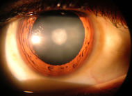
- CataractCataractA cataract is a clouding that develops in the crystalline lens of the eye or in its envelope, varying in degree from slight to complete opacity and obstructing the passage of light...
- ConjunctivitisConjunctivitisConjunctivitis refers to inflammation of the conjunctiva...
- Corneal injury such as corneal ulcerCorneal ulcerA corneal ulcer, or ulcerative keratitis, is an inflammatory condition of the cornea involving loss of its outer layer. It is very common in dogs and is sometimes seen in cats...
or corneal swelling - Diabetic retinopathyDiabetic retinopathyDiabetic retinopathy is retinopathy caused by complications of diabetes mellitus, which can eventually lead to blindness....
- Fuchs' dystrophyFuchs' dystrophyFuchs' dystrophy, also known as Fuchs' endothelial dystrophy, is a slowly progressing corneal disease that usually affects both eyes and is slightly more common in women than in men...
- KeratoconusKeratoconusKeratoconus , is a degenerative disorder of the eye in which structural changes within the cornea cause it to thin and change to a more conical shape than its normal gradual curve....
(Fleischer ringFleischer ringFleischer rings are pigmented rings in the peripheral cornea, resulting from iron deposition in basal epithelial cells, in the form of hemosiderin...
) - Macular degenerationMacular degenerationAge-related macular degeneration is a medical condition which usually affects older adults and results in a loss of vision in the center of the visual field because of damage to the retina. It occurs in “dry” and “wet” forms. It is a major cause of blindness and visual impairment in older adults...
- PresbyopiaPresbyopiaPresbyopia is a condition where the eye exhibits a progressively diminished ability to focus on near objects with age. Presbyopia’s exact mechanisms are not known with certainty; the research evidence most strongly supports a loss of elasticity of the crystalline lens, although changes in the...
- Retinal detachmentRetinal detachmentRetinal detachment is a disorder of the eye in which the retina peels away from its underlying layer of support tissue. Initial detachment may be localized, but without rapid treatment the entire retina may detach, leading to vision loss and blindness. It is a medical emergency.The retina is a...
- Retinal vessel occlusion
- Retinitis pigmentosaRetinitis pigmentosaRetinitis pigmentosa is a group of genetic eye conditions that leads to incurable blindness. In the progression of symptoms for RP, night blindness generally precedes tunnel vision by years or even decades. Many people with RP do not become legally blind until their 40s or 50s and retain some...
- Sjögren's syndromeSjögren's syndromeSjögren's syndrome , also known as "Mikulicz disease" and "Sicca syndrome", is a systemic autoimmune disease in which immune cells attack and destroy the exocrine glands that produce tears and saliva....
- ToxoplasmosisToxoplasmosisToxoplasmosis is a parasitic disease caused by the protozoan Toxoplasma gondii. The parasite infects most genera of warm-blooded animals, including humans, but the primary host is the felid family. Animals are infected by eating infected meat, by ingestion of feces of a cat that has itself...
- UveitisUveitisUveitis specifically refers to inflammation of the middle layer of the eye, termed the "uvea" but in common usage may refer to any inflammatory process involving the interior of the eye....
- Wilson's diseaseWilson's diseaseWilson's disease or hepatolenticular degeneration is an autosomal recessive genetic disorder in which copper accumulates in tissues; this manifests as neurological or psychiatric symptoms and liver disease...
(Kayser-Fleischer ringKayser-Fleischer ringKayser-Fleischer rings are dark rings that appear to encircle the iris of the eye. They are due to copper deposition as a result of particular liver diseases. They are named after Dr Bernhard Kayser and Dr Bruno Fleischer, the German doctors who first described them in 1902 and 1903...
)