
Rhodopsin
Encyclopedia
Rhodopsin, also known as visual purple, is a biological pigment
of the retina
that is responsible for both the formation of the photoreceptor cells and the first events in the perception of light
. Rhodopsins belong to the G-protein coupled receptor family and are extremely sensitive to light, enabling vision in low-light conditions. Exposed to light, the pigment immediately photobleaches
, and it takes about 30 minutes to regenerate fully in humans.
and a reversibly covalently bound cofactor
, retinal
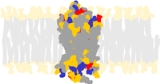
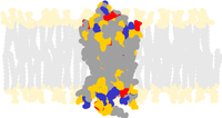
. Opsin, a bundle of seven transmembrane helices connected to each other by protein loops, binds retinal (a photoreactive chromophore
), which is located in a central pocket on the seventh helix at a lysine
residue. Retinal lies horizontally with relation to the membrane. Each outer segment disc contains thousands of visual pigment molecules. About half the opsin is within the lipid bilayer
. Retinal
is produced in the retina
from Vitamin A
, from dietary beta-carotene
. Isomerization of 11-cis-retinal into all-trans-retinal by light
induces a conformational change (bleaching) in opsin continuing with metarhodopsin II, which activates the associated G protein
transducin
and triggers a second messenger cascade.
Rhodopsin of the rod
s most strongly absorbs green-blue light and therefore appears reddish-purple, which is why it is also called "visual purple". It is responsible for monochromatic vision in the dark.
 Several closely related opsins exist that differ only in a few amino acid
s and in the wavelength
s of light that they absorb most strongly. Humans have four different other opsins beside rhodopsin. The photopsin
s are found in the different types of the cone cell
s of the retina and are the basis of color vision
. They have absorption maxima for yellowish-green (photopsin I), green (photopsin II), and bluish-violet (photopsin III) light. The remaining opsin (melanopsin
) is found in photosensitive ganglion cell
s and absorbs blue light most strongly.
The structure of rhodopsin has been studied in detail via x-ray crystallography
on rhodopsin crystals. The photoisomerization dynamics has been investigated with time-resolved IR spectroscopy and UV/Vis spectroscopy. A first photoproduct called photorhodopsin forms within 200 femtoseconds after irradiation followed within picosecond
s by a second one called bathorhodopsin with distorted all-trans bonds. This intermediate can be trapped and studied at cryogenic temperatures. Several models (e.g. the bicycle-pedal mechanism, hula-twist mechanism) attempt to explain how the retinal group can change its conformation without clashing with the enveloping rhodopsin protein pocket.
Recent data supports that it is a functional monomer as opposed to a dimer, which was the paradigm of G-coupled protein receptors for many years.
. The disease-causing protein generally aggregates with ubiquitin
in inclusion bodies, disrupts the intermediate filament network and impairs the ability of the cell to degrade non-functioning proteins which leads to photoreceptor apoptosis
. Other mutations on rhodopsin lead to X-linked congenital stationary night blindness
, mainly due to constitutive activation, when the mutations occur around the chromophore binding pocket of rhodopsin. Several other pathological states relating to rhodopsin have been discovered including poor post-Golgi trafficking, dysregulative activation, rod outer segment instability and arrestin binding.
s express proton pump
s called bacteriorhodopsin
, proteorhodopsin
, xanthorhodopsin to carry out phototrophy. Like rhodopsin, these contain retinal and have seven transmembrane alpha helices
; however they are not coupled to a G protein. Bacterial halorhodopsin
is a light-activated chloride pump. Finally, an alga is known to have an opsin that contains its own monolithic light-gated ion channel, channelrhodopsin
. While bacteriorhodopsin, halorhodopsin, and channelrhodopsin all have significant sequence homology to one another, they have no detectable sequence identity to the G-protein coupled receptor (GPCR) family to which rhodopsins belong. Nevertheless, bacterial rhodopsins and GPCR are possibly evolutionarily related, based on the similarity of their three-dimensional structures. Therefore, they have been assigned to the same superfamily in Structural Classification of Proteins
.
Biological pigment
Biological pigments, also known simply as pigments or biochromes are substances produced by living organisms that have a color resulting from selective color absorption. Biological pigments include plant pigments and flower pigments...
of the retina
Retina
The vertebrate retina is a light-sensitive tissue lining the inner surface of the eye. The optics of the eye create an image of the visual world on the retina, which serves much the same function as the film in a camera. Light striking the retina initiates a cascade of chemical and electrical...
that is responsible for both the formation of the photoreceptor cells and the first events in the perception of light
Light
Light or visible light is electromagnetic radiation that is visible to the human eye, and is responsible for the sense of sight. Visible light has wavelength in a range from about 380 nanometres to about 740 nm, with a frequency range of about 405 THz to 790 THz...
. Rhodopsins belong to the G-protein coupled receptor family and are extremely sensitive to light, enabling vision in low-light conditions. Exposed to light, the pigment immediately photobleaches
Photobleaching
Photobleaching is the photochemical destruction of a fluorophore. In microscopy, photobleaching may complicate the observation of fluorescent molecules, since they will eventually be destroyed by the light exposure necessary to stimulate them into fluorescing...
, and it takes about 30 minutes to regenerate fully in humans.
Structure
Rhodopsin consists of the protein moiety opsinOpsin
Opsins are a group of light-sensitive 35–55 kDa membrane-bound G protein-coupled receptors of the retinylidene protein family found in photoreceptor cells of the retina. Five classical groups of opsins are involved in vision, mediating the conversion of a photon of light into an electrochemical...
and a reversibly covalently bound cofactor
Cofactor (biochemistry)
A cofactor is a non-protein chemical compound that is bound to a protein and is required for the protein's biological activity. These proteins are commonly enzymes, and cofactors can be considered "helper molecules" that assist in biochemical transformations....
, retinal
Retinal
Retinal, also called retinaldehyde or vitamin A aldehyde, is one of the many forms of vitamin A . Retinal is a polyene chromophore, and bound to proteins called opsins, is the chemical basis of animal vision...
. Opsin, a bundle of seven transmembrane helices connected to each other by protein loops, binds retinal (a photoreactive chromophore
Chromophore
A chromophore is the part of a molecule responsible for its color. The color arises when a molecule absorbs certain wavelengths of visible light and transmits or reflects others. The chromophore is a region in the molecule where the energy difference between two different molecular orbitals falls...
), which is located in a central pocket on the seventh helix at a lysine
Lysine
Lysine is an α-amino acid with the chemical formula HO2CCH4NH2. It is an essential amino acid, which means that the human body cannot synthesize it. Its codons are AAA and AAG....
residue. Retinal lies horizontally with relation to the membrane. Each outer segment disc contains thousands of visual pigment molecules. About half the opsin is within the lipid bilayer
Lipid bilayer
The lipid bilayer is a thin membrane made of two layers of lipid molecules. These membranes are flat sheets that form a continuous barrier around cells. The cell membrane of almost all living organisms and many viruses are made of a lipid bilayer, as are the membranes surrounding the cell nucleus...
. Retinal
Retinal
Retinal, also called retinaldehyde or vitamin A aldehyde, is one of the many forms of vitamin A . Retinal is a polyene chromophore, and bound to proteins called opsins, is the chemical basis of animal vision...
is produced in the retina
Retina
The vertebrate retina is a light-sensitive tissue lining the inner surface of the eye. The optics of the eye create an image of the visual world on the retina, which serves much the same function as the film in a camera. Light striking the retina initiates a cascade of chemical and electrical...
from Vitamin A
Vitamin A
Vitamin A is a vitamin that is needed by the retina of the eye in the form of a specific metabolite, the light-absorbing molecule retinal, that is necessary for both low-light and color vision...
, from dietary beta-carotene
Beta-carotene
β-Carotene is a strongly-coloured red-orange pigment abundant in plants and fruits. It is an organic compound and chemically is classified as a hydrocarbon and specifically as a terpenoid , reflecting its derivation from isoprene units...
. Isomerization of 11-cis-retinal into all-trans-retinal by light
Light
Light or visible light is electromagnetic radiation that is visible to the human eye, and is responsible for the sense of sight. Visible light has wavelength in a range from about 380 nanometres to about 740 nm, with a frequency range of about 405 THz to 790 THz...
induces a conformational change (bleaching) in opsin continuing with metarhodopsin II, which activates the associated G protein
G protein
G proteins are a family of proteins involved in transmitting chemical signals outside the cell, and causing changes inside the cell. They communicate signals from many hormones, neurotransmitters, and other signaling factors. G protein-coupled receptors are transmembrane receptors...
transducin
Transducin
Transducin is a heterotrimeric G protein that is naturally expressed in vertebrate retina rods and cones .- Mechanism of action :...
and triggers a second messenger cascade.
Rhodopsin of the rod
Rod cell
Rod cells, or rods, are photoreceptor cells in the retina of the eye that can function in less intense light than can the other type of visual photoreceptor, cone cells. Named for their cylindrical shape, rods are concentrated at the outer edges of the retina and are used in peripheral vision. On...
s most strongly absorbs green-blue light and therefore appears reddish-purple, which is why it is also called "visual purple". It is responsible for monochromatic vision in the dark.
Amino acid
Amino acids are molecules containing an amine group, a carboxylic acid group and a side-chain that varies between different amino acids. The key elements of an amino acid are carbon, hydrogen, oxygen, and nitrogen...
s and in the wavelength
Wavelength
In physics, the wavelength of a sinusoidal wave is the spatial period of the wave—the distance over which the wave's shape repeats.It is usually determined by considering the distance between consecutive corresponding points of the same phase, such as crests, troughs, or zero crossings, and is a...
s of light that they absorb most strongly. Humans have four different other opsins beside rhodopsin. The photopsin
Photopsin
Photopsins are the photoreceptor proteins found in the cone cells of the retina that are the basis of color vision. Photopsins are very close analogs of the visual purple rhodopsin that is used in night vision...
s are found in the different types of the cone cell
Cone cell
Cone cells, or cones, are photoreceptor cells in the retina of the eye that are responsible for color vision; they function best in relatively bright light, as opposed to rod cells that work better in dim light. If the retina is exposed to an intense visual stimulus, a negative afterimage will be...
s of the retina and are the basis of color vision
Color vision
Color vision is the capacity of an organism or machine to distinguish objects based on the wavelengths of the light they reflect, emit, or transmit...
. They have absorption maxima for yellowish-green (photopsin I), green (photopsin II), and bluish-violet (photopsin III) light. The remaining opsin (melanopsin
Melanopsin
Melanopsin is a photopigment found in specialized photosensitive ganglion cells of the retina that are involved in the regulation of circadian rhythms, pupillary light reflex, and other non-visual responses to light. In structure, melanopsin is an opsin, a retinylidene protein variety of...
) is found in photosensitive ganglion cell
Photosensitive ganglion cell
Photosensitive ganglion cells, also called photosensitive Retinal Ganglion Cells , intrinsically photosensitive Retinal Ganglion Cells or melanopsin-containing ganglion cells, are a type of neuron in the retina of the mammalian eye.They were discovered in the early 1990sand are, unlike other...
s and absorbs blue light most strongly.
The structure of rhodopsin has been studied in detail via x-ray crystallography
X-ray crystallography
X-ray crystallography is a method of determining the arrangement of atoms within a crystal, in which a beam of X-rays strikes a crystal and causes the beam of light to spread into many specific directions. From the angles and intensities of these diffracted beams, a crystallographer can produce a...
on rhodopsin crystals. The photoisomerization dynamics has been investigated with time-resolved IR spectroscopy and UV/Vis spectroscopy. A first photoproduct called photorhodopsin forms within 200 femtoseconds after irradiation followed within picosecond
Picosecond
A picosecond is 10−12 of a second. That is one trillionth, or one millionth of one millionth of a second, or 0.000 000 000 001 seconds. A picosecond is to one second as one second is to 31,700 years....
s by a second one called bathorhodopsin with distorted all-trans bonds. This intermediate can be trapped and studied at cryogenic temperatures. Several models (e.g. the bicycle-pedal mechanism, hula-twist mechanism) attempt to explain how the retinal group can change its conformation without clashing with the enveloping rhodopsin protein pocket.
Recent data supports that it is a functional monomer as opposed to a dimer, which was the paradigm of G-coupled protein receptors for many years.
Rhodopsin and retinal disease
Mutation of the rhodopsin gene is a major contributor to various retinopathies such as retinitis pigmentosaRetinitis pigmentosa
Retinitis pigmentosa is a group of genetic eye conditions that leads to incurable blindness. In the progression of symptoms for RP, night blindness generally precedes tunnel vision by years or even decades. Many people with RP do not become legally blind until their 40s or 50s and retain some...
. The disease-causing protein generally aggregates with ubiquitin
Ubiquitin
Ubiquitin is a small regulatory protein that has been found in almost all tissues of eukaryotic organisms. Among other functions, it directs protein recycling.Ubiquitin can be attached to proteins and label them for destruction...
in inclusion bodies, disrupts the intermediate filament network and impairs the ability of the cell to degrade non-functioning proteins which leads to photoreceptor apoptosis
Apoptosis
Apoptosis is the process of programmed cell death that may occur in multicellular organisms. Biochemical events lead to characteristic cell changes and death. These changes include blebbing, cell shrinkage, nuclear fragmentation, chromatin condensation, and chromosomal DNA fragmentation...
. Other mutations on rhodopsin lead to X-linked congenital stationary night blindness
X-linked congenital stationary night blindness
X-linked congenital stationary night blindness is a rare X-linked non-progressive retinal disorder. It has two forms, complete, also known as type-1 , and incomplete, also known as type-2 , depending on severity. In the complete form , there is no measurable rod cell response to light, whereas...
, mainly due to constitutive activation, when the mutations occur around the chromophore binding pocket of rhodopsin. Several other pathological states relating to rhodopsin have been discovered including poor post-Golgi trafficking, dysregulative activation, rod outer segment instability and arrestin binding.
Microbial rhodopsins
Some prokaryoteProkaryote
The prokaryotes are a group of organisms that lack a cell nucleus , or any other membrane-bound organelles. The organisms that have a cell nucleus are called eukaryotes. Most prokaryotes are unicellular, but a few such as myxobacteria have multicellular stages in their life cycles...
s express proton pump
Proton pump
A proton pump is an integral membrane protein that is capable of moving protons across a cell membrane, mitochondrion, or other organelle. Mechanisms are based on conformational changes of the protein structure or on the Q cycle.-Function:...
s called bacteriorhodopsin
Bacteriorhodopsin
Bacteriorhodopsin is a protein used by Archaea, the most notable one being Halobacteria. It acts as a proton pump; that is, it captures light energy and uses it to move protons across the membrane out of the cell...
, proteorhodopsin
Proteorhodopsin
Proteorhodopsin is a photoactive retinylidene protein in marine bacterioplanktons and eukaryotes. Just like the homologous pigment bacteriorhodopsin found in some archaea, it consists of a transmembrane protein bound to a retinal molecule and functions as a light-driven proton pump. Some members...
, xanthorhodopsin to carry out phototrophy. Like rhodopsin, these contain retinal and have seven transmembrane alpha helices
Transmembrane helix
Transmembrane domain usually denotes a single transmembrane alpha helix of a transmembrane protein. It is called a "domain" because an alpha-helix in a membrane can fold independently from the rest of the protein, similar to domains of water-soluble proteins...
; however they are not coupled to a G protein. Bacterial halorhodopsin
Halorhodopsin
Halorhodopsin is a light-driven ion pump, specific for chloride ions, and found in phylogenetically ancient archaea, known as halobacteria...
is a light-activated chloride pump. Finally, an alga is known to have an opsin that contains its own monolithic light-gated ion channel, channelrhodopsin
Channelrhodopsin
Channelrhodopsins are a subfamily of opsin proteins that function as light-gated ion channels. They serve as sensory photoreceptors in unicellular green algae, controlling phototaxis, i.e. movement in response to light. Expressed in cells of other organisms, they enable the use of light to control...
. While bacteriorhodopsin, halorhodopsin, and channelrhodopsin all have significant sequence homology to one another, they have no detectable sequence identity to the G-protein coupled receptor (GPCR) family to which rhodopsins belong. Nevertheless, bacterial rhodopsins and GPCR are possibly evolutionarily related, based on the similarity of their three-dimensional structures. Therefore, they have been assigned to the same superfamily in Structural Classification of Proteins
Structural Classification of Proteins
The Structural Classification of Proteins database is a largely manual classification of protein structural domains based on similarities of their structures and amino acid sequences. A motivation for this classification is to determine the evolutionary relationship between proteins...
.
External links
- The Rhodopsin Protein
- Photoisomerization of rhodopsin, animation.
- Rhodopsin and the eye, summary with pictures. - Calculated spatial positions of rhodopsin-like proteins in membrane