
Atrial septal defect
Encyclopedia
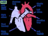
Atrial septal defect is a form of congenital heart defect
that enables blood flow between the left and right atria via the interatrial septum
. The interatrial septum is the tissue that divides the right
and left
atria. Without this septum, or if there is a defect in this septum, it is possible for blood to travel from the left side of the heart to the right side of the heart, or vice versa. This results in the mixing of arterial and venous blood, which may or may not be clinically significant. This mixture of blood may or may not result in what is known as a "shunt". The amount of shunting present, if any, dictates hemodynamic significance (see Pathophysiology below). A "right-to-left-shunt" typically poses the more dangerous scenario (see Pathophysiology below).
The right side of the heart contains venous blood
with a low oxygen
content, and the left side of the heart contains arterial
blood
with a high oxygen
content. A normal heart has an interatrial septum that prevents oxygen-rich blood and oxygen-deficient blood from mixing together.
During development of the fetus
, the interatrial septum develops to separate the left
and right
atrium. However, the foramen ovale
(icon) allows blood from the right atrium to the left atrium during fetal development. This opening allows blood to bypass the nonfunctional fetal lungs when the fetus obtains its oxygen from the placenta
. A layer of tissue called the septum primum acts as a valve over the foramen ovale during fetal development. After birth, the pressure in the pulmonary circulatory system drops, thus causing the foramen ovale to close entirely. In approximately 25% of adults, the foramen ovale does not entirely seal. In this case, elevation of pressure in the pulmonary circulatory system (i.e.: pulmonary hypertension
due to various causes, or transiently during a cough
) can cause the foramen ovale to remain open. This is known as a patent foramen ovale (PFO).
 In unaffected individuals, the chambers of the left side of the heart are under higher pressure than the chambers of the right side of the heart. This is because the left ventricle
has to produce enough pressure to pump blood throughout the entire body, while the right ventricle
only has to produce enough pressure to pump blood to the lung
s.
In the case of a large ASD (>9mm), which may result in a clinically remarkable left-to-right shunt
, blood will shunt from the left atrium
to the right atrium
. This extra blood from the left atrium may cause a volume overload of both the right atrium
and the right ventricle
. If untreated, this condition can result in enlargement of the right side of the heart and ultimately heart failure.
Any process that increases the pressure in the left ventricle
can cause worsening of the left-to-right shunt. This includes hypertension
, which increases the pressure that the left ventricle has to generate in order to open the aortic valve
during ventricular systole
, and coronary artery disease which increases the stiffness of the left ventricle, thereby increasing the filling pressure of the left ventricle during ventricular diastole
.
The right ventricle will have to push out more blood than the left ventricle due to the left-to-right shunt. This constant overload of the right side of the heart will cause an overload of the entire pulmonary vasculature. Eventually pulmonary hypertension
may develop.
The pulmonary hypertension will cause the right ventricle to face increased afterload
in addition to the increased preload that the shunted blood from the left atrium to the right atrium caused. The right ventricle will be forced to generate higher pressures to try to overcome the pulmonary hypertension. This may lead to right ventricular failure (dilatation and decreased systolic
function of the right ventricle) or elevations of the right sided pressures relative to left sided pressures.
When the pressure in the right atrium rises to the level in the left atrium, there will no longer be a pressure gradient between these heart chambers, and the left-to-right shunt will diminish or cease.
If left uncorrected, the pressure in the right side of the heart will be greater than the left side of the heart. This will cause the pressure in the right atrium to be higher than the pressure in the left atrium. This will reverse the pressure gradient across the ASD, and the shunt will reverse; a right-to-left shunt will exist. This phenomenon is known as Eisenmenger's syndrome
.
Once right-to-left shunting occurs, a portion of the oxygen-poor blood will get shunted to the left side of the heart and ejected to the peripheral vascular system. This will cause signs of cyanosis
.
The ostium secundum atrial septal defect accounts for 7% of all congenital heart lesions. This lesion shows a female preponderance, with a male : female ratio of 1:2.
 There are many types of atrial septal defects. They are differentiated from each other by whether they involve other structures of the heart and how they are formed during the developmental process during early fetal
development.
The secundum atrial septal defect usually arises from an enlarged foramen ovale
, inadequate growth of the septum secundum
, or excessive absorption of the septum primum
. Ten to twenty percent of individuals with ostium secundum ASDs also have mitral valve prolapse
.
s, and syncope.
Complications of an uncorrected secundum ASD include pulmonary hypertension
, right-sided heart failure
, atrial fibrillation
or flutter
, stroke, and Eisenmenger's syndrome
.
While pulmonary hypertension is unusual before 20 years of age, it is seen in 50% of individuals above the age of 40. Progression to Eisenmenger's syndrome
occurs in 5 to 10% of individuals late in the disease process.
. Clinically it is linked to decompression sickness
, paradoxical embolism and migraine
. On echocardiography, there may not be any shunting of blood noted except when the patient coughs.
There is debate within the neurology and cardiology communities about the role of a PFO in cryptogenic (i.e. of unknown cause) neurologic events such as strokes and transient ischemia attacks (TIAs) without any other potential cause. Some data suggested that PFOs may be involved in the pathogenesis of some migraine headaches. Several clinical trials are currently underway to investigate the role of PFO in these clinical situations.
A defect in the ostium primum
is occasionally classified as an atrial septal defect, but it is more commonly classified as an atrioventricular septal defect
.
or the inferior vena cava
.
A sinus venosus ASD that involves the superior vena cava makes up 2 to 3% of all interatrial communication. It is located at the junction of the superior vena cava and the right atrium. It is frequently associated with anomalous drainage of the right-sided pulmonary vein
s into the right atrium (instead of the normal drainage of the pulmonary veins into the left atrium).
or in early childhood with the use of ultrasonography or auscultation
of the heart sounds
during physical examination
.
Adults with an uncorrected ASD will present with symptoms of dyspnea on exertion (shortness of breath with minimal exercise), congestive heart failure
, or cerebrovascular accident (stroke). They may be noted on routine testing to have an abnormal chest x-ray
or an abnormal ECG and may have atrial fibrillation
.
that may be present in these individuals.
Upon auscultation
of the heart sounds
, there may be an ejection systolic murmur that is attributed to the pulmonic valve. This is due to the increased flow of blood through the pulmonic valve rather than any structural abnormality of the valve leaflets.
In unaffected individuals, there are respiratory variations in the splitting of the second heart sound
(S2). During respiratory inspiration, the negative intrathoracic pressure causes increased blood return into the right side of the heart. The increased blood volume in the right ventricle causes the pulmonic valve to stay open longer during ventricular systole
. This causes a normal delay in the P2 component of S2. During expiration, the positive intrathoracic pressure causes decreased blood return to the right side of the heart. The reduced volume in the right ventricle allows the pulmonic valve to close earlier at the end of ventricular systole, causing P2 to occur earlier.
In individuals with an ASD, there is a fixed splitting of S2. The reason that there is a fixed splitting of the second heart sound is that the extra blood return during inspiration gets equalized between the left and right atrium due to the communication that exists between the atria in individuals with ASD.
The right ventricle can be thought of as continuously overloaded because of the left to right shunt, producing a widely split S2. Because the atria are linked via the atrial septal defect, inspiration produces no net pressure change between them, and has no effect on the splitting of S2. Thus, S2 is split to the same degree during inspiration as expiration, and is said to be “fixed.”
, an atrial septal defect may be seen on color flow imaging as a jet of blood from the left atrium to the right atrium.
If agitated saline is injected into a peripheral vein
during echocardiography, small air bubbles can be seen on echocardiographic imaging. It may be possible to see bubbles travel across an ASD either at rest or during a cough. (Bubbles will only flow from right atrium to left atrium if the RA pressure is greater than LA).
Because better visualization of the atria is achieved with transesophageal echocardiography, this test may be performed in individuals with a suspected ASD which is not visualized on transthoracic imaging.
Newer techniques to visualize these defects involve intracardiac imaging with special catheters that are typically placed in the venous system and advanced to the level of the heart. This type of imaging is becoming more common and involves only mild sedation for the patient typically.
If the individual has adequate echocardiographic windows, it is possible to use the echocardiogram to measure the cardiac output of the left ventricle and the right ventricle independently. In this way, it is possible to estimate the shunt fraction using echocardiograpy.
). The prolongation of the PR interval is probably due to the enlargement of the atria that is common in ASDs and the increased distance due to the defect itself. Both of these can cause an increased distance of internodal conduction from the SA node to the AV node.
In addition to the PR prolongation, individuals with a primum ASD have a left axis deviation of the QRS complex while those with a secundum ASD have a right axis deviation of the QRS complex. Individuals with a sinus venosus ASD exhibit a left axis deviation of the P wave (not the QRS complex).
A common finding in the ECG is the presence of incomplete RBBB (Right Bundle Branch Block). In fact this finding is so characteristic that if it is absent, the diagnosis of ASD should be revised.
Surgical mortality due to closure of an ASD is lowest when the procedure is performed prior to the development of significant pulmonary hypertension. The lowest mortality rates are achieved in individuals with a pulmonary artery systolic pressure of less than 40 mmHg.
If Eisenmenger's syndrome
has occurred, there is significant risk of mortality regardless of the method of closure of the ASD. In individuals who have developed Eisenmenger's syndrome, the pressure in the right ventricle has raised high enough to reverse the shunt in the atria. If the ASD is then closed, the afterload
that the right ventricle has to act against has suddenly increased. This may cause immediate right ventricular failure, since it may not be able to pump the blood against the pulmonary hypertension.
Closure of an ASD in individuals under age 25 has been shown to have a low risk of complications, and individuals have a normal lifespan (comparable to a healthy age-matched population). Closure of an ASD in individuals between the ages of 25 and 40 who are asymptomatic but have a clinically significant shunt is controversial. Those that perform the procedure believe that they are preventing long-term deterioration in cardiac function and preventing the progression of pulmonary hypertension.
Methods of closure of an ASD include surgical closure and percutaneous closure.
If pulmonary hypertension is present, the evaluation may include a right heart catheterization. This involves placing a catheter in the venous system of the heart and measuring pressures and oxygen saturations in the SVC
, IVC
, right atrium
, right ventricle
, pulmonary artery
, and in the wedge position. Individuals with a pulmonary vascular resistance (PVR) of less than 7 wood units show regression of symptoms (including NYHA functional class
). On the other hand, individuals with a PVR of greater than 15 wood units have increased mortality associated with closure of the ASD.
If the pulmonary arterial pressure is more than 2/3 the systemic systolic pressure, there should be a net left-to-right shunt of at least 1.5:1 or evidence of reversibility of the shunt when given pulmonary artery vasodilators prior to surgery. (If eisenmenger's physiology has set in, it must be proven that the right-to-left shunt is reversible with pulmonary artery vasodilators prior to surgery.)
During the procedure, the doctor inserts a catheter (a thin, flexible tube) into a vein in the groin (upper thigh) and threads it to the heart's septum. The catheter has a tiny umbrella-like device folded up inside it.When the catheter reaches the septum, the device is pushed out of the catheter and positioned so that it plugs the hole between the atria. The device is secured in place and the catheter is withdrawn from the body.Within 6 months, normal tissue grows in and over the device. There is no need to replace the closure device as the child grows.Doctors often use echocardiography (echo) or transesophageal (tranz-ih-sof-uh-JEE-ul) echo (TEE) as well as angiography (an-jee-OG-ra-fee) to guide them in threading the catheter to the heart and closing the defect. TEE is a special type of echo that takes pictures of the heart through the esophagus (the passage leading from the mouth to the stomach).Catheter procedures are much easier on patients than surgery because they involve only a needle puncture in the skin where the catheter is inserted. This means that recovery is faster and easier.The outlook for children having this procedure is excellent. Closures are successful in more than 9 out of 10 patients, with no significant leakage. Rarely, a defect is too large for catheter closure and surgery is needed.
and closing the defect with a patch under direct visualization.
, IVC
, or the tricuspid
or mitral
valves. The Amplatzer
Septal Occluder (ASO) is commonly used to close ASDs. The ASO consists of two self-expandable round discs connected to each other with a 4 mm waist, made up of 0.004–0.005´´ Nitinol wire mesh filled with Dacron fabric. Implantation of the device is relatively easy. The prevalence of residual defect is low. The disadvantages are a thick profile of the device and concern related to a large amount of nitinol (a nickel-titanium compound) in the device and consequent potential for nickel toxicity.
Percutaneous closure is the method of choice in most centres.
in divers because a proportion of venous blood carrying inert gases, such as helium
or nitrogen
does not pass through the lungs.
The only way to release the excess inert gases from the body is to pass the blood carrying the inert gases through the lung
s to be exhaled. If some of the inert gas-laden blood passes through the PFO, it avoids the lungs and the inert gas is more likely to form large bubbles in the arterial blood stream causing decompression sickness.
(clots in the vein
s) are quite common. Embolization (dislodgement of thrombi) normally go to the lung and cause pulmonary emboli
. In an individual with ASD, these emboli can potentially enter the arterial system. This can cause any phenomenon that is attributed to acute loss of blood to a portion of the body, including cerebrovascular accident (stroke), infarction of the spleen
or intestine
s, or even a distal extremity .......(i.e.: finger or toe).
This is known as a paradoxical embolus because the clot material paradoxially enters the arterial system instead of going to the lungs.
may be caused by patent foramen ovale. While the exact mechanism remains unclear, closure of a PFO can reduce symptoms in certain cases. This remains controversial. 20% of the general population have a PFO, which for the most part, is asymptomatic. 20% of the female population have migraines. And, the placebo effect
in migraine typically averages around 40%. The high frequency of these facts makes statistically significant relationships between PFO and migraine difficult (i.e., the relationship may just be chance or coincidence). In a large randomized controlled trial the higher prevalence of patent foramen ovale in migraine patients was confirmed, but migraine headache cessation was not more prevalent in the group of migraine patients that underwent closure of their patent foramen ovale.
which can lead to viral meningitis
. (This claims requires supporting evidence)
Congenital heart defect
A congenital heart defect is a defect in the structure of the heart and great vessels which is present at birth. Many types of heart defects exist, most of which either obstruct blood flow in the heart or vessels near it, or cause blood to flow through the heart in an abnormal pattern. Other...
that enables blood flow between the left and right atria via the interatrial septum
Interatrial septum
The interatrial septum is the wall of tissue that separates the right and left atria of the heart.-Development:The interatrial septum forms during the first and second months of fetal development. Formation of the septum occurs in several stages...
. The interatrial septum is the tissue that divides the right
Right atrium
The right atrium is one of four chambers in the hearts of mammals and archosaurs...
and left
Left atrium
The left atrium is one of the four chambers in the human heart. It receives oxygenated blood from the pulmonary veins, and pumps it into the left ventricle, via the mitral valve.-Foramen ovale:...
atria. Without this septum, or if there is a defect in this septum, it is possible for blood to travel from the left side of the heart to the right side of the heart, or vice versa. This results in the mixing of arterial and venous blood, which may or may not be clinically significant. This mixture of blood may or may not result in what is known as a "shunt". The amount of shunting present, if any, dictates hemodynamic significance (see Pathophysiology below). A "right-to-left-shunt" typically poses the more dangerous scenario (see Pathophysiology below).
The right side of the heart contains venous blood
Venous blood
Venous blood is deoxygenated blood in the circulatory system. It runs in the systemic veins from the organs to the heart. Deoxygenated blood is then pumped by the heart to lungs via the pulmonary arteries, one of the few arteries in the body that carries deoxygenated blood .Venous blood is...
with a low oxygen
Oxygen
Oxygen is the element with atomic number 8 and represented by the symbol O. Its name derives from the Greek roots ὀξύς and -γενής , because at the time of naming, it was mistakenly thought that all acids required oxygen in their composition...
content, and the left side of the heart contains arterial
Artery
Arteries are blood vessels that carry blood away from the heart. This blood is normally oxygenated, exceptions made for the pulmonary and umbilical arteries....
blood
Blood
Blood is a specialized bodily fluid in animals that delivers necessary substances such as nutrients and oxygen to the cells and transports metabolic waste products away from those same cells....
with a high oxygen
Oxygen
Oxygen is the element with atomic number 8 and represented by the symbol O. Its name derives from the Greek roots ὀξύς and -γενής , because at the time of naming, it was mistakenly thought that all acids required oxygen in their composition...
content. A normal heart has an interatrial septum that prevents oxygen-rich blood and oxygen-deficient blood from mixing together.
During development of the fetus
Fetus
A fetus is a developing mammal or other viviparous vertebrate after the embryonic stage and before birth.In humans, the fetal stage of prenatal development starts at the beginning of the 11th week in gestational age, which is the 9th week after fertilization.-Etymology and spelling variations:The...
, the interatrial septum develops to separate the left
Left atrium
The left atrium is one of the four chambers in the human heart. It receives oxygenated blood from the pulmonary veins, and pumps it into the left ventricle, via the mitral valve.-Foramen ovale:...
and right
Right atrium
The right atrium is one of four chambers in the hearts of mammals and archosaurs...
atrium. However, the foramen ovale
Foramen ovale (heart)
In the fetal heart, the foramen ovale , also ostium secundum of Born or falx septi, allows blood to enter the left atrium from the right atrium. It is one of two fetal cardiac shunts, the other being the ductus arteriosus...
(icon) allows blood from the right atrium to the left atrium during fetal development. This opening allows blood to bypass the nonfunctional fetal lungs when the fetus obtains its oxygen from the placenta
Placenta
The placenta is an organ that connects the developing fetus to the uterine wall to allow nutrient uptake, waste elimination, and gas exchange via the mother's blood supply. "True" placentas are a defining characteristic of eutherian or "placental" mammals, but are also found in some snakes and...
. A layer of tissue called the septum primum acts as a valve over the foramen ovale during fetal development. After birth, the pressure in the pulmonary circulatory system drops, thus causing the foramen ovale to close entirely. In approximately 25% of adults, the foramen ovale does not entirely seal. In this case, elevation of pressure in the pulmonary circulatory system (i.e.: pulmonary hypertension
Pulmonary hypertension
In medicine, pulmonary hypertension is an increase in blood pressure in the pulmonary artery, pulmonary vein, or pulmonary capillaries, together known as the lung vasculature, leading to shortness of breath, dizziness, fainting, and other symptoms, all of which are exacerbated by exertion...
due to various causes, or transiently during a cough
Cough
A cough is a sudden and often repetitively occurring reflex which helps to clear the large breathing passages from secretions, irritants, foreign particles and microbes...
) can cause the foramen ovale to remain open. This is known as a patent foramen ovale (PFO).
Pathophysiology
Left ventricle
The left ventricle is one of four chambers in the human heart. It receives oxygenated blood from the left atrium via the mitral valve, and pumps it into the aorta via the aortic valve.-Shape:...
has to produce enough pressure to pump blood throughout the entire body, while the right ventricle
Right ventricle
The right ventricle is one of four chambers in the human heart. It receives deoxygenated blood from the right atrium via the tricuspid valve, and pumps it into the pulmonary artery via the pulmonary valve and pulmonary trunk....
only has to produce enough pressure to pump blood to the lung
Lung
The lung is the essential respiration organ in many air-breathing animals, including most tetrapods, a few fish and a few snails. In mammals and the more complex life forms, the two lungs are located near the backbone on either side of the heart...
s.
In the case of a large ASD (>9mm), which may result in a clinically remarkable left-to-right shunt
Cardiac shunt
Cardiac shunts is when the blood flow follows a pattern in the heart that deviates from the normal circuit of the circulatory system. It may be described as right-left, left-to-right or bidirectional, or as systemic-to-pulmonary or pulmonary-to-systemic. The direction may be controlled by left...
, blood will shunt from the left atrium
Left atrium
The left atrium is one of the four chambers in the human heart. It receives oxygenated blood from the pulmonary veins, and pumps it into the left ventricle, via the mitral valve.-Foramen ovale:...
to the right atrium
Right atrium
The right atrium is one of four chambers in the hearts of mammals and archosaurs...
. This extra blood from the left atrium may cause a volume overload of both the right atrium
Right atrium
The right atrium is one of four chambers in the hearts of mammals and archosaurs...
and the right ventricle
Right ventricle
The right ventricle is one of four chambers in the human heart. It receives deoxygenated blood from the right atrium via the tricuspid valve, and pumps it into the pulmonary artery via the pulmonary valve and pulmonary trunk....
. If untreated, this condition can result in enlargement of the right side of the heart and ultimately heart failure.
Any process that increases the pressure in the left ventricle
Left ventricle
The left ventricle is one of four chambers in the human heart. It receives oxygenated blood from the left atrium via the mitral valve, and pumps it into the aorta via the aortic valve.-Shape:...
can cause worsening of the left-to-right shunt. This includes hypertension
Hypertension
Hypertension or high blood pressure is a cardiac chronic medical condition in which the systemic arterial blood pressure is elevated. What that means is that the heart is having to work harder than it should to pump the blood around the body. Blood pressure involves two measurements, systolic and...
, which increases the pressure that the left ventricle has to generate in order to open the aortic valve
Aortic valve
The aortic valve is one of the valves of the heart. It is normally tricuspid , although in 1% of the population it is found to be congenitally bicuspid . It lies between the left ventricle and the aorta....
during ventricular systole
Systole (medicine)
Systole is the contraction of the heart. Used alone, it usually means the contraction of the left ventricle.In all mammals, the heart has 4 chambers. The left and right ventricles pump together. The atria and ventricles pump in sequence...
, and coronary artery disease which increases the stiffness of the left ventricle, thereby increasing the filling pressure of the left ventricle during ventricular diastole
Diastole
Diastole is the period of time when the heart fills with blood after systole . Ventricular diastole is the period during which the ventricles are relaxing, while atrial diastole is the period during which the atria are relaxing...
.
The right ventricle will have to push out more blood than the left ventricle due to the left-to-right shunt. This constant overload of the right side of the heart will cause an overload of the entire pulmonary vasculature. Eventually pulmonary hypertension
Pulmonary hypertension
In medicine, pulmonary hypertension is an increase in blood pressure in the pulmonary artery, pulmonary vein, or pulmonary capillaries, together known as the lung vasculature, leading to shortness of breath, dizziness, fainting, and other symptoms, all of which are exacerbated by exertion...
may develop.
The pulmonary hypertension will cause the right ventricle to face increased afterload
Afterload
Afterload is the tension or stress developed in the wall of the left ventricle during ejection. Following Laplace's law, the tension upon the muscle fibers in the heart wall is the product of the pressure within the ventricle, multiplied by the volume within the ventricle, divided by the wall...
in addition to the increased preload that the shunted blood from the left atrium to the right atrium caused. The right ventricle will be forced to generate higher pressures to try to overcome the pulmonary hypertension. This may lead to right ventricular failure (dilatation and decreased systolic
Systole (medicine)
Systole is the contraction of the heart. Used alone, it usually means the contraction of the left ventricle.In all mammals, the heart has 4 chambers. The left and right ventricles pump together. The atria and ventricles pump in sequence...
function of the right ventricle) or elevations of the right sided pressures relative to left sided pressures.
When the pressure in the right atrium rises to the level in the left atrium, there will no longer be a pressure gradient between these heart chambers, and the left-to-right shunt will diminish or cease.
If left uncorrected, the pressure in the right side of the heart will be greater than the left side of the heart. This will cause the pressure in the right atrium to be higher than the pressure in the left atrium. This will reverse the pressure gradient across the ASD, and the shunt will reverse; a right-to-left shunt will exist. This phenomenon is known as Eisenmenger's syndrome
Eisenmenger's syndrome
Eisenmenger's syndrome is defined as the process in which a left-to-right shunt caused by a congenital heart defect causes increased flow through the pulmonary vasculature, causing pulmonary hypertension, which in turn, causes increased pressures in the right side of the heart and reversal of the...
.
Once right-to-left shunting occurs, a portion of the oxygen-poor blood will get shunted to the left side of the heart and ejected to the peripheral vascular system. This will cause signs of cyanosis
Cyanosis
Cyanosis is the appearance of a blue or purple coloration of the skin or mucous membranes due to the tissues near the skin surface being low on oxygen. The onset of cyanosis is 2.5 g/dL of deoxyhemoglobin. The bluish color is more readily apparent in those with high hemoglobin counts than it is...
.
Epidemiology
As a group, atrial septal defects are detected in 1 child per 1500 live births. PFO are quite common (appearing in 10 - 20% of adults) but asymptomatic and therefore undiagnosed. ASDs make up 30 to 40% of all congenital heart disease that is seen in adults.The ostium secundum atrial septal defect accounts for 7% of all congenital heart lesions. This lesion shows a female preponderance, with a male : female ratio of 1:2.
Types of atrial septal defects
Fetus
A fetus is a developing mammal or other viviparous vertebrate after the embryonic stage and before birth.In humans, the fetal stage of prenatal development starts at the beginning of the 11th week in gestational age, which is the 9th week after fertilization.-Etymology and spelling variations:The...
development.
Ostium secundum atrial septal defect
The ostium secundum atrial septal defect is the most common type of atrial septal defect, and comprises 6-10% of all congenital heart diseases.The secundum atrial septal defect usually arises from an enlarged foramen ovale
Foramen ovale (heart)
In the fetal heart, the foramen ovale , also ostium secundum of Born or falx septi, allows blood to enter the left atrium from the right atrium. It is one of two fetal cardiac shunts, the other being the ductus arteriosus...
, inadequate growth of the septum secundum
Septum secundum
The septum secundum, semilunar in shape, grows downward from the upper wall of the atrium immediately to the right of the primary septum and ostium secundum....
, or excessive absorption of the septum primum
Septum primum
In the developing heart, the cavity of the primitive atrium becomes subdivided into right and left chambers by a septum, the septum primum, which grows downward into the cavity. The increasingly smaller gap below it is known as the ostium primum...
. Ten to twenty percent of individuals with ostium secundum ASDs also have mitral valve prolapse
Mitral valve prolapse
Mitral valve prolapse is a valvular heart disease characterized by the displacement of an abnormally thickened mitral valve leaflet into the left atrium during systole. There are various types of MVP, broadly classified as classic and nonclassic. In its nonclassic form, MVP carries a low risk of...
.
Natural history
Most individuals with an uncorrected secundum ASD do not have significant symptoms through early adulthood. About 70% develop symptoms by the time they are in their 40s. Symptoms are typically decreased exercise tolerance, easy fatigueability, palpitationPalpitation
A palpitation is an abnormality of heartbeat that causes a conscious awareness of its beating, whether it is too slow, too fast, irregular, or at its normal frequency. The word may also refer to this sensation itself...
s, and syncope.
Complications of an uncorrected secundum ASD include pulmonary hypertension
Pulmonary hypertension
In medicine, pulmonary hypertension is an increase in blood pressure in the pulmonary artery, pulmonary vein, or pulmonary capillaries, together known as the lung vasculature, leading to shortness of breath, dizziness, fainting, and other symptoms, all of which are exacerbated by exertion...
, right-sided heart failure
Congestive heart failure
Heart failure often called congestive heart failure is generally defined as the inability of the heart to supply sufficient blood flow to meet the needs of the body. Heart failure can cause a number of symptoms including shortness of breath, leg swelling, and exercise intolerance. The condition...
, atrial fibrillation
Atrial fibrillation
Atrial fibrillation is the most common cardiac arrhythmia . It is a common cause of irregular heart beat, identified clinically by taking a pulse. Chaotic electrical activity in the two upper chambers of the heart result in the muscle fibrillating , instead of achieving coordinated contraction...
or flutter
Atrial flutter
Atrial flutter is an abnormal heart rhythm that occurs in the atria of the heart. When it first occurs, it is usually associated with a fast heart rate or tachycardia , and falls into the category of supra-ventricular tachycardias. While this rhythm occurs most often in individuals with...
, stroke, and Eisenmenger's syndrome
Eisenmenger's syndrome
Eisenmenger's syndrome is defined as the process in which a left-to-right shunt caused by a congenital heart defect causes increased flow through the pulmonary vasculature, causing pulmonary hypertension, which in turn, causes increased pressures in the right side of the heart and reversal of the...
.
While pulmonary hypertension is unusual before 20 years of age, it is seen in 50% of individuals above the age of 40. Progression to Eisenmenger's syndrome
Eisenmenger's syndrome
Eisenmenger's syndrome is defined as the process in which a left-to-right shunt caused by a congenital heart defect causes increased flow through the pulmonary vasculature, causing pulmonary hypertension, which in turn, causes increased pressures in the right side of the heart and reversal of the...
occurs in 5 to 10% of individuals late in the disease process.
Patent foramen ovale
A patent foramen ovale (PFO) is a small channel that has little hemodynamic consequence; it is a remnant of the fetal foramen ovaleForamen ovale (heart)
In the fetal heart, the foramen ovale , also ostium secundum of Born or falx septi, allows blood to enter the left atrium from the right atrium. It is one of two fetal cardiac shunts, the other being the ductus arteriosus...
. Clinically it is linked to decompression sickness
Decompression sickness
Decompression sickness describes a condition arising from dissolved gases coming out of solution into bubbles inside the body on depressurization...
, paradoxical embolism and migraine
Migraine
Migraine is a chronic neurological disorder characterized by moderate to severe headaches, and nausea...
. On echocardiography, there may not be any shunting of blood noted except when the patient coughs.
There is debate within the neurology and cardiology communities about the role of a PFO in cryptogenic (i.e. of unknown cause) neurologic events such as strokes and transient ischemia attacks (TIAs) without any other potential cause. Some data suggested that PFOs may be involved in the pathogenesis of some migraine headaches. Several clinical trials are currently underway to investigate the role of PFO in these clinical situations.
Ostium primum atrial septal defect
A defect in the ostium primum
Ostium primum
In the developing heart, the atria initially communicate with each other by an opening between the free edge of the septum primum and the AV cushions, known as the primary interatrial foramen or ostium primum , below the free margin of the septum.-Closing of ostium primum:This opening is closed by...
is occasionally classified as an atrial septal defect, but it is more commonly classified as an atrioventricular septal defect
Atrioventricular septal defect
Atrioventricular septal defect or atrioventricular canal defect , previously known as "common atrioventricular canal" or "endocardial cushion defect", is characterized by a deficiency of the atrioventricular septum of the heart...
.
Sinus venosus atrial septal defect
A sinus venosus ASD is a type of atrial septum defect in which the defect in the septum involves the venous inflow of either the superior vena cavaSuperior vena cava
The superior vena cava is truly superior, a large diameter, yet short, vein that carries deoxygenated blood from the upper half of the body to the heart's right atrium...
or the inferior vena cava
Inferior vena cava
The inferior vena cava , also known as the posterior vena cava, is the large vein that carries de-oxygenated blood from the lower half of the body into the right atrium of the heart....
.
A sinus venosus ASD that involves the superior vena cava makes up 2 to 3% of all interatrial communication. It is located at the junction of the superior vena cava and the right atrium. It is frequently associated with anomalous drainage of the right-sided pulmonary vein
Pulmonary vein
The pulmonary veins are large blood vessels that carry blood from the lungs to the left atrium of the heart. In humans there are four pulmonary veins, two from each lung...
s into the right atrium (instead of the normal drainage of the pulmonary veins into the left atrium).
Common or single atrium
Common (or single) atrium is a failure of development of the embryologic components that contribute to the atrial septal complex. It is frequently associated with heterotaxy syndrome.Mixed Atrial septal defect
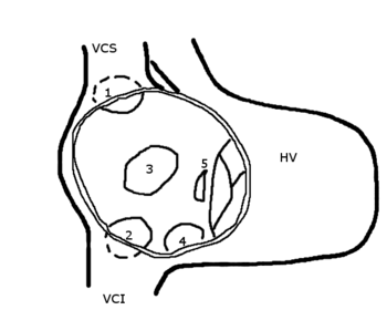
The inter atrial septum can be divided in to 5 septal zones. If the defect involves 2 or more of the 5 septal zones, then the defect is termed a mixed atrial septal defect.Diagnosis in children
Most individuals with a significant ASD are diagnosed in uteroIn utero
In utero is a Latin term literally meaning "in the womb". In biology, the phrase describes the state of an embryo or fetus. In legal contexts, the phrase is used to refer to unborn children. Under common law, unborn children are still considered to exist for property transfer purposes.-See also:*...
or in early childhood with the use of ultrasonography or auscultation
Auscultation
Auscultation is the term for listening to the internal sounds of the body, usually using a stethoscope...
of the heart sounds
Heart sounds
Heart sounds, or heartbeats, are the noises generated by the beating heart and the resultant flow of blood through it...
during physical examination
Physical examination
Physical examination or clinical examination is the process by which a doctor investigates the body of a patient for signs of disease. It generally follows the taking of the medical history — an account of the symptoms as experienced by the patient...
.
Diagnosis in adults
Some individuals with an ASD will have undergone surgical correction of their ASD during childhood. The development of signs and symptoms due to an ASD are related to the size of the intracardiac shunt. Individuals with a larger shunt tend to present with symptoms at a younger age.Adults with an uncorrected ASD will present with symptoms of dyspnea on exertion (shortness of breath with minimal exercise), congestive heart failure
Congestive heart failure
Heart failure often called congestive heart failure is generally defined as the inability of the heart to supply sufficient blood flow to meet the needs of the body. Heart failure can cause a number of symptoms including shortness of breath, leg swelling, and exercise intolerance. The condition...
, or cerebrovascular accident (stroke). They may be noted on routine testing to have an abnormal chest x-ray
Chest X-ray
In medicine, a chest radiograph, commonly called a chest X-ray , is a projection radiograph of the chest used to diagnose conditions affecting the chest, its contents, and nearby structures...
or an abnormal ECG and may have atrial fibrillation
Atrial fibrillation
Atrial fibrillation is the most common cardiac arrhythmia . It is a common cause of irregular heart beat, identified clinically by taking a pulse. Chaotic electrical activity in the two upper chambers of the heart result in the muscle fibrillating , instead of achieving coordinated contraction...
.
Physical exam auscultation of the heart
The physical findings in an adult with an ASD include those related directly to the intracardiac shunt, and those that are secondary to the right heart failureCongestive heart failure
Heart failure often called congestive heart failure is generally defined as the inability of the heart to supply sufficient blood flow to meet the needs of the body. Heart failure can cause a number of symptoms including shortness of breath, leg swelling, and exercise intolerance. The condition...
that may be present in these individuals.
Upon auscultation
Auscultation
Auscultation is the term for listening to the internal sounds of the body, usually using a stethoscope...
of the heart sounds
Heart sounds
Heart sounds, or heartbeats, are the noises generated by the beating heart and the resultant flow of blood through it...
, there may be an ejection systolic murmur that is attributed to the pulmonic valve. This is due to the increased flow of blood through the pulmonic valve rather than any structural abnormality of the valve leaflets.
In unaffected individuals, there are respiratory variations in the splitting of the second heart sound
Split S2
A split S2 is a finding upon auscultation of the S2 heart sound.It is caused when the closure of the aortic valve and the closure of the pulmonary valve are not synchronized.-Physiologic split:...
(S2). During respiratory inspiration, the negative intrathoracic pressure causes increased blood return into the right side of the heart. The increased blood volume in the right ventricle causes the pulmonic valve to stay open longer during ventricular systole
Systole (medicine)
Systole is the contraction of the heart. Used alone, it usually means the contraction of the left ventricle.In all mammals, the heart has 4 chambers. The left and right ventricles pump together. The atria and ventricles pump in sequence...
. This causes a normal delay in the P2 component of S2. During expiration, the positive intrathoracic pressure causes decreased blood return to the right side of the heart. The reduced volume in the right ventricle allows the pulmonic valve to close earlier at the end of ventricular systole, causing P2 to occur earlier.
In individuals with an ASD, there is a fixed splitting of S2. The reason that there is a fixed splitting of the second heart sound is that the extra blood return during inspiration gets equalized between the left and right atrium due to the communication that exists between the atria in individuals with ASD.
The right ventricle can be thought of as continuously overloaded because of the left to right shunt, producing a widely split S2. Because the atria are linked via the atrial septal defect, inspiration produces no net pressure change between them, and has no effect on the splitting of S2. Thus, S2 is split to the same degree during inspiration as expiration, and is said to be “fixed.”
Echocardiography
In transthoracic echocardiographyEchocardiography
An echocardiogram, often referred to in the medical community as a cardiac ECHO or simply an ECHO, is a sonogram of the heart . Also known as a cardiac ultrasound, it uses standard ultrasound techniques to image two-dimensional slices of the heart...
, an atrial septal defect may be seen on color flow imaging as a jet of blood from the left atrium to the right atrium.
If agitated saline is injected into a peripheral vein
Vein
In the circulatory system, veins are blood vessels that carry blood towards the heart. Most veins carry deoxygenated blood from the tissues back to the heart; exceptions are the pulmonary and umbilical veins, both of which carry oxygenated blood to the heart...
during echocardiography, small air bubbles can be seen on echocardiographic imaging. It may be possible to see bubbles travel across an ASD either at rest or during a cough. (Bubbles will only flow from right atrium to left atrium if the RA pressure is greater than LA).
Because better visualization of the atria is achieved with transesophageal echocardiography, this test may be performed in individuals with a suspected ASD which is not visualized on transthoracic imaging.
Newer techniques to visualize these defects involve intracardiac imaging with special catheters that are typically placed in the venous system and advanced to the level of the heart. This type of imaging is becoming more common and involves only mild sedation for the patient typically.
If the individual has adequate echocardiographic windows, it is possible to use the echocardiogram to measure the cardiac output of the left ventricle and the right ventricle independently. In this way, it is possible to estimate the shunt fraction using echocardiograpy.
Transcranial Doppler (TCD) Bubble study
A less invasive method for detecting a PFO or other ASDs than transesophagal ultrasound is Transcranial Doppler with bubble contrast. This method reveals the cerebral impact of the ASD or PFO.Electrocardiogram
The ECG findings in atrial septal defect vary with the type of defect the individual has. Individuals with atrial septal defects may have a prolonged PR interval (a first degree heart blockFirst degree heart block
First-degree AV block, or PR prolongation, is a disease of the electrical conduction system of the heart in which the PR interval is lengthened beyond 0.20 seconds....
). The prolongation of the PR interval is probably due to the enlargement of the atria that is common in ASDs and the increased distance due to the defect itself. Both of these can cause an increased distance of internodal conduction from the SA node to the AV node.
In addition to the PR prolongation, individuals with a primum ASD have a left axis deviation of the QRS complex while those with a secundum ASD have a right axis deviation of the QRS complex. Individuals with a sinus venosus ASD exhibit a left axis deviation of the P wave (not the QRS complex).
A common finding in the ECG is the presence of incomplete RBBB (Right Bundle Branch Block). In fact this finding is so characteristic that if it is absent, the diagnosis of ASD should be revised.
Treatment
Once someone is found to have an atrial septal defect, a determination of whether it should be corrected has to be made.Surgical mortality due to closure of an ASD is lowest when the procedure is performed prior to the development of significant pulmonary hypertension. The lowest mortality rates are achieved in individuals with a pulmonary artery systolic pressure of less than 40 mmHg.
If Eisenmenger's syndrome
Eisenmenger's syndrome
Eisenmenger's syndrome is defined as the process in which a left-to-right shunt caused by a congenital heart defect causes increased flow through the pulmonary vasculature, causing pulmonary hypertension, which in turn, causes increased pressures in the right side of the heart and reversal of the...
has occurred, there is significant risk of mortality regardless of the method of closure of the ASD. In individuals who have developed Eisenmenger's syndrome, the pressure in the right ventricle has raised high enough to reverse the shunt in the atria. If the ASD is then closed, the afterload
Afterload
Afterload is the tension or stress developed in the wall of the left ventricle during ejection. Following Laplace's law, the tension upon the muscle fibers in the heart wall is the product of the pressure within the ventricle, multiplied by the volume within the ventricle, divided by the wall...
that the right ventricle has to act against has suddenly increased. This may cause immediate right ventricular failure, since it may not be able to pump the blood against the pulmonary hypertension.
Closure of an ASD in individuals under age 25 has been shown to have a low risk of complications, and individuals have a normal lifespan (comparable to a healthy age-matched population). Closure of an ASD in individuals between the ages of 25 and 40 who are asymptomatic but have a clinically significant shunt is controversial. Those that perform the procedure believe that they are preventing long-term deterioration in cardiac function and preventing the progression of pulmonary hypertension.
Methods of closure of an ASD include surgical closure and percutaneous closure.
Evaluation prior to correction
Prior to correction of an ASD, an evaluation is made of the severity of the individual's pulmonary hypertension (If present at all) and whether it is reversible (Closure of an ASD may be recommended for prevention purposes, to avoid such a complication in the first place. Pulmomary hypertension is not always present in adults that are diagnosed with an ASD in adulthood).If pulmonary hypertension is present, the evaluation may include a right heart catheterization. This involves placing a catheter in the venous system of the heart and measuring pressures and oxygen saturations in the SVC
Superior vena cava
The superior vena cava is truly superior, a large diameter, yet short, vein that carries deoxygenated blood from the upper half of the body to the heart's right atrium...
, IVC
Inferior vena cava
The inferior vena cava , also known as the posterior vena cava, is the large vein that carries de-oxygenated blood from the lower half of the body into the right atrium of the heart....
, right atrium
Right atrium
The right atrium is one of four chambers in the hearts of mammals and archosaurs...
, right ventricle
Right ventricle
The right ventricle is one of four chambers in the human heart. It receives deoxygenated blood from the right atrium via the tricuspid valve, and pumps it into the pulmonary artery via the pulmonary valve and pulmonary trunk....
, pulmonary artery
Pulmonary artery
The pulmonary arteries carry deoxygenated blood from the heart to the lungs. They are the only arteries that carry deoxygenated blood....
, and in the wedge position. Individuals with a pulmonary vascular resistance (PVR) of less than 7 wood units show regression of symptoms (including NYHA functional class
New York Heart Association Functional Classification
The New York Heart Association Functional Classification provides a simple way of classifying the extent of heart failure. It places patients in one of four categories based on how much they are limited during physical activity; the limitations/symptoms are in regards to normal breathing and...
). On the other hand, individuals with a PVR of greater than 15 wood units have increased mortality associated with closure of the ASD.
If the pulmonary arterial pressure is more than 2/3 the systemic systolic pressure, there should be a net left-to-right shunt of at least 1.5:1 or evidence of reversibility of the shunt when given pulmonary artery vasodilators prior to surgery. (If eisenmenger's physiology has set in, it must be proven that the right-to-left shunt is reversible with pulmonary artery vasodilators prior to surgery.)
Catheter Procedure
Until the early 1990s, surgery was the usual method for closing all ASDs. Now, thanks to medical advances, doctors can use catheter procedures to close secundum ASDs, the most common type of ASD. For this procedure, the patient is given medicine so he or she will sleep through it and not feel any pain.During the procedure, the doctor inserts a catheter (a thin, flexible tube) into a vein in the groin (upper thigh) and threads it to the heart's septum. The catheter has a tiny umbrella-like device folded up inside it.When the catheter reaches the septum, the device is pushed out of the catheter and positioned so that it plugs the hole between the atria. The device is secured in place and the catheter is withdrawn from the body.Within 6 months, normal tissue grows in and over the device. There is no need to replace the closure device as the child grows.Doctors often use echocardiography (echo) or transesophageal (tranz-ih-sof-uh-JEE-ul) echo (TEE) as well as angiography (an-jee-OG-ra-fee) to guide them in threading the catheter to the heart and closing the defect. TEE is a special type of echo that takes pictures of the heart through the esophagus (the passage leading from the mouth to the stomach).Catheter procedures are much easier on patients than surgery because they involve only a needle puncture in the skin where the catheter is inserted. This means that recovery is faster and easier.The outlook for children having this procedure is excellent. Closures are successful in more than 9 out of 10 patients, with no significant leakage. Rarely, a defect is too large for catheter closure and surgery is needed.
Surgical ASD closure
Surgical closure of an ASD involves opening up at least one atriumAtrium (anatomy)
In anatomy, the atrium , sometimes called auricle , refers to a chamber or space. For example, the term is used for a portion of the lateral ventricle in the brain and the blood collection chamber of the heart...
and closing the defect with a patch under direct visualization.
Percutaneous ASD closure
Percutaneous closure of an ASD is currently only indicated for the closure of secundum ASDs with a sufficient rim of tissue around the septal defect so that the closure device does not impinge upon the SVCSuperior vena cava
The superior vena cava is truly superior, a large diameter, yet short, vein that carries deoxygenated blood from the upper half of the body to the heart's right atrium...
, IVC
Inferior vena cava
The inferior vena cava , also known as the posterior vena cava, is the large vein that carries de-oxygenated blood from the lower half of the body into the right atrium of the heart....
, or the tricuspid
Tricuspid valve
The tricuspid valve, or right atrioventricular valve, is on the right dorsal side of the mammalian heart, between the right atrium and the right ventricle. The normal tricuspid valve usually has three leaflets and three papillary muscles. They are connected to the papillary muscles by the chordae...
or mitral
Mitral valve
The mitral valve is a dual-flap valve in the heart that lies between the left atrium and the left ventricle...
valves. The Amplatzer
Kurt Amplatz
Dr. Kurt Amplatz is an Austrian radiologist and medical device inventor. He is best known for the invention of the Amplatzer Septal Occluder as well as the Amplatzer Cribriform Occluder, which is used for closing atrial septal defect, a common congenital heart defect found in infants...
Septal Occluder (ASO) is commonly used to close ASDs. The ASO consists of two self-expandable round discs connected to each other with a 4 mm waist, made up of 0.004–0.005´´ Nitinol wire mesh filled with Dacron fabric. Implantation of the device is relatively easy. The prevalence of residual defect is low. The disadvantages are a thick profile of the device and concern related to a large amount of nitinol (a nickel-titanium compound) in the device and consequent potential for nickel toxicity.
Percutaneous closure is the method of choice in most centres.
Associated conditions
Due to the communication between the atria that occurs in ASDs, disease entities or complications from the condition, are possible.Decompression sickness
ASDs, and particularly PFOs, are a predisposing risk factor for decompression sicknessDecompression sickness
Decompression sickness describes a condition arising from dissolved gases coming out of solution into bubbles inside the body on depressurization...
in divers because a proportion of venous blood carrying inert gases, such as helium
Helium
Helium is the chemical element with atomic number 2 and an atomic weight of 4.002602, which is represented by the symbol He. It is a colorless, odorless, tasteless, non-toxic, inert, monatomic gas that heads the noble gas group in the periodic table...
or nitrogen
Nitrogen
Nitrogen is a chemical element that has the symbol N, atomic number of 7 and atomic mass 14.00674 u. Elemental nitrogen is a colorless, odorless, tasteless, and mostly inert diatomic gas at standard conditions, constituting 78.08% by volume of Earth's atmosphere...
does not pass through the lungs.
The only way to release the excess inert gases from the body is to pass the blood carrying the inert gases through the lung
Lung
The lung is the essential respiration organ in many air-breathing animals, including most tetrapods, a few fish and a few snails. In mammals and the more complex life forms, the two lungs are located near the backbone on either side of the heart...
s to be exhaled. If some of the inert gas-laden blood passes through the PFO, it avoids the lungs and the inert gas is more likely to form large bubbles in the arterial blood stream causing decompression sickness.
Paradoxical emboli
Venous thrombiThrombus
A thrombus , or blood clot, is the final product of the blood coagulation step in hemostasis. It is achieved via the aggregation of platelets that form a platelet plug, and the activation of the humoral coagulation system...
(clots in the vein
Vein
In the circulatory system, veins are blood vessels that carry blood towards the heart. Most veins carry deoxygenated blood from the tissues back to the heart; exceptions are the pulmonary and umbilical veins, both of which carry oxygenated blood to the heart...
s) are quite common. Embolization (dislodgement of thrombi) normally go to the lung and cause pulmonary emboli
Pulmonary embolism
Pulmonary embolism is a blockage of the main artery of the lung or one of its branches by a substance that has travelled from elsewhere in the body through the bloodstream . Usually this is due to embolism of a thrombus from the deep veins in the legs, a process termed venous thromboembolism...
. In an individual with ASD, these emboli can potentially enter the arterial system. This can cause any phenomenon that is attributed to acute loss of blood to a portion of the body, including cerebrovascular accident (stroke), infarction of the spleen
Spleen
The spleen is an organ found in virtually all vertebrate animals with important roles in regard to red blood cells and the immune system. In humans, it is located in the left upper quadrant of the abdomen. It removes old red blood cells and holds a reserve of blood in case of hemorrhagic shock...
or intestine
Intestine
In human anatomy, the intestine is the segment of the alimentary canal extending from the pyloric sphincter of the stomach to the anus and, in humans and other mammals, consists of two segments, the small intestine and the large intestine...
s, or even a distal extremity .......(i.e.: finger or toe).
This is known as a paradoxical embolus because the clot material paradoxially enters the arterial system instead of going to the lungs.
Migraine
Some recent research has suggested that a proportion of cases of migraineMigraine
Migraine is a chronic neurological disorder characterized by moderate to severe headaches, and nausea...
may be caused by patent foramen ovale. While the exact mechanism remains unclear, closure of a PFO can reduce symptoms in certain cases. This remains controversial. 20% of the general population have a PFO, which for the most part, is asymptomatic. 20% of the female population have migraines. And, the placebo effect
Placebo effect
Placebo effect may refer to:* Placebo effect, the tendency of any medication or treatment, even an inert or ineffective one, to exhibit results simply because the recipient believes that it will work...
in migraine typically averages around 40%. The high frequency of these facts makes statistically significant relationships between PFO and migraine difficult (i.e., the relationship may just be chance or coincidence). In a large randomized controlled trial the higher prevalence of patent foramen ovale in migraine patients was confirmed, but migraine headache cessation was not more prevalent in the group of migraine patients that underwent closure of their patent foramen ovale.
Viral Meningitis
It's likely that a virus can pass through PFO, therefore not being filtered by the lungs, and sent directly to the brain. This can cause a type of sinusitisSinusitis
Sinusitis is inflammation of the paranasal sinuses, which may be due to infection, allergy, or autoimmune issues. Most cases are due to a viral infection and resolve over the course of 10 days...
which can lead to viral meningitis
Viral meningitis
Viral meningitis refers to meningitis caused by a viral infection. It is sometimes referred to as "aseptic meningitis" in contrast to meningitis caused by bacteria.An example is lymphocytic choriomeningitis....
. (This claims requires supporting evidence)
See also
- Atrioventricular septal defectAtrioventricular septal defectAtrioventricular septal defect or atrioventricular canal defect , previously known as "common atrioventricular canal" or "endocardial cushion defect", is characterized by a deficiency of the atrioventricular septum of the heart...
- Cardiac outputCardiac outputCardiac output is the volume of blood being pumped by the heart, in particular by a left or right ventricle in the time interval of one minute. CO may be measured in many ways, for example dm3/min...
- Congenital heart disease
- Heart soundsHeart soundsHeart sounds, or heartbeats, are the noises generated by the beating heart and the resultant flow of blood through it...
- Pulmonary hypertensionPulmonary hypertensionIn medicine, pulmonary hypertension is an increase in blood pressure in the pulmonary artery, pulmonary vein, or pulmonary capillaries, together known as the lung vasculature, leading to shortness of breath, dizziness, fainting, and other symptoms, all of which are exacerbated by exertion...
- Vascular resistanceVascular resistanceVascular resistance is a term used to define the resistance to flow that must be overcome to push blood through the circulatory system. The resistance offered by the peripheral circulation is known as the systemic vascular resistance , while the resistance offered by the vasculature of the lungs...
- Pulmonary vascular resistance
- Ventricular septal defectVentricular septal defectA ventricular septal defect is a defect in the ventricular septum, the wall dividing the left and right ventricles of the heart.The ventricular septum consists of an inferior muscular and superior membranous portion and is extensively innervated with conducting cardiomyocytes.The membranous...
- Illnesses of Ariel Sharon
- Minimally Invasive Heart Surgery