
Ventricular septal defect
Encyclopedia
Ventricle (heart)
In the heart, a ventricle is one of two large chambers that collect and expel blood received from an atrium towards the peripheral beds within the body and lungs. The Atria primes the Pump...
of the heart
Heart
The heart is a myogenic muscular organ found in all animals with a circulatory system , that is responsible for pumping blood throughout the blood vessels by repeated, rhythmic contractions...
.
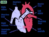
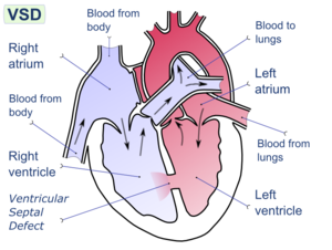
The ventricular septum consists of an inferior muscular and superior membranous portion and is extensively innervated with conducting cardiomyocytes.
The membranous portion, which is close to the atrioventricular node
Atrioventricular node
The atrioventricular node is a part of the electrical control system of the heart that coordinates heart rate. It electrically connects atrial and ventricular chambers...
, is most commonly affected in adults and older children in the United States. It is also the type that will most commonly require surgical intervention, comprising over 80% of cases.
Membranous ventricular septal defects are more common than muscular ventricular septal defects, and are the most common congenital cardiac anomaly.
Diagnosis
A VSD can be detected by cardiac auscultationAuscultation
Auscultation is the term for listening to the internal sounds of the body, usually using a stethoscope...
. Classically, a VSD causes a pathognomonic holo- or pansystolic murmur
Heart murmur
Murmurs are extra heart sounds that are produced as a result of turbulent blood flow that is sufficient to produce audible noise. Most murmurs can only be heard with the assistance of a stethoscope ....
. Auscultation is generally considered sufficient for detecting a significant VSD. The murmur depends on the abnormal flow of blood from the left ventricle, through the VSD, to the right ventricle. If there is not much difference in pressure between the left and right ventricles, then the flow of blood through the VSD will not be very great and the VSD may be silent. This situation occurs a) in the fetus (when the right and left ventricular pressures are essentially equal), b) for a short time after birth (before the right ventricular pressure has decreased), and c) as a late complication of unrepaired VSD. Confirmation of cardiac auscultation can be obtained by non-invasive cardiac ultrasound
Medical ultrasonography
Diagnostic sonography is an ultrasound-based diagnostic imaging technique used for visualizing subcutaneous body structures including tendons, muscles, joints, vessels and internal organs for possible pathology or lesions...
(echocardiography
Echocardiography
An echocardiogram, often referred to in the medical community as a cardiac ECHO or simply an ECHO, is a sonogram of the heart . Also known as a cardiac ultrasound, it uses standard ultrasound techniques to image two-dimensional slices of the heart...
). To more accurately measure ventricular pressures, cardiac catheterization
Cardiac catheterization
Cardiac catheterization is the insertion of a catheter into a chamber or vessel of the heart. This is done for both investigational and interventional purposes...
, can be performed.
Pathophysiology
During ventricular contraction, or systole, some of the blood from the left ventricle leaks into the right ventricle, passes through the lungs and reenters the left ventricle via the pulmonary veins and left atrium. This has two net effects. First, the circuitous refluxing of blood causes volume overload on the left ventricle. Second, because the left ventricle normally has a much higher systolic pressure (~120 mm Hg) than the right ventricle (~20 mm Hg), the leakage of blood into the right ventricle therefore elevates right ventricular pressure and volume, causing pulmonary hypertensionPulmonary hypertension
In medicine, pulmonary hypertension is an increase in blood pressure in the pulmonary artery, pulmonary vein, or pulmonary capillaries, together known as the lung vasculature, leading to shortness of breath, dizziness, fainting, and other symptoms, all of which are exacerbated by exertion...
with its associated symptoms.
In serious cases, the pulmonary arterial pressure can reach levels that equal the systemic pressure. This reverses the left to right shunt, so that blood then flows from the right ventricle into the left ventricle, resulting in cyanosis, as blood is by-passing the lungs for oxygenation.
This effect is more noticeable in patients with larger defects, who may present with breathlessness, poor feeding and failure to thrive in infancy. Patients with smaller defects may be asymptomatic. Four different septal defects exist, with perimembranous most common, outlet, atrioventricular, and muscular less commonly.
Signs and symptoms
Ventricular septal defect is usually symptomless at birth. It usually manifests a few weeks after birth.Symptoms
VSD is an acyanotic congenital heart defect, aka a Left-to-right shunt, so there are no signs of cyanosis.Signs
- Pansystolic (Holosystolic) murmur (depending upon the size of the defect) +/- palpable thrill (palpable turbulence of blood flow). Heart sounds are normal. Larger VSDs may cause a parasternal heave, a displaced apex beat (the palpable heartbeat moves laterally over time, as the heart enlarges). An infant with a large VSD will fail to thrive and become sweaty and tachypnoeic (breathe faster) with feeds.
CAUSES: The cause of VSD ( ventricular septal defect) includes the incomplete looping of the heart during days 24-28 of development. Faults with NKX2.5 gene can cause this.
Treatment
Some cases may necessitate surgical intervention, i.e. with the following indications:
1. Failure of congestive cardiac failure to respond to medications
2. VSD with pulmonic stenosis
Pulmonic stenosis
Pulmonic stenosis, also known as Pulmonary stenosis, is a dynamic or fixed obstruction to flow from the right ventricle of the heart to the pulmonary artery. It is usually first diagnosed in childhood....
3. Large VSD with pulmonary hypertension
Pulmonary hypertension
In medicine, pulmonary hypertension is an increase in blood pressure in the pulmonary artery, pulmonary vein, or pulmonary capillaries, together known as the lung vasculature, leading to shortness of breath, dizziness, fainting, and other symptoms, all of which are exacerbated by exertion...
4. VSD with aortic regurgitation
For the surgical procedure, a heart-lung machine
Heart-lung machine
Cardiopulmonary bypass is a technique that temporarily takes over the function of the heart and lungs during surgery, maintaining the circulation of blood and the oxygen content of the body. The CPB pump itself is often referred to as a heart–lung machine or "the pump"...
is required and a median sternotomy
Median sternotomy
Median sternotomy is a type of surgical procedure in which a vertical inline incision is made along the sternum, after which the sternum itself is divided, or "cracked"...
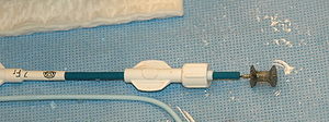
is performed. Percutaneous endovascular
Percutaneous
In surgery, percutaneous pertains to any medical procedure where access to inner organs or other tissue is done via needle-puncture of the skin, rather than by using an "open" approach where inner organs or tissue are exposed .The percutaneous approach is commonly used in vascular procedures...
procedures are less invasive and can be done on a beating heart, but are only suitable for certain patients. Repair of most VSDs is complicated by the fact that the conducting system of the heart
Electrical conduction system of the heart
The normal intrinsic electrical conduction of the heart allows electrical propagation to be transmitted from the Sinoatrial Node through both atria and forward to the Atrioventricular Node. Normal/baseline physiology allows further propagation from the AV node to the ventricle or Purkinje Fibers...
is in the immediate vicinity.
Ventricular septum defect in infants is initially treated medically with cardiac glycoside
Cardiac glycoside
Cardiac glycosides are drugs used in the treatment of congestive heart failure and cardiac arrhythmia. These glycosides are found as secondary metabolites in several plants, but also in some animals, such as the milkweed butterflies. -Function:...
s (e.g., digoxin
Digoxin
Digoxin INN , also known as digitalis, is a purified cardiac glycoside and extracted from the foxglove plant, Digitalis lanata. Its corresponding aglycone is digoxigenin, and its acetyl derivative is acetyldigoxin...
10-20mcg/kg per day), loop diuretic
Loop diuretic
Loop diuretics are diuretics that act on the ascending loop of Henle in the kidney. They are primarily used in medicine to treat hypertension and edema often due to congestive heart failure or renal insufficiency...
s (e.g., furosemide
Furosemide
Furosemide or frusemide is a loop diuretic used in the treatment of congestive heart failure and edema. It is most commonly marketed by Sanofi-Aventis under the brand name Lasix...
1–3 mg/kg per day) and ACE inhibitor
ACE inhibitor
ACE inhibitors or angiotensin-converting enzyme inhibitors are a group of drugs used primarily for the treatment of hypertension and congestive heart failure...
s (e.g., captopril
Captopril
Captopril is an angiotensin-converting enzyme inhibitor used for the treatment of hypertension and some types of congestive heart failure. Captopril was the first ACE inhibitor developed and was considered a breakthrough both because of its novel mechanism of action and also because of the...
0.5–2 mg/kg per day).
Surgical technique for Repair of Perimembranous VSD
a) Surgical closure of a Perimembranous VSD is performed on cardiopulmonary bypass with ischemic arrest. Patients are usually cooled to 28 degrees. Percutaneous Device closure of these defects is rarely performed in the United States because of the reported incidence of both early and late onset complete heart block after device closure, presumably secondary to device trauma to the AV node.b) Surgical exposure is achieved through the right atrium. The tricuspid valve septal leaflet is retracted or incised to expose the defect margins.
c) Several patch materials are available, including native pericardium, bovine pericardium, PTFE (Gore-Tex
Gore-Tex
Gore-Tex is a waterproof/breathable fabric, and a registered trademark of W. L. Gore and Associates. It was co-invented by Wilbert L. Gore, Rowena Taylor, and Gore's son, Robert W. Gore. Robert Gore was granted on April 27, 1976, for a porous form of polytetrafluoroethylene with a...
or Impra), or Dacron.
d) Suture techniques include horizontal pledgeted mattress sutures, and running polypropylene suture.
e) Critical attention is necessary to avoid injury to the conduction system located on the left ventricular side of the interventricular septum near the papillary muscle of the conus.
f) Care is taken to avoid injury to the aortic valve with sutures.
g) Once the repair is complete, the heart is extensively deaired by venting blood through the aortic cardioplegia site, and by infusing Carbon Dioxide into the operative field to displace air.
h) Intraoperative transesophageal echocardiography is used to confirm secure closure of the VSD, normal function of the aortic and tricuspid valves, good ventricular function, and the elimination of all air from the left side of the heart.
i) The sternum, fascia and skin are closed, with potential placement of a local anesthetic infusion catheter under the fascia, to enhance postoperative pain control.
j) A video of Perimembranous VSD repair, including the operative technique, and the daily postoperative recovery, can be seen here: VSD Repair, Perimembranous Ventricular Septal Defect
Epidemiology and Etiology
VSDs are the most common congenital cardiac anomalies. They are found in 30-60% of all newborns with a congenital heart defect, or about 2-6 per 1000 births. During heart formation, when the heart begins life as a hollow tube, it begins to partition, forming septa. If this does not occur properly it can lead to an opening being left within the ventricular septum. It is debatable whether all those defects are true heart defects, or if some of them are normal phenomena, since most of the trabecular VSDs close spontaneously. Prospective studies give a prevalence of 2-5 per 100 births of trabecular VSDs that closes shortly after birth in 80-90% of the cases.Congenital VSDs are frequently associated with other congenital conditions, such as Down syndrome
Down syndrome
Down syndrome, or Down's syndrome, trisomy 21, is a chromosomal condition caused by the presence of all or part of an extra 21st chromosome. It is named after John Langdon Down, the British physician who described the syndrome in 1866. The condition was clinically described earlier in the 19th...
.
A VSD can also form a few days after a myocardial infarction
Myocardial infarction
Myocardial infarction or acute myocardial infarction , commonly known as a heart attack, results from the interruption of blood supply to a part of the heart, causing heart cells to die...
(heart attack) due to mechanical tearing of the septal
Septum
In anatomy, a septum is a wall, dividing a cavity or structure into smaller ones.-In human anatomy:...
wall, before scar tissue
Myocardial scarring
Myocardial scarring is fibrous tissue that replaces normal tissue destroyed by injury or disease pertaining to the muscular tissue of the heart....
forms, when macrophage
Macrophage
Macrophages are cells produced by the differentiation of monocytes in tissues. Human macrophages are about in diameter. Monocytes and macrophages are phagocytes. Macrophages function in both non-specific defense as well as help initiate specific defense mechanisms of vertebrate animals...
s start remodeling the dead heart tissue.
See also
- Atrial septal defectAtrial septal defectAtrial septal defect is a form of congenital heart defect that enables blood flow between the left and right atria via the interatrial septum. The interatrial septum is the tissue that divides the right and left atria...
- Atrioventricular septal defectAtrioventricular septal defectAtrioventricular septal defect or atrioventricular canal defect , previously known as "common atrioventricular canal" or "endocardial cushion defect", is characterized by a deficiency of the atrioventricular septum of the heart...
- Cardiac outputCardiac outputCardiac output is the volume of blood being pumped by the heart, in particular by a left or right ventricle in the time interval of one minute. CO may be measured in many ways, for example dm3/min...
- Congenital heart disease
- Heart soundsHeart soundsHeart sounds, or heartbeats, are the noises generated by the beating heart and the resultant flow of blood through it...
- Pulmonary hypertensionPulmonary hypertensionIn medicine, pulmonary hypertension is an increase in blood pressure in the pulmonary artery, pulmonary vein, or pulmonary capillaries, together known as the lung vasculature, leading to shortness of breath, dizziness, fainting, and other symptoms, all of which are exacerbated by exertion...
External links
- Pediatric Heart Surgery
- The Congenital Heart Surgery Video Project
- VSD Repair, Perimembranous Ventricular Septal Defect
- VSD Repair Powerpoint™ Presentation
- Ventricular septal defect - Children's Hospital Boston
- Ventricular septal defect - American Heart AssociationAmerican Heart AssociationThe American Heart Association is a non-profit organization in the United States that fosters appropriate cardiac care in an effort to reduce disability and deaths caused by cardiovascular disease and stroke. It is headquartered in Dallas, Texas...
- Ventricular septal defect - medlineplus.org
- Ventricular Septal Defect information from Seattle Children's Hospital Heart Center
- Animation of ventricular septal defect from AboutKidsHealth.ca
- Perimembranous VSD - emedicine.com
- Supracristal VSD - emedicine.com
- Down's Heart Group Easy to understand diagram and explanation of VSD.
- C.S. Mott Children's Hospital, Congenital Heart Center: Ventricular Septal Defect at umich.edu
- Ventricular Septal Defect Cove Point Foundation
- VSD repair: Perimembranous The Congenital Heart Surgery Video Project