

Split S2
Encyclopedia
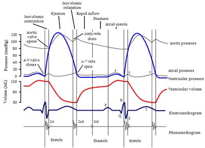
It is caused when the closure of the aortic valve
Aortic valve
The aortic valve is one of the valves of the heart. It is normally tricuspid , although in 1% of the population it is found to be congenitally bicuspid . It lies between the left ventricle and the aorta....
(A2) and the closure of the pulmonary valve
Pulmonary valve
The pulmonary valve is the semilunar valve of the heart that lies between the right ventricle and the pulmonary artery and has three cusps. Similar to the aortic valve, the pulmonary valve opens in ventricular systole, when the pressure in the right ventricle rises above the pressure in the...
(P2) are not synchronized.
Physiologic split
During inspiration, the chest wall expands and causes the intrathoracic pressure to become more negative (think of a vacuum). The increased negative pressure allows the lungs to fill with air and expand. While doing so, it also induces an increase in venous blood return from the body into the right atrium via the superior and inferior vena cavae, and into the right ventricle by increasing the pressure gradient (blood is being pulled by the vacuum from the body and towards the right side of the heart). Simultaneously, there is a reduction in blood volume returning from the lungs into the left ventricle (the blood wants to stay in the lungs because of the vacuum surrounding the lungs). Since there is an increase in blood volume in the right ventricle, the pulmonary valve (P2 component of S2) stays open longer during ventricular systole due to an increase in ventricular emptying time, whereas the aortic valve (A2 component of S2) closes slightly earlier due to a reduction in left ventricular volume and ventricular emptying time. Thus the P2 component of S2 is delayed relative to that of the A2 component. This delay in P2 versus A2 is heard as a slight broadening or even "splitting" of the second heart sound; though it is usually only heard in the pulmonic area of the chest because the P2 is soft and not heard in other areas. During expiration, the chest wall collapses and decreases the negative intrathoracic pressure (compared to inspiration). Therefore, there is no longer an increase in blood return to the right ventricle versus the left ventricle and the right ventricle volume is no longer increased. This allows the pulmonary valve to close earlier such that it overlaps the closing of the aortic valve, and the split is no longer heard.It is physiologically normal to hear a "splitting" of the second heart tone in younger people, during inspiration and in the "pulmonary area", i.e. the 2nd ICS (intercostal space) at the left edge of the sternum.
Steps
- Chest wall expands during inspiration
- Intrathoracic pressure becomes more negative to form a vacuum
- Venous return from the body to the right heart increases, venous return from the lungs to the left heart decreases
- Right ventricular volume and emptying time increases, left ventricular volume and emptying time decreases
- Pulmonic valve closure is delayed, aortic valve closure is advanced
- S2 splits into A2 and P2 respectively
- Expiration equalizes filling and emptying times on both sides of the heart eliminating the splitting of S2
Analysis of pressure
According to Harrison's Principles of Internal Medicine, "Normally, blood pressure falls during inspiration (equal or less than 10 mmHg), due to an increase in blood flow into the right ventricle with displacement of the interventricular septum to the left, decreasing left ventricular filling and cardiac output".The pressure in the right ventricle tries to open the pulmonary valve. The pressure in the pulmonary artery tries to close the pulmonary valve. Remember that the higher pressure will "win". Hence, the closure of the pulmonary valve (P2) will be delayed since the pressure in the right ventricle is increased in inspiration, opposing the pressure in the pulmonary artery and keeping it open longer than in expiration. The change in A2 is not that evident. Thus P2 appears after A2 in inspiration.
Pathologic split
The different types of split S2 can be associated with medical conditions:- Split during inspiration: normal. (See above)
- Split during expiration: Reverse splitting indicates pathology. Aortic stenosis, hypertrophic cardiomyopathyHypertrophic cardiomyopathyHypertrophic cardiomyopathy is a disease of the myocardium in which a portion of the myocardium is hypertrophied without any obvious cause...
, left bundle branch blockLeft bundle branch blockLeft bundle branch block is a cardiac conduction abnormality seen on the electrocardiogram . In this condition, activation of the left ventricle is delayed, which results in the left ventricle contracting later than the right ventricle....
(LBBB), and a ventricular pacemaker could all cause a reverse splitting of the second heart sound.
- Split during both inspiration and expiration:
- If splitting does not vary with inspiration, it is termed a "fixed split S2" and is usually due to a septal defect, such as an atrial septal defectAtrial septal defectAtrial septal defect is a form of congenital heart defect that enables blood flow between the left and right atria via the interatrial septum. The interatrial septum is the tissue that divides the right and left atria...
(ASD) or ventricular septal defectVentricular septal defectA ventricular septal defect is a defect in the ventricular septum, the wall dividing the left and right ventricles of the heart.The ventricular septum consists of an inferior muscular and superior membranous portion and is extensively innervated with conducting cardiomyocytes.The membranous...
(VSD). The ASD or VSD creates a left to right shunt that increases the blood flow to the right side of the heart, thereby causing the pulmonary valve to close later than the aortic valve independent of inspiration/expiration. - A bundle branch block either LBBB or RBBB, (although RBBB is known to be associated only with S1 split), will produce continuous splitting but the degree of splitting will still vary with respiration.
- If splitting does not vary with inspiration, it is termed a "fixed split S2" and is usually due to a septal defect, such as an atrial septal defect
When the pulmonary valve closes before the aortic valve, this is known as a "paradoxically split S2".