
Ciliary ganglion
Encyclopedia
The ciliary ganglion is a parasympathetic
ganglion
located in the posterior orbit
. It measures 1–2 millimeters in diameter and contains approximately 2,500 neuron
s. Preganglionic axon
s from the Edinger-Westphal nucleus
travel along the oculomotor nerve
and form synapse
s with these cells
. The postganglionic axons run in the short ciliary nerves and innervate two eye
muscles:
Both of these muscles are involuntary – they are controlled by the autonomic nervous system
.
It is one of four parasympathetic ganglia of the head and neck. (The others are the submandibular ganglion
, pterygopalatine ganglion
, and otic ganglion
).
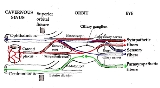
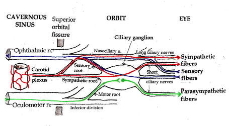 Three types of nerve fibers run through the ciliary ganglion: parasympathetic fibers, sympathetic fibers and sensory fibers. Only parasympathetic fibers form synapses in the ganglion. The other two types of nerve fibers simply pass through. In classical anatomy, the ciliary ganglion is said to have three “roots:”
).
When a patient with an Adie pupil attempts to focus on a nearby object, the pupil (which would normally constrict rapidly) constricts slowly. On close inspection, the constricted pupil is not perfectly round. When the patient focuses on a more distant object (say the far side of the room), the pupil (which would normally dilate immediately) remains constricted for several minutes, and then slowly dilates back to the expected size.
Tonic pupils are fairly common – they are seen in roughly 1 out of every 500 people. A patient with anisocoria (one pupil bigger than the other) whose pupil does not react to light (does not constrict when exposed to bright light) most likely has Adie syndrome – idiopathic degeneration of the ciliary ganglion.
Loewenfeld’s theory is now generally accepted. It explains the defining features of a tonic pupil:
The pupil does not react to light. The original light-reaction neurons have been destroyed.
Tonic constriction with attempted near vision. Aberrant regeneration of nerve fibers intended for the ciliary muscle causes abnormal, tonic contraction of the pupil with accommodation.
Segmental iris constriction. When carefully examined under magnification, the iris does not constrict uniformly with attempted near vision. Only the re-innervated segments contract, producing a slightly irregular contour to the pupil.
[Denervation supersensitivity]. Like any denervated muscle, the iris becomes supersensitive to its normal neurotransmitter (in this case, acetylcholine). Very weak solutions of cholinergic substances such as pilocarpine (that have no effect on the normal iris) cause the denervated iris to constrict.
Tonic pupils are usually due to Adie syndrome, but other diseases can denervate the ciliary ganglion. Peripheral neuropathies (such as diabetic neuropathy) occasionally produce tonic pupils. Herpes zoster virus can attack the ciliary ganglion. Trauma to the orbit can damage the short ciliary nerves. Anything that denervates the ciliary ganglion will produce a tonic pupil due to aberrant nerve regeneration.
Early in the course of Adie syndrome (when the cells of the ciliary ganglion have been destroyed, but before regeneration has occurred) the pupil will be fixed and dilated. The sphincter pupillae will be paralyzed. There will be no response to accommodation – the ciliary muscle is also paralyzed.
With aberrant nerve regeneration, the pupil will remain fixed, but it will constrict with attempted near vision. The constriction will be abnormal (“tonic”).
Late in the course of Adie syndrome, the pupil becomes small (as all pupils do with old age). It will still be “fixed” (it will not constrict to bright light) and it will continue to show abnormal, tonic constriction with attempted near vision.
The brainstem causes of light-near dissociation include Argyll Robertson pupil
and Parinaud syndrome. These are discussed elsewhere in Wikipedia.
Parasympathetic nervous system
The parasympathetic nervous system is one of the two main divisions of the autonomic nervous system . The ANS is responsible for regulation of internal organs and glands, which occurs unconsciously...
ganglion
Ganglion
In anatomy, a ganglion is a biological tissue mass, most commonly a mass of nerve cell bodies. Cells found in a ganglion are called ganglion cells, though this term is also sometimes used to refer specifically to retinal ganglion cells....
located in the posterior orbit
Orbit (anatomy)
In anatomy, the orbit is the cavity or socket of the skull in which the eye and its appendages are situated. "Orbit" can refer to the bony socket, or it can also be used to imply the contents...
. It measures 1–2 millimeters in diameter and contains approximately 2,500 neuron
Neuron
A neuron is an electrically excitable cell that processes and transmits information by electrical and chemical signaling. Chemical signaling occurs via synapses, specialized connections with other cells. Neurons connect to each other to form networks. Neurons are the core components of the nervous...
s. Preganglionic axon
Axon
An axon is a long, slender projection of a nerve cell, or neuron, that conducts electrical impulses away from the neuron's cell body or soma....
s from the Edinger-Westphal nucleus
Edinger-Westphal nucleus
The Edinger-Westphal nucleus is the accessory parasympathetic cranial nerve nucleus of the oculomotor nerve , supplying the constricting muscles of the iris...
travel along the oculomotor nerve
Oculomotor nerve
The oculomotor nerve is the 3rd of 12 paired cranial nerves. It enters the orbit via the superior orbital fissure and controls most of the eye's movements, including constriction of the pupil and maintaining an open eyelid by innervating the Levator palpebrae superiors muscle. The optic nerve is...
and form synapse
Synapse
In the nervous system, a synapse is a structure that permits a neuron to pass an electrical or chemical signal to another cell...
s with these cells
Cell (biology)
The cell is the basic structural and functional unit of all known living organisms. It is the smallest unit of life that is classified as a living thing, and is often called the building block of life. The Alberts text discusses how the "cellular building blocks" move to shape developing embryos....
. The postganglionic axons run in the short ciliary nerves and innervate two eye
Human eye
The human eye is an organ which reacts to light for several purposes. As a conscious sense organ, the eye allows vision. Rod and cone cells in the retina allow conscious light perception and vision including color differentiation and the perception of depth...
muscles:
- the sphincter pupillae constricts the pupilPupilThe pupil is a hole located in the center of the iris of the eye that allows light to enter the retina. It appears black because most of the light entering the pupil is absorbed by the tissues inside the eye. In humans the pupil is round, but other species, such as some cats, have slit pupils. In...
, a movement known as MiosisMiosisMiosis is the constriction of the pupil of the eye to two millimeters or less...
. The opposite, MydriasisMydriasisMydriasis is a dilation of the pupil due to disease, trauma or the use of drugs. Normally, the pupil dilates in the dark and constricts in the light to respectively improve vividity at night and to protect the retina from sunlight damage during the day...
, is the dilation of the pupil. - the ciliaris muscleMuscleMuscle is a contractile tissue of animals and is derived from the mesodermal layer of embryonic germ cells. Muscle cells contain contractile filaments that move past each other and change the size of the cell. They are classified as skeletal, cardiac, or smooth muscles. Their function is to...
contracts, releasing tension on the Zonular Fibers, making the lensLens (anatomy)The crystalline lens is a transparent, biconvex structure in the eye that, along with the cornea, helps to refract light to be focused on the retina. The lens, by changing shape, functions to change the focal distance of the eye so that it can focus on objects at various distances, thus allowing a...
more convex, also known as accommodationAccommodation (eye)Accommodation is the process by which the vertebrate eye changes optical power to maintain a clear image on an object as its distance changes....
.
Both of these muscles are involuntary – they are controlled by the autonomic nervous system
Autonomic nervous system
The autonomic nervous system is the part of the peripheral nervous system that acts as a control system functioning largely below the level of consciousness, and controls visceral functions. The ANS affects heart rate, digestion, respiration rate, salivation, perspiration, diameter of the pupils,...
.
It is one of four parasympathetic ganglia of the head and neck. (The others are the submandibular ganglion
Submandibular ganglion
The submandibular ganglion is part of the human autonomic nervous system. It is one of four parasympathetic ganglia of the head and neck...
, pterygopalatine ganglion
Pterygopalatine ganglion
The pterygopalatine ganglion is a parasympathetic ganglion found in the pterygopalatine fossa. It is one of four parasympathetic ganglia of the head and neck....
, and otic ganglion
Otic ganglion
The otic ganglion is a small, oval shaped, flattened parasympathetic ganglion of a reddish-gray color, located immediately below the foramen ovale in the infratemporal fossa. It gives innervation to the parotid gland for salivation....
).
Anatomy
- a parasympathetic root of ciliary ganglionParasympathetic root of ciliary ganglionThe parasympathetic root of ciliary ganglion provides parasympathetic innervation to the ciliary ganglion.The ciliary ganglion is a parasympathetic ganglion. Incoming parasympathetic nerve fibers form synapses with the dendrites of nerve cells within the ganglion...
(or motor root) - a sympathetic root of ciliary ganglionSympathetic root of ciliary ganglionThe sympathetic root of ciliary ganglion is one of three roots of the ciliary ganglion, a tissue mass behind the eye. It contains postganglionic sympathetic fibers whose cell bodies are located in the superior cervical ganglion. Their axons ascend with the internal carotid artery as a plexus of...
- a sensory root of ciliary ganglionSensory root of ciliary ganglionSensory fibers from the eyeball run posteriorly through the short ciliary nerves and pass through the ciliary ganglion without forming synapses...
Adie tonic pupil
Diseases of the ciliary ganglion produce a tonic pupil. This is a pupil that does not react to light (it is “fixed”) and has an abnormally slow and prolonged response to attempted near vision (accommodationAccommodation (eye)
Accommodation is the process by which the vertebrate eye changes optical power to maintain a clear image on an object as its distance changes....
).
When a patient with an Adie pupil attempts to focus on a nearby object, the pupil (which would normally constrict rapidly) constricts slowly. On close inspection, the constricted pupil is not perfectly round. When the patient focuses on a more distant object (say the far side of the room), the pupil (which would normally dilate immediately) remains constricted for several minutes, and then slowly dilates back to the expected size.
Tonic pupils are fairly common – they are seen in roughly 1 out of every 500 people. A patient with anisocoria (one pupil bigger than the other) whose pupil does not react to light (does not constrict when exposed to bright light) most likely has Adie syndrome – idiopathic degeneration of the ciliary ganglion.
Physiology
The strange behavior of tonic pupils was first explained by Irene Loewenfeld in 1979. The ciliary ganglion contain many more nerve fibers directed to the ciliary muscle than nerve fibers directed to the constrictor pupillae – roughly twenty times more. The ciliary muscle is also more massive than the constrictor pupillae, again by a factor of twenty. Based on these observations, Loewenfeld proposed an explanation of the tonic pupil. She noted that pathological destruction of nerve cells in the ciliary ganglion that is found in all cases of Adie pupil. In her own words :- Let’s say that in a given fresh Adie’s pupil, a random 70% of the cells in the ciliary ganglion stop working; and that, in a couple of months, these neurons re-grow and randomly re-innervate both intraocular sphincters (the ciliary muscle and the iris sphincter). Some parasympathetic light-reaction neurons that were originally destined for the iris sphincter will end up innervating the ciliary muscle. But there will not be enough of them to budge that big muscle, so there will be no detectable accommodation with exposure to light. The other way around, it is a different story. There will be plenty of accommodative neurons re-growing into the iris sphincter, and it won’t take very many of them to make a little muscle like the iris sphincter contract. This means that every time the patient accommodates her gaze to a near object, some of the innervation to the ciliary muscle will spill over into the iris and constrict the pupil.
Loewenfeld’s theory is now generally accepted. It explains the defining features of a tonic pupil:
The pupil does not react to light. The original light-reaction neurons have been destroyed.
Tonic constriction with attempted near vision. Aberrant regeneration of nerve fibers intended for the ciliary muscle causes abnormal, tonic contraction of the pupil with accommodation.
Segmental iris constriction. When carefully examined under magnification, the iris does not constrict uniformly with attempted near vision. Only the re-innervated segments contract, producing a slightly irregular contour to the pupil.
[Denervation supersensitivity]. Like any denervated muscle, the iris becomes supersensitive to its normal neurotransmitter (in this case, acetylcholine). Very weak solutions of cholinergic substances such as pilocarpine (that have no effect on the normal iris) cause the denervated iris to constrict.
Tonic pupils are usually due to Adie syndrome, but other diseases can denervate the ciliary ganglion. Peripheral neuropathies (such as diabetic neuropathy) occasionally produce tonic pupils. Herpes zoster virus can attack the ciliary ganglion. Trauma to the orbit can damage the short ciliary nerves. Anything that denervates the ciliary ganglion will produce a tonic pupil due to aberrant nerve regeneration.
Adie syndrome
Adie syndrome is tonic pupil plus absent deep tendon reflexes. Adie syndrome is a fairly common, benign, idiopathic neuropathy that selectively affects the ciliary ganglion and the spinal cord neurons involved in deep tendon reflex arcs. It usually develops in middle age, although it can occur in children. A variant of Adie syndrome, Ross syndrome, affects sweating as well.Early in the course of Adie syndrome (when the cells of the ciliary ganglion have been destroyed, but before regeneration has occurred) the pupil will be fixed and dilated. The sphincter pupillae will be paralyzed. There will be no response to accommodation – the ciliary muscle is also paralyzed.
With aberrant nerve regeneration, the pupil will remain fixed, but it will constrict with attempted near vision. The constriction will be abnormal (“tonic”).
Late in the course of Adie syndrome, the pupil becomes small (as all pupils do with old age). It will still be “fixed” (it will not constrict to bright light) and it will continue to show abnormal, tonic constriction with attempted near vision.
Light-near dissociation
The Adie pupil does not react to light, but it does react to accommodation. This is an example of “light-near dissociation”. All other causes of light-near dissociation involve the brainstem. They do not involve the ciliary ganglion, and they do not produce a tonic pupil. Irene Loewenfeld is generally credited for being the first physiologist to make this distinction.The brainstem causes of light-near dissociation include Argyll Robertson pupil
Argyll Robertson pupil
Argyll Robertson pupils are bilateral small pupils that constrict when the patient focuses on a near object , but do not constrict when exposed to bright light . They are a highly specific sign of neurosyphilis. In general, pupils that “accommodate but do not react” are said to show light-near...
and Parinaud syndrome. These are discussed elsewhere in Wikipedia.