

Dental anatomy
Encyclopedia
Anatomy
Anatomy is a branch of biology and medicine that is the consideration of the structure of living things. It is a general term that includes human anatomy, animal anatomy , and plant anatomy...
dedicated to the study of human tooth structures. The development, appearance, and classification of teeth fall within its purview. (The function of teeth as they contact one another falls elsewhere, under dental occlusion.) Tooth formation begins before birth, and teeth's eventual morphology
Morphology (biology)
In biology, morphology is a branch of bioscience dealing with the study of the form and structure of organisms and their specific structural features....
is dictated during this time. Dental anatomy is also a taxonomical
Taxonomy
Taxonomy is the science of identifying and naming species, and arranging them into a classification. The field of taxonomy, sometimes referred to as "biological taxonomy", revolves around the description and use of taxonomic units, known as taxa...
science: it is concerned with the naming of teeth and the structures of which they are made, this information serving a practical purpose in dental treatment.
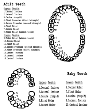
Usually, there are 20 primary ("baby") teeth and 28 to 32 permanent teeth, the last four being third molars or "wisdom teeth", each of which may or may not grow in. Among primary teeth, 10 usually are found in the maxilla
Maxilla
The maxilla is a fusion of two bones along the palatal fissure that form the upper jaw. This is similar to the mandible , which is also a fusion of two halves at the mental symphysis. Sometimes The maxilla (plural: maxillae) is a fusion of two bones along the palatal fissure that form the upper...
(upper jaw) and the other 10 in the mandible (lower jaw). Among permanent teeth, 16 are found in the maxilla and the other 16 in the mandible. Most of the teeth have distinguishing features.
Tooth development

Embryo
An embryo is a multicellular diploid eukaryote in its earliest stage of development, from the time of first cell division until birth, hatching, or germination...
nic cells
Cell (biology)
The cell is the basic structural and functional unit of all known living organisms. It is the smallest unit of life that is classified as a living thing, and is often called the building block of life. The Alberts text discusses how the "cellular building blocks" move to shape developing embryos....
, grow
Cell growth
The term cell growth is used in the contexts of cell development and cell division . When used in the context of cell division, it refers to growth of cell populations, where one cell grows and divides to produce two "daughter cells"...
, and erupt into the mouth. Although many diverse species
Species
In biology, a species is one of the basic units of biological classification and a taxonomic rank. A species is often defined as a group of organisms capable of interbreeding and producing fertile offspring. While in many cases this definition is adequate, more precise or differing measures are...
have teeth, non-human tooth development is largely the same as in humans. For human teeth to have a healthy oral environment, enamel
Tooth enamel
Tooth enamel, along with dentin, cementum, and dental pulp is one of the four major tissues that make up the tooth in vertebrates. It is the hardest and most highly mineralized substance in the human body. Tooth enamel is also found in the dermal denticles of sharks...
, dentin
Dentin
Dentine is a calcified tissue of the body, and along with enamel, cementum, and pulp is one of the four major components of teeth. Usually, it is covered by enamel on the crown and cementum on the root and surrounds the entire pulp...
, cementum
Cementum
Cementum is a specialized calcified substance covering the root of a tooth. Cementum is excreted by cells called cementoblasts within the root of the tooth and is thickest at the root apex. These cementoblasts develop from undifferentiated mesenchymal cells in the connective tissue of the dental...
, and the periodontium
Periodontium
Periodontium refers to the specialized tissues that both surround and support the teeth, maintaining them in the maxillary and mandibular bones. The word comes from the Greek terms peri-, meaning "around" and -odons, meaning "tooth." Literally taken, it means that which is "around the tooth"...
must all develop during appropriate stages of fetal development
Fetal development
Prenatal or antenatal development is the process in which a human embryo or fetus gestates during pregnancy, from fertilization until birth. Often, the terms fetal development, foetal development, or embryology are used in a similar sense.After fertilization the embryogenesis starts...
. Primary (baby) teeth
Deciduous teeth
Deciduous teeth, otherwise known as reborner teeth, baby teeth, temporary teeth and primary teeth, are the first set of teeth in the growth development of humans and many other mammals. In some Asian countries they are referred to as fall teeth as they will eventually fall out, while in almost all...
start to form between the sixth and eighth weeks in utero
In utero
In utero is a Latin term literally meaning "in the womb". In biology, the phrase describes the state of an embryo or fetus. In legal contexts, the phrase is used to refer to unborn children. Under common law, unborn children are still considered to exist for property transfer purposes.-See also:*...
, and permanent teeth
Permanent teeth
Permanent teeth are the second set of teeth formed in humans. There are thirty-two permanent teeth, consisting of six maxillary and six mandibular molars, four maxillary and four mandibular premolars, two maxillary and two mandibular canines, four maxillary and four mandibular incisors.The first...
begin to form in the twentieth week in utero. If teeth do not start to develop at or near these times, they will not develop at all.
A significant amount of research has focused on determining the processes that initiate tooth development. It is widely accepted that there is a factor within the tissues of the first branchial arch
Branchial arch
In the development of vertebrate animals, the pharyngeal arches are anlage for a multitude of structures. In humans, they develop during the fourth week in utero as a series of mesodermal outpouchings on the left and right sides of the developing pharynx...
that is necessary for the development of teeth. The tooth bud (sometimes called the tooth germ) is an aggregation of cells that eventually forms a tooth and is organized into three parts: the enamel organ
Enamel organ
The enamel organ, also known as dental organ, is a cellular aggregation seen in histologic sections of a developing tooth. It lies above a condensation of ectomesenchymal cells called the dental papilla...
, the dental papilla
Dental papilla
The dental papilla is a condensation of ectomesenchymal cells called odontoblasts, seen in histologic sections of a developing tooth. It lies below a cellular aggregation known as the enamel organ. The dental papilla appears after 8-10 weeks intra uteral life...
and the dental follicle
Dental follicle
The dental follicle is a sac containing the developing tooth and its odontogenic organ. The dental follicle differentiates into the periodontal ligament. In addition, it may be the precursor of other cells of the periodontium, including osteoblasts and cementoblasts...
.
The enamel organ is composed of the outer enamel epithelium
Outer enamel epithelium
The outer enamel epithelium, also known as the external enamel epithelium, is a layer of cuboidal cells located on the periphery of the enamel organ in a developing tooth. This layer is first seen during the bell stage....
, inner enamel epithelium
Inner enamel epithelium
The inner enamel epithelium, also known as the internal enamel epithelium, is a layer of cells located on the rim nearest the dental papilla of the enamel organ in a developing tooth. This layer is first seen during the bell stage....
, stellate reticulum
Stellate reticulum
The stellate reticulum is a group of cells located in the center of the enamel organ of a developing tooth. These cells are star shaped and synthesize glycosaminoglycans. As glycosamingoglycans are produced, water is drawn in between the cells and stretch them apart...
and stratum intermedium
Stratum intermedium
The stratum intermedium in a developing tooth is a layer of two or three cells between the inner enamel epithelium and the newly forming cells of the stellate reticulum. It first appears during the early bell stage of tooth development, at around the 14th week of intrauterine life. The stratum...
. These cells give rise to ameloblast
Ameloblast
Ameloblasts are cells, present only during tooth development, that deposit tooth enamel, the hard outermost layer of the tooth that forms the chewing surface....
s, which produce enamel and the reduced enamel epithelium
Reduced enamel epithelium
The reduced enamel epithelium, sometimes called reduced dental epithelium, overlies a developing tooth and is formed by two layers: a layer of ameloblast cells and the adjacent layer of cuboidal cells from the dental lamina. As the cells of the reduced enamel epithelium degenerate, the tooth is...
. The growth of cervical loop
Cervical loop
The cervical loop is the location on an enamel organ in a developing tooth where the outer enamel epithelium and the inner enamel epithelium join. The cervical loop is a histologic term indicating a specific epithelial structure at the apical side of the tooth germ, consisting of loosely aggregated...
cells into the deeper tissues forms Hertwig's Epithelial Root Sheath
Hertwig's epithelial root sheath
The Hertwig's epithelial root sheath is a proliferation of epithelial cells located at the cervical loop of the enamel organ in a developing tooth. Hertwig's epithelial root sheath initiates the formation of dentin in the root of a tooth by causing the differentiation of odontoblasts from the...
, which determines the root shape of the tooth. The dental papilla contains cells that develop into odontoblast
Odontoblast
In vertebrates, an odontoblast is a biological cell of neural crest origin that is part of the outer surface of the dental pulp, and whose biological function is dentinogenesis, which is the creation of dentin, the substance under the tooth enamel....
s, which are dentin-forming cells. Additionally, the junction between the dental papilla and inner enamel epithelium determines the crown shape of a tooth. The dental follicle gives rise to three important entities: cementoblast
Cementoblast
A cementoblast is a biological cell that forms from the follicular cells around the root of a tooth, and whose biological function is cementogenesis, which is the creation of cementum ....
s, osteoblast
Osteoblast
Osteoblasts are mononucleate cells that are responsible for bone formation; in essence, osteoblasts are specialized fibroblasts that in addition to fibroblastic products, express bone sialoprotein and osteocalcin.Osteoblasts produce a matrix of osteoid, which is composed mainly of Type I collagen...
s, and fibroblast
Fibroblast
A fibroblast is a type of cell that synthesizes the extracellular matrix and collagen, the structural framework for animal tissues, and plays a critical role in wound healing...
s. Cementoblasts form the cementum of a tooth. Osteoblasts give rise to the alveolar bone around the roots of teeth. Fibroblasts develop the periodontal ligament
Periodontal ligament
The periodontal fiber or periodontal ligament, commonly abbreviated as the PDL, is a group of specialized connective tissue fibers that essentially attach a tooth to the alveolar bone within which it sits...
s which connect teeth to the alveolar bone through cementum.
Tooth development is commonly divided into the following stages: the bud stage, the cap, the bell, and finally maturation. The staging of tooth development is an attempt to categorize changes that take place along a continuum; frequently it is difficult to decide what stage should be assigned to a particular developing tooth. This determination is further complicated by the varying appearance of different histologic sections of the same developing tooth, which can appear to be different stages.
Nomenclature
Teeth are named by their set, arch, class, type, and side. Teeth can belong to one of two sets of teeth: primary ("baby") teeth or permanent teeth. Often, "deciduous" may be used in place of "primary", and "adult" may be used for "permanent". "Succedaneous" refers to those teeth of the permanent dentitionDentition
Dentition pertains to the development of teeth and their arrangement in the mouth. In particular, the characteristic arrangement, kind, and number of teeth in a given species at a given age...
that replace primary teeth (incisors, canines, and premolars of the permanent dentition). Succedaneous would refer to these teeth as a group. Further, the name depends upon which arch the tooth is found in. The term, "maxillary", is given to teeth in the upper jaw and "mandibular" to those in the lower jaw. There are four classes of teeth: incisor
Incisor
Incisors are the first kind of tooth in heterodont mammals. They are located in the premaxilla above and mandible below.-Function:...
s, canines
Canine tooth
In mammalian oral anatomy, the canine teeth, also called cuspids, dogteeth, fangs, or eye teeth, are relatively long, pointed teeth...
, premolar
Premolar
The premolar teeth or bicuspids are transitional teeth located between the canine and molar teeth. In humans, there are two premolars per quadrant, making eight premolars total in the mouth. They have at least two cusps. Premolars can be considered as a 'transitional tooth' during chewing, or...
s, and molar
Molar (tooth)
Molars are the rearmost and most complicated kind of tooth in most mammals. In many mammals they grind food; hence the Latin name mola, "millstone"....
s. Premolars are found only in permanent teeth; there are no premolars in deciduous teeth. Within each class, teeth may be classified into different traits. Incisors are divided further into central and lateral incisors. Among premolars and molars, there are 1st and 2nd premolars, and 1st, 2nd, and 3rd molars. The side of the mouth in which a tooth is found may also be included in the name. For example, a specific name for a tooth may be "primary maxillary left lateral incisor."
Numbering systems
There are several different dental notation systems for associating information to a specific tooth. The three most commons systems are the FDI World Dental Federation notationFDI World Dental Federation notation
FDI World Dental Federation notation is widely used by dentists internationally to associate information to a specific tooth.Developed by the FDI World Dental Federation, World Dental Federation notation is also known as ISO 3950 notation....
, Universal numbering system (dental)
Universal numbering system (dental)
The Universal numbering system is a dental notation system for associating information to a specific tooth, and is commonly used in the United States.The designations "left" and "right" on the chart correspond to the patient's left and right, respectively....
, and Palmer notation method. The FDI system is used worldwide, and the universal is used widely in the USA.
Although the Palmer notation was supposedly superseded by the FDI World Dental Federation notation, it overwhelmingly continues to be the preferred method used by dental students and practitioners in the United Kingdom
United Kingdom
The United Kingdom of Great Britain and Northern IrelandIn the United Kingdom and Dependencies, other languages have been officially recognised as legitimate autochthonous languages under the European Charter for Regional or Minority Languages...
. It was originally termed the "Zsigmondy system" after the Austrian dentist Adolf Zsigmondy
Adolf Zsigmondy
Adolf Zsigmondy, aka Adolph Zsigmondy was a dentist of Hungarian origin who lived in Vienna. It was he who conceived the idea of charting teeth on the Zsigmondy-cross...
who developed the idea in 1861, using a Zsigmondy cross to record quadrants of tooth positions.. The Palmer notation consists of a symbol (┘└ ┐┌) designating in which quadrant the tooth is found and a number indicating the position from the midline. Permanent teeth are numbered 1 to 8, and primary teeth are indicated by a letter A to E. The universal numbering system uses a unique letter or number for each tooth. The uppercase letters A through T are used for primary teeth and the numbers 1 - 32 are used for permanent teeth. The tooth designated "1" is the right maxillary third molar and the count continues along the upper teeth to the left side. Then the count begins at the left mandibular third molar, designated number 17, and continues along the bottom teeth to the right side. The FDI system uses a two-digit numbering system in which the first number represents a tooth's quadrant and the second number represents the number of the tooth from the midline of the face. For permanent teeth, the upper right teeth begin with the number, "1". The upper left teeth begin with the number, "2". The lower left teeth begin with the number, "3". The lower right teeth begin with the number, "4". For primary teeth, the sequence of numbers goes 5, 6, 7, and 8 for the teeth in the upper right, upper left, lower left, and lower right respectively.
As a result, any given tooth has three different ways to identify it, depending on which notation system is used. The permanent right maxillary central incisor is identified by the number "8" in the universal system. In the FDI system, the same tooth is identified by the number "11". The palmer system uses the number and symbol, 1┘, to identify the tooth. Further confusion may result if a number is given on a tooth without assuming (or specifying) a common notation method. Since the number, "12", may signify the permanent left maxillary first premolar in the universal system or the permanent right maxillary lateral incisor in the FDI system, the notation being used must be clear to prevent confusion.
Victor Haderup of Denmark in 1891 devised a variant of eight tooth quadrant system in which plus(+) and minus(-) were used to differentiate between upper and lower quadrants, and between right and left quadrants (e.g., +1=upper right central incisor; 1-=lower left central incisor). Primary teeth were numbered as upper right (05+ to 01+), lower left (-01 to -05). This system is still taught in Denmark.
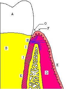
Crown and root
Crown (tooth)
In dentistry, crown refers to the anatomical area of teeth, usually covered by enamel. The crown is usually visible in the mouth after developing below the gingiva and then erupting into place.-References:...
of a tooth can be used to describe two situations. The anatomic crown of a tooth is designated by the area above the cementoenamel junction
Cementoenamel junction
The cementoenamel junction, frequently abbreviated as the CEJ, is an anatomical border identified on a tooth. It is the location where the enamel, which covers the anatomical crown of a tooth, and the cementum, which covers the anatomical root of a tooth, meet...
(CEJ) and is consequently covered in enamel. Also, it is possible to describe the clinical crown of a tooth as any parts visible in the mouth, but frequently the anatomic crown is meant when the term is used. The majority of the crown is composed of dentin, with the pulp chamber found in the center. The crown is only found within bone before eruption
Tooth eruption
Tooth eruption is a process in tooth development in which the teeth enter the mouth and become visible. It is currently believed that the periodontal ligaments play an important role in tooth eruption...
into the mouth. Afterwards, it is almost always visible.
The anatomic root is found below the cementoenamel junction and is covered with cementum, whereas the clinical root is any part of a tooth not visible in the mouth. Similarly, the anatomic root is assumed in most circumstances. Dentin composes most of the root, which normally has pulp canals. The roots of teeth may be single in number (single-rooted teeth) or multiple. Canines and most premolars, except for maxillary first premolars, usually have one root. Maxillary first premolars and mandibular molars usually have two roots. Maxillary molars usually have three roots. The tooth is supported in bone by an attachment apparatus, known as the periodontium, which interacts with the root.
Surfaces
Surfaces that are nearest the cheekCheek
Cheeks constitute the area of the face below the eyes and between the nose and the left or right ear. They may also be referred to as jowls. "Buccal" means relating to the cheek. In humans, the region is innervated by the buccal nerve...
s or lip
Lip
Lips are a visible body part at the mouth of humans and many animals. Lips are soft, movable, and serve as the opening for food intake and in the articulation of sound and speech...
s are referred to as facial
Commonly used terms of relationship and comparison in dentistry
There are numerous commonly used terms of relationship and comparison that refer to different aspects of teeth and are frequently utilized in articles about dentistry...
, and those nearest the tongue
Tongue
The tongue is a muscular hydrostat on the floors of the mouths of most vertebrates which manipulates food for mastication. It is the primary organ of taste , as much of the upper surface of the tongue is covered in papillae and taste buds. It is sensitive and kept moist by saliva, and is richly...
are known as lingual. Facial surfaces can be subdivided into buccal (when found on posterior teeth nearest the cheeks) and labial (when found on anterior teeth nearest the lips). Lingual surfaces can also be described as palatal when found on maxillary teeth beside the hard palate
Hard palate
The hard palate is a thin horizontal bony plate of the skull, located in the roof of the mouth. It spans the arch formed by the upper teeth.It is formed by the palatine process of the maxilla and horizontal plate of palatine bone....
.
Surfaces that aid in chewing are known as occlusal on posterior teeth and incisal on anterior teeth. Surfaces nearest the junction of the crown and root are referred to as cervical, and those closest to the apex of the root are referred to as apical. The words mesial and distal are also used as descriptions. "Mesial" signifies a surface closer to the median line of the face, which is located on a vertical axis between the eyes, down the nose, and between the contact of the central incisors. Surfaces further away from the median line are described as distal.
Cusp
A cusp is an elevation on an occlusal surface of posterior teeth and canines. It contributes to a significant portion of the tooth's surface. Canines have one cusp. Maxillary premolars and the mandibular first premolars usually have two cusps. Mandibular second premolars frequently have three cusps--- one buccal and two lingual. Maxillary molars have two buccal cusps and two lingual cusps. A fifth cusp that may form on the maxillary first molar is known as the cusp of CarabelliCusp of Carabelli
The cusp of Carabelli, or Carabelli's tubercle, or tuberculum anomale of Georg Carabelli is a small additional cusp at the mesiopalatal line angle of maxillary first molars. This extra cusp is usually found on the first molar, and becomes progressively less likely in the second, third molars...
. Mandibular molars may have five or four cusps.
Cingulum
A cingulumCingulum (tooth)
In dentistry, cingulum refers to an anatomical feature of the anterior teeth . It refers to the portion of the teeth, occurring on the lingual or palatal aspects, that forms a convex protuberance at the cervical third of the anatomic crown. It represents the lingual or palatal developmental lobe...
is a convexity found on the lingual surface of anterior teeth. It is frequently identifiable as an inverted V-shaped ridge, and its appearance is comparable to a girdle. All anterior teeth are formed from four centers of development, referred to as lobes. Three are located on the facial side of the tooth, and one on the lingual side. The cingulum forms from this lingual lobe of development. The majority of a lingual surface's cervical third is made up of the cingulum. On lower incisors, a cingulum usually is poorly developed or absent. Maxillary canines have a large, well-developed cingulum, where as the cingulum of mandibular canines is smoother and rounded.
Ridges
Ridges are any linear, flat elevations on teeth, and they are named according to their location. The buccal ridge runs cervico-occlusally in approximately the center of the buccal surface of premolars. The labial ridge is one that runs cervico-incisally in approximately the center of the labial surface of canines. The lingual ridge extends from the cingulum to the cusp tip on the lingual surface of most canines. The cervical ridge runs mesiodistally on the cervical third of the buccal surface of the crown. These are found on all primary teeth but only on the permanent molars.Cusp ridges are ridges that radiate from cusp tips. There are two marginal ridges, mesial and distal, present on all teeth. On anterior teeth, they are located on the mesial and distal borders of the lingual surface; on posterior teeth, they are located on the mesial and distal borders of the occlusal surface. Triangular ridges are those that project from the cusp tips of premolar and molars to the central groove. Transverse ridges are formed by the union of two triangular ridges on posterior teeth. The joining of buccal and lingual triangular ridges is usually named as an example. The oblique ridge is found on the occlusal surfaces of maxillary molars. It is formed by the union of the distal cusp ridge of the mesiolingual cusp and the triangular ridge of the distobuccal cusp. The oblique ridges usually forms the distal boundary of the central fossa.
Developmental groove
The teeth demonstrating the least number of developmental grooves are the mandibular central and lateral incisors.However, the canines show the most prominent developmental grooves, because they have strong anchorage to the bone.
Embrasures
Embrasures are triangularly shaped spaces located between the proximal surfaces of adjacent teeth. The borders of embrasures are formed by the interdental papilla of the gingiva, the adjacent teeth, and the contact point where the two teeth meet. There are four embrasures for every contact area: facial (also called labial or buccal), lingual (or palatal), occlusal or incisal, and cervical or interproximal space. The cervical embrasure usually is filled by the interdental papilla from the gingiva.Embrasures have three functions. They form spillways between teeth to direct food away from the gingiva. Also, they provide a mechanism for teeth to be more self cleansing. Lastly, they protect the gingiva from undue frictional trauma but also providing the proper degree of stimulation to the tissues.
Mammelons
MammelonMammelon
In dentistry, mammelons refer to an anatomical feature on the incisal edges of teeth. Frequently found on erupting central and lateral incisors, mammelons appear as three little bumps. Since this part of the tooth is the first to wear away from attrition, mammelons may not visible on teeth of...
s are usually found as three small bumps on the incisal edges of anterior teeth. They are the remnants of three lobes of formation of these teeth, the fourth lobe represented by the cingulum. Since this surface of the tooth is the first to wear away from attrition
Attrition (dental)
Attrition is the loss of teeth structure by mechanical forces from opposing teeth. Attrition initially affects the enamel and, if unchecked, may proceed to the underlying dentin. Once past the enamel, attrition quickly destroys the softer dentin. Erosion is a very important contributing factor to...
, mammelons may not be visible on teeth of older people. Instead, the best chance to see this characteristic is soon after eruption of the tooth into the mouth.
Incisor
8 incisors are anterior teeth, 4 in the upper arch and 4 in the lower. Their function is for shearingShearing (physics)
Shearing in continuum mechanics refers to the occurrence of a shear strain, which is a deformation of a material substance in which parallel internal surfaces slide past one another. It is induced by a shear stress in the material...
or cutting food during chewing
Mastication
Mastication or chewing is the process by which food is crushed and ground by teeth. It is the first step of digestion and it increases the surface area of foods to allow more efficient break down by enzymes. During the mastication process, the food is positioned between the teeth for grinding by...
. There are no cusps on the teeth. Instead, the surface area of the tooth used in eating is called the incisal ridge or incisal edge. Though similar, there are some minor differences between the primary and permanent incisors.
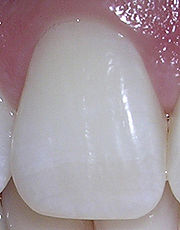
Maxillary central incisor
Maxillary lateral incisor
The Maxillary lateral incisor is a tooth that is located distally from both maxillary central incisors of the mouth and mesially from both maxillary canines. As with all incisors, their function is for shearing or cutting food during mastication, commonly known as chewing. There are no cusps on...
.The overall length of the deciduous maxillary central incisor is 16 mm on average, with the crown being 6 mm and the root being 10 mm. In comparison to the permanent maxillary central incisor, the ratio of the root length to the crown length is greater in the deciduous tooth. The diameter of the crown mesiodistally is greater than the length cervicoincisally, which makes the tooth appear wider rather than taller from a labial viewpoint.
The permanent maxillary central incisor is the widest tooth mesiodistally in comparison to any other anterior tooth. It is larger than the neighboring lateral incisor and is usually not as convex on its labial surface. As a result, the central incisor appears to be more rectangular or square in shape. The mesial incisal angle is sharper than the distal incisal angle. When this tooth is newly erupted into the mouth, the incisal edges have three rounded features called mammelons. Mammelons disappear with time as the enamel wears away by friction.