
Branchial arch
Encyclopedia
In the development
of vertebrate animals
, the pharyngeal arches (which develop into the branchial arches or gill arches in fish) are anlage for a multitude of structures. In humans, they develop during the fourth week in utero
as a series of mesodermal outpouchings on the left and right
sides of the developing pharynx
. In fish, the branchial arches support the gills.
from the pericardium
. By differential growth the neck elongates and new arches form, so the pharynx has six arches ultimately.
Each pharyngeal arch has a cartilaginous
stick, a muscle
component which differentiates from the cartilaginous tissue, an artery, and a cranial nerve.
Each of these is surrounded by mesenchyme. Arches do not develop simultaneously, but instead possess a "staggered" development.
s (or clefts) form from the lateral ectodermal surface of the neck
region to separate the arches.
The pouches line up with the clefts, and these thin segments become gill
s in fish.
In mammals the endoderm
and ectoderm
not only remain intact, but continue to be separated by a mesoderm
layer.
More is known about the fate of the first arch than the remaining four. The first three contribute to structures above the larynx, while the last two contribute to the larynx
and trachea
.
landmark
with which to establish the precise stage of embryonic development. Their formation and development corresponds to Carnegie stages
10 to 16 in mammals, and Hamburger-Hamilton stages
14 to 28 in the chicken.
Embryogenesis
Embryogenesis is the process by which the embryo is formed and develops, until it develops into a fetus.Embryogenesis starts with the fertilization of the ovum by sperm. The fertilized ovum is referred to as a zygote...
of vertebrate animals
Vertebrate
Vertebrates are animals that are members of the subphylum Vertebrata . Vertebrates are the largest group of chordates, with currently about 58,000 species described. Vertebrates include the jawless fishes, bony fishes, sharks and rays, amphibians, reptiles, mammals, and birds...
, the pharyngeal arches (which develop into the branchial arches or gill arches in fish) are anlage for a multitude of structures. In humans, they develop during the fourth week in utero
In utero
In utero is a Latin term literally meaning "in the womb". In biology, the phrase describes the state of an embryo or fetus. In legal contexts, the phrase is used to refer to unborn children. Under common law, unborn children are still considered to exist for property transfer purposes.-See also:*...
as a series of mesodermal outpouchings on the left and right
Left and Right
Left and Right: A Journal of Libertarian Thought was a libertarian journal published between 1965 and 1968. Founded by Murray N. Rothbard, Karl Hess, George Resch, and Leonard P...
sides of the developing pharynx
Pharynx
The human pharynx is the part of the throat situated immediately posterior to the mouth and nasal cavity, and anterior to the esophagus and larynx. The human pharynx is conventionally divided into three sections: the nasopharynx , the oropharynx , and the laryngopharynx...
. In fish, the branchial arches support the gills.
Development
These grow and join in the ventral midline. The first arch, as the first to form, separates the mouth pit or stomodeumStomodeum
The stomodeum, also called stomatodeum or stomatodaeum, is a depression between the brain and the pericardium in an embryo, and is the precursor of the mouth and the anterior lobe of the pituitary gland.-Structure:...
from the pericardium
Pericardium
The pericardium is a double-walled sac that contains the heart and the roots of the great vessels.-Layers:...
. By differential growth the neck elongates and new arches form, so the pharynx has six arches ultimately.
Each pharyngeal arch has a cartilaginous
Cartilage
Cartilage is a flexible connective tissue found in many areas in the bodies of humans and other animals, including the joints between bones, the rib cage, the ear, the nose, the elbow, the knee, the ankle, the bronchial tubes and the intervertebral discs...
stick, a muscle
Muscle
Muscle is a contractile tissue of animals and is derived from the mesodermal layer of embryonic germ cells. Muscle cells contain contractile filaments that move past each other and change the size of the cell. They are classified as skeletal, cardiac, or smooth muscles. Their function is to...
component which differentiates from the cartilaginous tissue, an artery, and a cranial nerve.
Each of these is surrounded by mesenchyme. Arches do not develop simultaneously, but instead possess a "staggered" development.
Relations
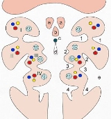
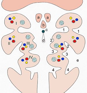
Pharyngeal pouches (or branchial pouches) form on the endodermal side between the arches, and pharyngeal groovePharyngeal groove
A pharyngeal groove is the counterpart of the branchial pouch on the ectodermal side.The first pharyngeal groove produces the external auditory meatus...
s (or clefts) form from the lateral ectodermal surface of the neck
Neck
The neck is the part of the body, on many terrestrial or secondarily aquatic vertebrates, that distinguishes the head from the torso or trunk. The adjective signifying "of the neck" is cervical .-Boner anatomy: The cervical spine:The cervical portion of the human spine comprises seven boney...
region to separate the arches.
The pouches line up with the clefts, and these thin segments become gill
Gill
A gill is a respiratory organ found in many aquatic organisms that extracts dissolved oxygen from water, afterward excreting carbon dioxide. The gills of some species such as hermit crabs have adapted to allow respiration on land provided they are kept moist...
s in fish.
In mammals the endoderm
Endoderm
Endoderm is one of the three primary germ cell layers in the very early embryo. The other two layers are the ectoderm and mesoderm , with the endoderm as the intermost layer...
and ectoderm
Ectoderm
The "ectoderm" is one of the three primary germ cell layers in the very early embryo. The other two layers are the mesoderm and endoderm , with the ectoderm as the most exterior layer...
not only remain intact, but continue to be separated by a mesoderm
Mesoderm
In all bilaterian animals, the mesoderm is one of the three primary germ cell layers in the very early embryo. The other two layers are the ectoderm and endoderm , with the mesoderm as the middle layer between them.The mesoderm forms mesenchyme , mesothelium, non-epithelial blood corpuscles and...
layer.
Specific arches
There are six pharyngeal arches, but in humans the fifth arch only exists transiently during embryologic growth and development. Since no human structures result from the fifth arch, the arches in humans are I, II, III, IV, and VI.More is known about the fate of the first arch than the remaining four. The first three contribute to structures above the larynx, while the last two contribute to the larynx
Larynx
The larynx , commonly called the voice box, is an organ in the neck of amphibians, reptiles and mammals involved in breathing, sound production, and protecting the trachea against food aspiration. It manipulates pitch and volume...
and trachea
Vertebrate trachea
In tetrapod anatomy the trachea, or windpipe, is a tube that connects the pharynx or larynx to the lungs, allowing the passage of air. It is lined with pseudostratified ciliated columnar epithelium cells with goblet cells that produce mucus...
.
| Pharyngeal arch | Muscular contributions | Skeletal contributions | Nerve | Artery Aortic arches The aortic arches or pharyngeal arch arteries are a series of six paired embryological vascular structures which give rise to several major arteries... >- | 1st (also called "mandibular arch") |
Muscles of mastication Muscles of mastication During mastication, four muscles of mastication are responsible for adduction and lateral motion of the jaw. Other muscles, usually associated with the hyoid such as the sternohyomastoid, are responsible for opening the jaw.-Muscles:*The masseter... , anterior belly of the digastric, mylohyoid Mylohyoid Mylohyoid can refer to:* Mylohyoid muscle* Mylohyoid line* Mylohyoid nerve* Mylohyoid branch of inferior alveolar artery* Mylohyoid groove... , tensor tympani Tensor tympani The tensor tympani, the larger of the two muscles of the tympanic cavity, is contained in the bony canal above the osseous portion of the auditory tube... , tensor veli palatini |
Maxilla Maxilla The maxilla is a fusion of two bones along the palatal fissure that form the upper jaw. This is similar to the mandible , which is also a fusion of two halves at the mental symphysis. Sometimes The maxilla (plural: maxillae) is a fusion of two bones along the palatal fissure that form the upper... , mandible (only as a model for mandible not actual formation of mandible), the incus Incus The incus or anvil is the anvil-shaped small bone or ossicle in themiddle ear. It connects the malleus to the stapes. It was first described by Alessandro Achillini of Bologna.The incus transmits sound vibrations from the malleus to the stapes.... and malleus Malleus The malleus or hammer is a hammer-shaped small bone or ossicle of the middle ear which connects with the incus and is attached to the inner surface of the eardrum... of the middle ear, also Meckel's cartilage Meckel's cartilage The cartilaginous bar of the mandibular arch is formed by what are known as Meckel’s cartilages also known as Meckelian cartilages; above this the incus and malleus are developed.... |
Trigeminal nerve Trigeminal nerve The trigeminal nerve contains both sensory and motor fibres. It is responsible for sensation in the face and certain motor functions such as biting, chewing, and swallowing. Sensory information from the face and body is processed by parallel pathways in the central nervous system... (V2 and V3) |
external carotid artery External carotid artery In human anatomy, the external carotid artery is a major artery of the head and neck. It arises from the common carotid artery when it bifurcates into the external and internal carotid artery.-Course:... >- | 2nd (also called the "hyoid arch Hyoid arch The second pharyngeal arch or hyoid arch assists in forming the side and front of the neck.-Skeletal elements:From the cartilage of the second arch arises*Stapes,*Styloid process,*Stylohyoid ligament, and... ") |
Muscles of facial expression, buccinator Buccinator The buccinator muscle is a muscle at the side of the face.Buccinator may also refer to:* Buccinator artery * Buccinator lymph node* Buccinator nerve * An ancient Roman buccina player... , platysma, stapedius Stapedius The stapedius is the smallest skeletal muscle in the human body. At just over one millimeter in length, its purpose is to stabilize the smallest bone in the body, the stapes.... , stylohyoid, posterior belly of the digastric |
Stapes, styloid process Styloid process In anatomy, a styloid process , usually serving as points of attachment for muscles, refers to the slender, pointed process of :* temporal bone of the skull - Temporal styloid process... , hyoid (lesser horn and upper part of body), Reichert's cartilage |
Facial nerve Facial nerve The facial nerve is the seventh of twelve paired cranial nerves. It emerges from the brainstem between the pons and the medulla, and controls the muscles of facial expression, and functions in the conveyance of taste sensations from the anterior two-thirds of the tongue and oral cavity... (VII) |
>- | Stylopharyngeus | Hyoid (greater horn and lower part of body), thymus Thymus The thymus is a specialized organ of the immune system. The thymus produces and "educates" T-lymphocytes , which are critical cells of the adaptive immune system.... , inferior parathyroids |
Glossopharyngeal nerve Glossopharyngeal nerve The glossopharyngeal nerve is the ninth of twelve pairs of cranial nerves . It exits the brainstem out from the sides of the upper medulla, just rostral to the vagus nerve... (IX) |
>- | Cricothyroid muscle Cricothyroid muscle The cricothyroid muscle is the only tensor muscle of the larynx, aiding with phonation. It attaches to the anterolateral aspect of the cricoid and the inferior cornu and lower lamina of the thyroid cartilage, and its action tilts the thyroid forward to help tense the vocal cords... , all intrinsic muscles of soft palate Soft palate The soft palate is the soft tissue constituting the back of the roof of the mouth. The soft palate is distinguished from the hard palate at the front of the mouth in that it does not contain bone.... including levator veli palatini Levator veli palatini The levator veli palatini is the elevator muscle of the soft palate in the human body. During swallowing, it contracts, elevating the soft palate to help prevent food from entering the nasopharynx... |
Thyroid cartilage Thyroid cartilage The thyroid cartilage is the largest of the nine cartilages that make up the laryngeal skeleton, the cartilage structure in and around the trachea that contains the larynx.... , superior parathyroids, epiglottic cartilage |
Vagus nerve Vagus nerve The vagus nerve , also called pneumogastric nerve or cranial nerve X, is the tenth of twelve paired cranial nerves... (X) Superior laryngeal nerve Superior laryngeal nerve The superior laryngeal nerve is a branch of the vagus nerve. It arises from the middle of the ganglion nodosum and in its course receives a branch from the superior cervical ganglion of the sympathetic.... |
subclavian artery Subclavian artery In human anatomy, the subclavian arteries are two major arteries of the upper thorax , below the clavicle . They receive blood from the top of the aorta... Left 4th aortic arch: aortic arch Aortic arch The arch of the aorta or the transverse aortic arch is the part of the aorta that begins at the level of the upper border of the second sternocostal articulation of the right side, and runs at first upward, backward, and to the left in front of the trachea; it is then directed backward on the left... >- | 6th |
All intrinsic muscles of larynx Larynx The larynx , commonly called the voice box, is an organ in the neck of amphibians, reptiles and mammals involved in breathing, sound production, and protecting the trachea against food aspiration. It manipulates pitch and volume... except the cricothyroid muscle Cricothyroid muscle The cricothyroid muscle is the only tensor muscle of the larynx, aiding with phonation. It attaches to the anterolateral aspect of the cricoid and the inferior cornu and lower lamina of the thyroid cartilage, and its action tilts the thyroid forward to help tense the vocal cords... |
Cricoid cartilage, arytenoid cartilage Arytenoid cartilage The arytenoid cartilages are a pair of small three-sided pyramids which form part of the larynx, to which the vocal folds are attached... s, corniculate cartilage |
Vagus nerve Vagus nerve The vagus nerve , also called pneumogastric nerve or cranial nerve X, is the tenth of twelve paired cranial nerves... (X) Recurrent laryngeal nerve Recurrent laryngeal nerve The recurrent laryngeal nerve is a branch of the vagus nerve that supplies motor function and sensation to the larynx . It travels within the endoneurium... |
Right 6th aortic arch: pulmonary artery Pulmonary artery The pulmonary arteries carry deoxygenated blood from the heart to the lungs. They are the only arteries that carry deoxygenated blood.... Left 6th aortic arch: Pulmonary artery Pulmonary artery The pulmonary arteries carry deoxygenated blood from the heart to the lungs. They are the only arteries that carry deoxygenated blood.... and ductus arteriosus Ductus arteriosus In the developing fetus, the ductus arteriosus , also called the ductus Botalli, is a shunt connecting the pulmonary artery to the aortic arch. It allows most of the blood from the right ventricle to bypass the fetus's fluid-filled lungs. Upon closure at birth, it becomes the ligamentum arteriosum... |
Use in staging
The development of the pharyngeal arches provide a useful morphologicalMorphology (biology)
In biology, morphology is a branch of bioscience dealing with the study of the form and structure of organisms and their specific structural features....
landmark
Landmark
This is a list of landmarks around the world.Landmarks may be split into two categories - natural phenomena and man-made features, like buildings, bridges, statues, public squares and so forth...
with which to establish the precise stage of embryonic development. Their formation and development corresponds to Carnegie stages
Carnegie stages
In embryology, Carnegie stages are a standardized system of 23 stages used to provide a unified developmental chronology of the vertebrate embryo....
10 to 16 in mammals, and Hamburger-Hamilton stages
Hamburger-Hamilton stages
In developmental biology, the Hamburger-Hamilton stages are a series of 46 chronological stages in chick development, starting from laying of the egg and ending with a newly hatched chick. It is named for its creators, Viktor Hamburger and Howard L. Hamilton.Chicken embryos are a useful model...
14 to 28 in the chicken.
See also
- Aortic archesAortic archesThe aortic arches or pharyngeal arch arteries are a series of six paired embryological vascular structures which give rise to several major arteries...
- Branchial pouchBranchial pouchIn the development of vertebrate animals, pharyngeal or branchial pouches form on the endodermal side between the branchial arches, and pharyngeal grooves form the lateral ectodermal surface of the neck region to separate the arches....
- Branchial cleft cystBranchial cleft cystA branchial cleft cyst is an oval, moderately movable cystic mass that develops under the skin in the neck between the sternocleidomastoid muscle and the pharynx.-Pathology:...
- Congenital cartilaginous rest of the neckCongenital cartilaginous rest of the neckCongenital cartilaginous rest of the neck is a cutaneous condition characterized by branchial arch remnants that are considered to be the cervical variant of accessory tragus....