

Blood film
Encyclopedia
A blood film or peripheral blood smear is a thin layer of blood
Blood
Blood is a specialized bodily fluid in animals that delivers necessary substances such as nutrients and oxygen to the cells and transports metabolic waste products away from those same cells....
smeared on a microscope slide
Microscope slide
A microscope slide is a thin flat piece of glass, typically 75 by 25 mm and about 1 mm thick, used to hold objects for examination under a microscope. Typically the object is placed or secured on the slide, and then both are inserted together in the microscope for viewing...
and then stained in such a way to allow the various blood cells to be examined microscopically. Blood films are usually examined to investigate hematological
Hematology
Hematology, also spelled haematology , is the branch of biology physiology, internal medicine, pathology, clinical laboratory work, and pediatrics that is concerned with the study of blood, the blood-forming organs, and blood diseases...
problems (disorders of the blood) and, occasionally, to look for parasites
Apicomplexa
The Apicomplexa are a large group of protists, most of which possess a unique organelle called apicoplast and an apical complex structure involved in penetrating a host's cell. They are unicellular, spore-forming, and exclusively parasites of animals. Motile structures such as flagella or...
within the blood such as malaria
Malaria
Malaria is a mosquito-borne infectious disease of humans and other animals caused by eukaryotic protists of the genus Plasmodium. The disease results from the multiplication of Plasmodium parasites within red blood cells, causing symptoms that typically include fever and headache, in severe cases...
and filaria
Filariasis
Filariasis is a parasitic disease and is considered an infectious tropical disease, that is caused by thread-like nematodes belonging to the superfamily Filarioidea, also known as "filariae"....
.
Preparation
Blood films are made by placing a drop of blood on one end of a slide, and using a spreader slide to disperse the blood over the slide's length. The aim is to get a region where the cells are spaced far enough apart to be counted and differentiated.The slide is left to air dry, after which the blood is fixed to the slide by immersing it briefly in methanol
Methanol
Methanol, also known as methyl alcohol, wood alcohol, wood naphtha or wood spirits, is a chemical with the formula CH3OH . It is the simplest alcohol, and is a light, volatile, colorless, flammable liquid with a distinctive odor very similar to, but slightly sweeter than, ethanol...
. The fixative is essential for good staining and presentation of cellular detail. After fixation, the slide is stained
Staining (biology)
Staining is an auxiliary technique used in microscopy to enhance contrast in the microscopic image. Stains and dyes are frequently used in biology and medicine to highlight structures in biological tissues for viewing, often with the aid of different microscopes...
to distinguish the cells from each other.
Disorders
Characteristic red blood cell abnormalities are anemiaAnemia
Anemia is a decrease in number of red blood cells or less than the normal quantity of hemoglobin in the blood. However, it can include decreased oxygen-binding ability of each hemoglobin molecule due to deformity or lack in numerical development as in some other types of hemoglobin...
, sickle cell anemia and spherocytosis
Spherocytosis
Spherocytosis is an auto-hemolytic anemia characterized by the production of red blood cells , or erythrocytes, that are sphere-shaped, rather than bi-concave disk shaped. Spherocytes are found in hereditary spherocytosis and autoimmune hemolytic anemia.It almost always refers to hereditary...
. Sometimes the microscopic investigation of the red cells can be essential to the diagnosis of life-threatening disease (e.g. TTP
Thrombotic thrombocytopenic purpura
Thrombotic thrombocytopenic purpura is a rare disorder of the blood-coagulation system, causing extensive microscopic thromboses to form in small blood vessels throughout the body...
).
White blood cells are classified according to their propensity to stain with particular substances, the shape of the nuclei and the granular inclusions.
- Neutrophil granulocyteNeutrophil granulocyteNeutrophil granulocytes are the most abundant type of white blood cells in mammals and form an essential part of the innate immune system. They are generally referred to as either neutrophils or polymorphonuclear neutrophils , and are subdivided into segmented neutrophils and banded neutrophils...
s usually make up close to 80% of the white count. They have multilobate nuclei and lightly staining granules. They assist in destruction of foreign particles by the immune systemImmune systemAn immune system is a system of biological structures and processes within an organism that protects against disease by identifying and killing pathogens and tumor cells. It detects a wide variety of agents, from viruses to parasitic worms, and needs to distinguish them from the organism's own...
by phagocytosis and intracellular killing. - Eosinophil granulocyteEosinophil granulocyteEosinophil granulocytes, usually called eosinophils or eosinophiles , are white blood cells that are one of the immune system components responsible for combating multicellular parasites and certain infections in vertebrates. Along with mast cells, they also control mechanisms associated with...
s have granules that stain with eosinEosinEosin is a fluorescent red dye resulting from the action of bromine on fluorescein. It can be used to stain cytoplasm, collagen and muscle fibers for examination under the microscope. Structures that stain readily with eosin are termed eosinophilic....
and play a role in allergyAllergyAn Allergy is a hypersensitivity disorder of the immune system. Allergic reactions occur when a person's immune system reacts to normally harmless substances in the environment. A substance that causes a reaction is called an allergen. These reactions are acquired, predictable, and rapid...
and parasitic disease. Eos have a multilobate nucleusCell nucleusIn cell biology, the nucleus is a membrane-enclosed organelle found in eukaryotic cells. It contains most of the cell's genetic material, organized as multiple long linear DNA molecules in complex with a large variety of proteins, such as histones, to form chromosomes. The genes within these...
. - Basophil granulocyteBasophil granulocyteBasophil granulocytes, sometimes referred to as basophils, are the least common of the granulocytes, representing about 0.01% to 0.3% of circulating white blood cells....
s are only seen occasionally. They are polymorphonucleated and their granules stain dark with alkaline stains, such as haematoxylinHaematoxylinHaematoxylin, hematoxylin, Natural Black 1, or C.I. 75290 is extracted from the heartwood of the logwood tree. When oxidized it forms haematein, a compound that forms strongly coloured complexes with certain metal ions, the most notable ones being Fe and Al salts. Metal-haematein complexes are used...
. They are further characterised by the fact that the granules seem to overlie the nucleus. Basophils are similar if not identical in cell lineage to mast cells, although no conclusive evidence to this end has been shown. Mast cells are "tissue basophils" and mediate certain immune reactions to allergens. - LymphocyteLymphocyteA lymphocyte is a type of white blood cell in the vertebrate immune system.Under the microscope, lymphocytes can be divided into large lymphocytes and small lymphocytes. Large granular lymphocytes include natural killer cells...
s have very little cytoplasmCytoplasmThe cytoplasm is a small gel-like substance residing between the cell membrane holding all the cell's internal sub-structures , except for the nucleus. All the contents of the cells of prokaryote organisms are contained within the cytoplasm...
and a large nucleusCell nucleusIn cell biology, the nucleus is a membrane-enclosed organelle found in eukaryotic cells. It contains most of the cell's genetic material, organized as multiple long linear DNA molecules in complex with a large variety of proteins, such as histones, to form chromosomes. The genes within these...
(high NC ratio) and are responsible for antigenAntigenAn antigen is a foreign molecule that, when introduced into the body, triggers the production of an antibody by the immune system. The immune system will then kill or neutralize the antigen that is recognized as a foreign and potentially harmful invader. These invaders can be molecules such as...
-specific immune functions, either by antibodies (B cellB cellB cells are lymphocytes that play a large role in the humoral immune response . The principal functions of B cells are to make antibodies against antigens, perform the role of antigen-presenting cells and eventually develop into memory B cells after activation by antigen interaction...
) or by direct cytotoxicityCytotoxicityCytotoxicity is the quality of being toxic to cells. Examples of toxic agents are a chemical substance, an immune cell or some types of venom .-Cell physiology:...
(T cellT cellT cells or T lymphocytes belong to a group of white blood cells known as lymphocytes, and play a central role in cell-mediated immunity. They can be distinguished from other lymphocytes, such as B cells and natural killer cells , by the presence of a T cell receptor on the cell surface. They are...
). The distinction between B and T cells cannot be made by light microscopy. - Plasma cellPlasma cellPlasma cells, also called plasma B cells, plasmocytes, and effector B cells, are white blood cells which produce large volumes of antibodies. They are transported by the blood plasma and the lymphatic system...
s are mature B lymphocytes that engage in the production of one specific antibodyAntibodyAn antibody, also known as an immunoglobulin, is a large Y-shaped protein used by the immune system to identify and neutralize foreign objects such as bacteria and viruses. The antibody recognizes a unique part of the foreign target, termed an antigen...
. They are characterised by light basophilicBasophilicBasophilic is a technical term used by histologists. It describes the microscopic appearance of cells and tissues, as seen down the microscope, after a histological section has been stained with a basic dye. The most common such dye is haematoxylin....
staining and a very eccentric nucleusCell nucleusIn cell biology, the nucleus is a membrane-enclosed organelle found in eukaryotic cells. It contains most of the cell's genetic material, organized as multiple long linear DNA molecules in complex with a large variety of proteins, such as histones, to form chromosomes. The genes within these...
. - Other cells are white cell precursors. When these are very abundant it can be a feature of infectionInfectionAn infection is the colonization of a host organism by parasite species. Infecting parasites seek to use the host's resources to reproduce, often resulting in disease...
or leukemiaLeukemiaLeukemia or leukaemia is a type of cancer of the blood or bone marrow characterized by an abnormal increase of immature white blood cells called "blasts". Leukemia is a broad term covering a spectrum of diseases...
, although the most common types of leukemia (CML and CLL) are characterised by mature cells, and have more of an abnormal appearance on light microscopy (additional tests can aid the diagnosis).
Use in diagnosing Malaria
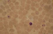
Plasmodium falciparum
Plasmodium falciparum is a protozoan parasite, one of the species of Plasmodium that cause malaria in humans. It is transmitted by the female Anopheles mosquito. Malaria caused by this species is the most dangerous form of malaria, with the highest rates of complications and mortality...
, P. vivax
Plasmodium vivax
Plasmodium vivax is a protozoal parasite and a human pathogen. The most frequent and widely distributed cause of recurring malaria, P. vivax is one of the four species of malarial parasite that commonly infect humans. It is less virulent than Plasmodium falciparum, which is the deadliest of the...
, P. ovale
Plasmodium ovale
Plasmodium ovale is a species of parasitic protozoa that causes tertian malaria in humans. It is closely related to Plasmodium falciparum and Plasmodium vivax, which are responsible for most malaria. It is rare compared to these two parasites, and substantially less dangerous than P...
, P. malariae
Plasmodium malariae
Plasmodium malariae is a parasitic protozoa that causes malaria in humans. It is closely related to Plasmodium falciparum and Plasmodium vivax which are responsible for most malarial infection. While found worldwide, it is a so-called "benign malaria" and is not nearly as dangerous as that...
.
The biggest pitfall in most laboratories in developed countries is leaving too great a delay between taking the blood sample and making the blood films. As blood cools to room temperature, male gametocytes will divide and release microgametes: these are long sinuous filamentous structures that can be mistaken for organisms such as Borrelia. If the blood is kept at warmer temperatures, schizonts will rupture and merozoites invading erythrocytes will mistakenly give the appearance of the accolé form of P. falciparum. If P. vivax or P. ovale is left for several hours in EDTA, the build up of acid in the sample will cause the parasitised erythrocytes to shrink and the parasite will roll up, simulating the appearance of P. malariae. This problem is made worse if anticoagulant
Anticoagulant
An anticoagulant is a substance that prevents coagulation of blood. A group of pharmaceuticals called anticoagulants can be used in vivo as a medication for thrombotic disorders. Some anticoagulants are used in medical equipment, such as test tubes, blood transfusion bags, and renal dialysis...
s such as heparin
Heparin
Heparin , also known as unfractionated heparin, a highly sulfated glycosaminoglycan, is widely used as an injectable anticoagulant, and has the highest negative charge density of any known biological molecule...
or citrate
Citrate
A citrate can refer either to the conjugate base of citric acid, , or to the esters of citric acid. An example of the former, a salt is trisodium citrate; an ester is triethyl citrate.-Other citric acid ions:...
are used. The anticoagulant that causes the least problems is EDTA
EDTA
Ethylenediaminetetraacetic acid, widely abbreviated as EDTA , is a polyamino carboxylic acid and a colourless, water-soluble solid. Its conjugate base is named ethylenediaminetetraacetate. It is widely used to dissolve limescale. Its usefulness arises because of its role as a hexadentate ligand...
. Romanowsky stain
Romanowsky stain
Romanowsky staining is a prototypical staining technique that was the forerunner of several distinct but similar methods, including Giemsa, Jenner, Wright, Field, and Leishman stains, which are used to differentiate cells in pathologic specimens....
or a variant stain
Staining (biology)
Staining is an auxiliary technique used in microscopy to enhance contrast in the microscopic image. Stains and dyes are frequently used in biology and medicine to highlight structures in biological tissues for viewing, often with the aid of different microscopes...
is usually used. Some laboratories mistakenly use the same staining pH as they do for routine haematology blood films (pH
PH
In chemistry, pH is a measure of the acidity or basicity of an aqueous solution. Pure water is said to be neutral, with a pH close to 7.0 at . Solutions with a pH less than 7 are said to be acidic and solutions with a pH greater than 7 are basic or alkaline...
6.8): malaria blood films must be stained at pH 7.2, or Schüffner's dots and James's dots will not be seen.
There are different kinds of blood smears that identifies many kinds of parasites in blood