
Abdominal ultrasonography
Encyclopedia


Medical ultrasonography
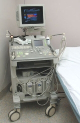
Diagnostic sonography is an ultrasound-based diagnostic imaging technique used for visualizing subcutaneous body structures including tendons, muscles, joints, vessels and internal organs for possible pathology or lesions...
(medical
Medicine
Medicine is the science and art of healing. It encompasses a variety of health care practices evolved to maintain and restore health by the prevention and treatment of illness....
application of ultrasound
Ultrasound
Ultrasound is cyclic sound pressure with a frequency greater than the upper limit of human hearing. Ultrasound is thus not separated from "normal" sound based on differences in physical properties, only the fact that humans cannot hear it. Although this limit varies from person to person, it is...
technology) to visualise abdominal
Abdomen
In vertebrates such as mammals the abdomen constitutes the part of the body between the thorax and pelvis. The region enclosed by the abdomen is termed the abdominal cavity...
anatomical
Anatomy
Anatomy is a branch of biology and medicine that is the consideration of the structure of living things. It is a general term that includes human anatomy, animal anatomy , and plant anatomy...
structures. It uses transmission and reflection of ultrasound waves to visualise internal organs through the abdominal wall (with the help of gel which helps transmission of the sound waves). For this reason, the procedure is also called a transabdominal ultrasound, in contrast with endoscopic ultrasound
Endoscopic ultrasound
Endoscopic ultrasound or echo-endoscopy is a medical procedure in endoscopy is combined with ultrasound to obtain images of the internal organs in the chest and abdomen. It can be used to visualize the wall of these organs, or to look at adjacent structures...
, the latter combining ultrasound with endoscopy
Endoscopy
Endoscopy means looking inside and typically refers to looking inside the body for medical reasons using an endoscope , an instrument used to examine the interior of a hollow organ or cavity of the body. Unlike most other medical imaging devices, endoscopes are inserted directly into the organ...
through visualize internal structures from within hollow organs.
Abdominal ultrasound examinations are performed by gastroenterologists or certain other specialists in internal medicine
Internal medicine
Internal medicine is the medical specialty dealing with the prevention, diagnosis, and treatment of adult diseases. Physicians specializing in internal medicine are called internists. They are especially skilled in the management of patients who have undifferentiated or multi-system disease processes...
, radiologists or sonographer
Sonographer
Sonographers are diagnostic medical professionals who operate ultrasonic imaging devices to produce diagnostic images, scans, videos, or 3D volumes of anatomy and diagnostic data. Sonography requires specialized education and skills to view, analyze and modify the scan to optimize the information...
s trained for this procedure.
Usage
Abdominal ultrasound can be used to diagnose abnormalities in various internal organs, such as the kidneys, liverLiver
The liver is a vital organ present in vertebrates and some other animals. It has a wide range of functions, including detoxification, protein synthesis, and production of biochemicals necessary for digestion...
, gallbladder
Gallbladder
In vertebrates the gallbladder is a small organ that aids mainly in fat digestion and concentrates bile produced by the liver. In humans the loss of the gallbladder is usually easily tolerated....
, pancreas
Pancreas
The pancreas is a gland organ in the digestive and endocrine system of vertebrates. It is both an endocrine gland producing several important hormones, including insulin, glucagon, and somatostatin, as well as a digestive organ, secreting pancreatic juice containing digestive enzymes that assist...
, spleen
Spleen
The spleen is an organ found in virtually all vertebrate animals with important roles in regard to red blood cells and the immune system. In humans, it is located in the left upper quadrant of the abdomen. It removes old red blood cells and holds a reserve of blood in case of hemorrhagic shock...
and abdominal aorta
Abdominal aorta
The abdominal aorta is the largest artery in the abdominal cavity. As part of the aorta, it is a direct continuation of the descending aorta .-Path:...
. If Doppler imaging
Doppler imaging
Inhomogeneous structures on stellar surfaces, i.e. temperature differences, chemical composition or magnetic fields, create characteristic distortions in the spectral lines due to the Doppler effect. These distortions will move across spectral line profiles due to the stellar rotation...
is added, the blood flow
Blood flow
Blood flow is the continuous running of blood in the cardiovascular system.The human body is made up of several processes all carrying out various functions. We have the gastrointestinal system which aids the digestion and the absorption of food...
inside blood vessel
Blood vessel
The blood vessels are the part of the circulatory system that transports blood throughout the body. There are three major types of blood vessels: the arteries, which carry the blood away from the heart; the capillaries, which enable the actual exchange of water and chemicals between the blood and...
s can be evaluated as well (for example, to look for renal artery stenosis
Renal artery stenosis
Renal artery stenosis is the narrowing of the renal artery, most often caused by atherosclerosis or fibromuscular dysplasia. This narrowing of the renal artery can impede blood flow to the target kidney...
).
Through the abdominal wall, organs inside the pelvis can be seen, such as the as urinary bladder
Urinary bladder
The urinary bladder is the organ that collects urine excreted by the kidneys before disposal by urination. A hollow muscular, and distensible organ, the bladder sits on the pelvic floor...
or the ovaries and uterus
Uterus
The uterus or womb is a major female hormone-responsive reproductive sex organ of most mammals including humans. One end, the cervix, opens into the vagina, while the other is connected to one or both fallopian tubes, depending on the species...
in women. Because water is an excellent conductor for ultrasound waves, visualizing these structures often requires a well-filled urinary bladder (this means the patients has to drink plenty of water before the examination).
Abdominal ultrasound is commonly used in the setting of abdominal pain
Abdominal pain
Abdominal pain can be one of the symptoms associated with transient disorders or serious disease. Making a definitive diagnosis of the cause of abdominal pain can be difficult, because many diseases can result in this symptom. Abdominal pain is a common problem...
or an acute abdomen
Acute abdomen
The term acute abdomen refers to a sudden, severe abdominal pain of unclear etiology that is less than 24 hours in duration. It is in many cases a medical emergency, requiring urgent and specific diagnosis...
(sudden and/or severe abdominal pain syndrome in which surgical intervention might be necessary), in which it can diagnose appendicitis
Appendicitis
Appendicitis is a condition characterized by inflammation of the appendix. It is classified as a medical emergency and many cases require removal of the inflamed appendix, either by laparotomy or laparoscopy. Untreated, mortality is high, mainly because of the risk of rupture leading to...
or cholecystitis
Cholecystitis
-Signs and symptoms:Cholecystitis usually presents as a pain in the right upper quadrant. This is known as biliary colic. This is initially intermittent, but later usually presents as a constant, severe pain. During the initial stages, the pain may be felt in an area totally separate from the site...
.
In patients with deranged liver function tests, ultrasound may show increased liver size (hepatomegaly
Hepatomegaly
Hepatomegaly is the condition of having an enlarged liver. It is a nonspecific medical sign having many causes, which can broadly be broken down into infection, direct toxicity, hepatic tumours, or metabolic disorder. Often, hepatomegaly will present as an abdominal mass...
), increased reflectiveness (which might, for example, indicate cholestasis
Cholestasis
In medicine, cholestasis is a condition where bile cannot flow from the liver to the duodenum. The two basic distinctions are an obstructive type of cholestasis where there is a mechanical blockage in the duct system such as can occur from a gallstone or malignancy and metabolic types of...
), gallbladder or bile duct
Bile duct
A bile duct is any of a number of long tube-like structures that carry bile.Bile, required for the digestion of food, is excreted by the liver into passages that carry bile toward the hepatic duct, which joins with the cystic duct to form the common bile duct, which opens into the intestine.The...
diseases, or a tumor
Tumor
A tumor or tumour is commonly used as a synonym for a neoplasm that appears enlarged in size. Tumor is not synonymous with cancer...
in the liver. The same is true for patients with an abnormal kidney function or pancreatic enzymes (pancreatic amylase and pancreatic lipase
Pancreatic lipase
Pancreatic lipase, also known as pancreatic triacylglycerol lipase, is secreted from the pancreas, and is the primary lipase that hydrolyzes dietary fat molecules in the human digestive system, converting triglyceride substrates found in ingested oils to monoglycerides and free fatty acids.Bile...
), in which ultrasound can be used for additional anatomical information.
Ultrasound can also be used if there is suspicion of enlargement of one or more organs, such as used in screening for abdominal aortic aneurysm
Abdominal aortic aneurysm
Abdominal aortic aneurysm is a localized dilatation of the abdominal aorta exceeding the normal diameter by more than 50 percent, and is the most common form of aortic aneurysm...
, investigation for splenomegaly
Splenomegaly
Splenomegaly is an enlargement of the spleen. The spleen usually lies in the left upper quadrant of the human abdomen. It is one of the four cardinal signs of hypersplenism, some reduction in the number of circulating blood cells affecting granulocytes, erythrocytes or platelets in any...
or urinary retention
Urinary retention
Urinary retention, also known as ischuria, is a lack of ability to urinate. It is a common complication of benign prostatic hyperplasia , although it can also be caused by nerve dysfunction, constipation, infection, or medications...
.
Ultrasound imaging is useful for detecting stones, for example kidney stone
Kidney stone
A kidney stone, also known as a renal calculus is a solid concretion or crystal aggregation formed in the kidneys from dietary minerals in the urine...
s or gallstone
Gallstone
A gallstone is a crystalline concretion formed within the gallbladder by accretion of bile components. These calculi are formed in the gallbladder, but may pass distally into other parts of the biliary tract such as the cystic duct, common bile duct, pancreatic duct, or the ampulla of...
s, because they create a clearly visible ultrasound shadow behind the stone.
Ultrasonography can be used to guide procedures such as treatment for kidney stones with Extracorporeal shock wave lithotripsy, needle biopsies
Biopsy
A biopsy is a medical test involving sampling of cells or tissues for examination. It is the medical removal of tissue from a living subject to determine the presence or extent of a disease. The tissue is generally examined under a microscope by a pathologist, and can also be analyzed chemically...
or paracentesis
Paracentesis
Paracentesis is a medical procedure involving needle drainage of fluid from a body cavity, most commonly the peritoneal cavity in the abdomen.A related procedure is thoracocentesis, which is needle drainage of the chest cavity...
(needle drainage of free fluid inside the abdominal cavity
Abdominal cavity
The abdominal cavity is the body cavity of the human body that holds the bulk of the viscera. It is located below the thoracic cavity, and above the pelvic cavity. Its dome-shaped roof is the thoracic diaphragm , and its oblique floor is the pelvic inlet...
).
Advantages and disadvantages
Advantages of ultrasound imaging of abdominal structures are that the procedure can be performed quickly, bed-side, involves no exposure to X-rayX-ray
X-radiation is a form of electromagnetic radiation. X-rays have a wavelength in the range of 0.01 to 10 nanometers, corresponding to frequencies in the range 30 petahertz to 30 exahertz and energies in the range 120 eV to 120 keV. They are shorter in wavelength than UV rays and longer than gamma...
s (which makes it useful in pregnant patients, for example) and is inexpensive compared to other often-used techniques such as computed tomography
Computed tomography
X-ray computed tomography or Computer tomography , is a medical imaging method employing tomography created by computer processing...
(CT scan) of the abdomen. Disadvantages are troublesome imaging if a lot of gas is present inside the bowels, if there is a lot of abdominal fat, and that the quality of the imaging depends on the experience of the person performing it.
The imaging occurs real-time and without sedation, so that the influence of movements can be assessed quickly. For example, by pressing the ultrasound probe against the gallbladder
Gallbladder
In vertebrates the gallbladder is a small organ that aids mainly in fat digestion and concentrates bile produced by the liver. In humans the loss of the gallbladder is usually easily tolerated....
, a radiological Murphy's sign
Murphy's sign
In medicine, Murphy's sign refers to a maneuver during a physical examination as part of the abdominal examination and a finding elicited in ultrasonography. It is useful for differentiating pain in the right upper quadrant...
can be elicited.
See also
- EchocardiographyEchocardiographyAn echocardiogram, often referred to in the medical community as a cardiac ECHO or simply an ECHO, is a sonogram of the heart . Also known as a cardiac ultrasound, it uses standard ultrasound techniques to image two-dimensional slices of the heart...
, ultrasound of the heart - Gynecologic ultrasonographyGynecologic ultrasonographyGynecologic ultrasonography or Gynecologic sonography refers to the application of medical ultrasonography to the female pelvic organs, specifically the uterus, the ovaries, the Fallopian tubes, as well as the bladder, the adnexa, the Pouch of Douglas, and any findings in the pelvis of relevance...
, ultrasound of female organs - Obstetric ultrasonographyObstetric ultrasonographyObstetric sonography is the application of medical ultrasonography to obstetrics, in which sonography is used to visualize the embryo or foetus in its mother's uterus...
, ultrasound during pregnancy - Ultrasound in ophthalmologyOphthalmologyOphthalmology is the branch of medicine that deals with the anatomy, physiology and diseases of the eye. An ophthalmologist is a specialist in medical and surgical eye problems...
: see A-scan ultrasonography and B-scan ultrasonographyB-scan ultrasonographyB-scan ultrasonography, or B-scan, is a diagnostic test used in ophthalmology to produce a two-dimensional, cross-sectional view of the eye and the orbit.It is otherwise called brightness scan.-External links:*... - Intravascular ultrasoundIntravascular ultrasoundIntravascular ultrasound is a medical imaging methodology using a specially designed catheter with a miniaturized ultrasound probe attached to the distal end of the catheter. The proximal end of the catheter is attached to computerized ultrasound equipment...
- Contrast-enhanced ultrasound
External links
- Abdominal Ultrasound, information for patients from the American College of RadiologyAmerican College of RadiologyThe American College of Radiology , founded in 1923, is a non-profit professional medical association composed of diagnostic radiologists, radiation oncologists, interventional radiologists, nuclear medicine physicians, and medical physicists. It is based in Reston, Virginia, with offices in...
and the Radiological Society of North AmericaRadiological Society of North AmericaThe Radiological Society of North America, Inc. is a professional membership society committed to excellence in patient care through education and research...
. - Abdominal ultrasound from MedlinePlusMedlinePlusMedlinePlus is a free Web site that provides consumer health information for patients, families, and Health care providers. The site brings together information from the United States National Library of Medicine, the National Institutes of Health , other U.S. government agencies, and...
.