TGF beta signaling pathway
Encyclopedia
The Transforming growth factor beta (TGFβ) signaling pathway is involved in many cellular processes in both the adult organism and the developing embryo
including cell growth
, cell differentiation, apoptosis
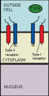
, cellular homeostasis and other cellular functions. In spite of the wide range of cellular processes that the TGFβ signaling pathway regulates, the process is relatively simple. TGFβ superfamily ligands bind to a type II receptor, which recruits and phosphorylates
a type I receptor. The type I receptor then phosphorylates receptor-regulated SMADs (R-SMAD
s) which can now bind the coSMAD SMAD4. R-SMAD/coSMAD complexes accumulate in the nucleus where they act as transcription factor
s and participate in the regulation of target gene expression.
 The TGF Beta superfamily of ligands include: Bone morphogenetic proteins (BMPs), Growth and differentiation factors (GDFs), Anti-müllerian hormone (AMH)
, Activin
, Nodal and TGFβ
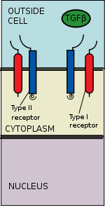
's. Signaling begins with the binding of a TGF beta superfamily ligand to a TGF beta type II receptor. The type II receptor is a serine/threonine receptor kinase, which catalyzes the phosphorylation
of the Type I receptor. Each class of ligand binds to a specific type II receptor.In mammals there are seven known type I receptors and five type II receptors.
There are three activins: Activin A
, Activin B
and Activin AB
. Activins are involved in embryogenesis and osteogenesis. They also regulate many hormones including pituitary, gonadal and hypothalamic hormones as well as insulin
. They are also nerve cell survival factors.
The BMPs bind to the Bone morphogenetic protein receptor type-2
(BMPR2). They are involved in a multitude of cellular functions including osteogenesis, cell differentiation, anterior/posterior axis specification, growth, and homeostasis.
The TGF beta family include: TGFβ1, TGFβ2, TGFβ3. Like the BMPS, TGF betas are involved in embryogenesis and cell differentiation, but they are also involved in apoptosis, as well as other functions. They bind to TGF-beta receptor type-2 (TGFBR2).
Nodal binds to activin A receptor, type IIB ACVR2B
. It can then either form a receptor complex with activin A receptor, type IB (ACVR1B
) or with activin A receptor, type IC (ACVR1C
).
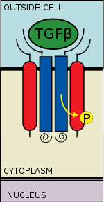 The TGF beta ligand binds to a type II receptor dimer, which recruits a type I receptor dimer forming a hetero-tetrameric complex with the ligand. These receptors are serine/threonine kinase receptors. They have a cysteine
rich extracellular domain, a transmembrane domain and a cytoplasmic serine/threonine rich domain. The GS domain of the type I receptor consists of a series of about thirty serine
-glycine
repeats. The binding of a TGF beta family ligand causes the rotation of the receptors so that their cytoplasmic kinase domains are arranged in a catalytically favorable orientation. The Type II receptor phosphorylates
serine
residues of the Type I receptor, which activates the protein.
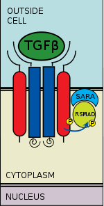 There are five receptor regulated SMADs: SMAD1, SMAD2, SMAD3, SMAD5, and SMAD9 (sometimes referred to as SMAD8). There are essentially two intracellular pathways involving these R-SMAD
s. TGF beta's, Activins, Nodals and some GDFs are mediated by SMAD2 and SMAD3, while BMPs, AMH and a few GDFs are mediated by SMAD1, SMAD5 and SMAD9. The binding of the R-SMAD to the type I receptor is mediated by a zinc double finger FYVE domain containing protein. Two such proteins that mediate the TGF beta pathway include SARA (The SMAD anchor for receptor activation) and HGS (Hepatocyte growth factor-regulated tyrosine kinase substrate).
SARA is present in an early endosome
which, by clathrin-mediated endocytosis, internalizes the receptor complex. SARA recruits an R-SMAD
. SARA permits the binding of the R-SMAD to the L45 region of the Type I receptor. SARA orients the R-SMAD such that serine residue on its C-terminus faces the catalytic region of the Type I receptor. The Type I receptor phosphorylates
the serine residue of the R-SMAD. Phosphorylation induces a conformational change in the MH2 domain of the R-SMAD and its subsequent dissociation from the receptor complex and SARA.
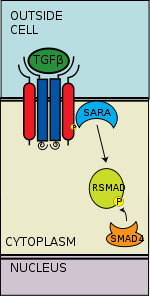 The phosphorylated RSMAD has a high affinity for a coSMAD (e.g. SMAD4) and forms a complex with one. The phosphate group does not act as a docking site for coSMAD, rather the phosphorylation opens up an amino acid stretch allowing interaction.
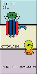 The phosphorylated RSMAD/coSMAD complex enters the nucleus where it binds transcription promoters/cofactors and causes the transcription of DNA.
Bone morphogenetic proteins cause the transcription of mRNAs involved in osteogenesis, neurogenesis
, and ventral mesoderm
specification.
TGF betas cause the transcription of mRNAs involved in apoptosis
, extracellular matrix
neogenesis and immunosuppression
. It is also involved in G1
arrest in the cell cycle
.
Activin causes the transcription of mRNAs involved in gonad
al growth, embryo differentiation and placenta formation.
Nodal causes the transcription of mRNAs involved in left and right axis specification, and mesoderm
and endoderm
induction.
and noggin
are antagonists
of BMPs. They bind BMPs preventing the binding of the ligand to the receptor. It has been demonstrated that Chordin and Noggin dorsalize mesoderm
. They are both found in the dorsal lip of Xenopus
and convert otherwise epidermis specified tissue into neural tissue (see neurulation
). Noggin plays a key role in cartilage and bone patterning. Mice Noggin-/- have excess cartilage and lacked joint formation.
Members of the DAN family of proteins also antagonize TGF beta family members. They include Cerberus
, DAN, and Gremlin
. These proteins contain nine conserved cysteine
s which can form disulfide bridges. It is believed that DAN antagonizes GDF5
, GDF6
and GDF7
.
Follistatin
inhibits Activin, which it binds. It directly affects follicle-stimulating hormone
(FSH) secretion. Follistatin also is implicated in prostate cancers where mutations in its gene may preventing it from acting on activin which has anti-proliferative properties.
Lefty
is a regulator of TGFβ and is involved in the axis patterning during embryogenesis. It is also a member of the TGF superfamily of proteins. It is asymmetrically expressed in the left side of murine embryos and subsequently plays a role in left-right specification. Lefty acts by preventing the phosphorylation of R-SMADs. It does so through a constitutively active TGFβ type I receptor and through a process downstream of its activation.
Drug-based antagonists have also been identified, such as SB431542, which selectively inhibits ALK4, ALK5, and ALK7.
BMP and Activin membrane bound inhibitor
(BAMBI), has a similar extracellular domain as type I receptors. It lacks an intracellular serine/threonine protein kinase domain and hence is a pseudoreceptor. It binds to the type I receptor preventing it from being activated. It serves as a negative regulator of TGF beta signaling and may limit tgf-beta expression during embryogeneis. It requires BMP signaling for its expression
FKBP12 binds the GS region of the type I receptor preventing phosphorylation of the receptor by the type II receptors. It is believed that FKBP12 and its homologs help to prevent type I receptor activation in the absence of a ligands, since ligand binding causes its dissociation.
s SMURF1
and SMURF2
regulate the levels of SMADs. They accept ubiquitin
from a E2 conjugating enzyme where they transfer ubiquitin to the RSMADs which causes their ubiquitination and subsequent proteosomal degradation. SMURF1 binds to SMAD1 and SMAD5 while SMURF2 binds SMAD1, SMAD2, SMAD3, SMAD6 and SMAD7. It enhances the inhibitory action of SMAD7 while reducing the transcriptional activities of SMAD2.
Embryo
An embryo is a multicellular diploid eukaryote in its earliest stage of development, from the time of first cell division until birth, hatching, or germination...
including cell growth
Cell growth
The term cell growth is used in the contexts of cell development and cell division . When used in the context of cell division, it refers to growth of cell populations, where one cell grows and divides to produce two "daughter cells"...
, cell differentiation, apoptosis
Apoptosis
Apoptosis is the process of programmed cell death that may occur in multicellular organisms. Biochemical events lead to characteristic cell changes and death. These changes include blebbing, cell shrinkage, nuclear fragmentation, chromatin condensation, and chromosomal DNA fragmentation...
, cellular homeostasis and other cellular functions. In spite of the wide range of cellular processes that the TGFβ signaling pathway regulates, the process is relatively simple. TGFβ superfamily ligands bind to a type II receptor, which recruits and phosphorylates
Phosphorylation
Phosphorylation is the addition of a phosphate group to a protein or other organic molecule. Phosphorylation activates or deactivates many protein enzymes....
a type I receptor. The type I receptor then phosphorylates receptor-regulated SMADs (R-SMAD
R-SMAD
R-SMAD stands for receptor-regulated SMAD. Smads are transcription factors that transduce extracellular TGF-ß superfamily ligand signaling from cell membrane bound TGF-ß receptors into the nucleus where they activate transcription TGF-ß target genes...
s) which can now bind the coSMAD SMAD4. R-SMAD/coSMAD complexes accumulate in the nucleus where they act as transcription factor
Transcription factor
In molecular biology and genetics, a transcription factor is a protein that binds to specific DNA sequences, thereby controlling the flow of genetic information from DNA to mRNA...
s and participate in the regulation of target gene expression.
Ligand Binding

Anti-müllerian hormone
Anti-Müllerian hormone also known as AMH is a protein that, in humans, is encoded by the AMH gene. It inhibits the development of the Müllerian ducts in the male embryo. It has also been called Müllerian inhibiting factor , Müllerian-inhibiting hormone , and Müllerian-inhibiting substance...
, Activin
Activin and inhibin
Activin and inhibin are two closely related protein complexes that have almost directly opposite biological effects. Activin enhances FSH biosynthesis and secretion, and participates in the regulation of the menstrual cycle...
, Nodal and TGFβ
TGF beta
Transforming growth factor beta is a protein that controls proliferation, cellular differentiation, and other functions in most cells. It plays a role in immunity, cancer, heart disease, diabetes, Marfan syndrome, and Loeys–Dietz syndrome....
's. Signaling begins with the binding of a TGF beta superfamily ligand to a TGF beta type II receptor. The type II receptor is a serine/threonine receptor kinase, which catalyzes the phosphorylation
Phosphorylation
Phosphorylation is the addition of a phosphate group to a protein or other organic molecule. Phosphorylation activates or deactivates many protein enzymes....
of the Type I receptor. Each class of ligand binds to a specific type II receptor.In mammals there are seven known type I receptors and five type II receptors.
There are three activins: Activin A
Activin and inhibin
Activin and inhibin are two closely related protein complexes that have almost directly opposite biological effects. Activin enhances FSH biosynthesis and secretion, and participates in the regulation of the menstrual cycle...
, Activin B
Activin and inhibin
Activin and inhibin are two closely related protein complexes that have almost directly opposite biological effects. Activin enhances FSH biosynthesis and secretion, and participates in the regulation of the menstrual cycle...
and Activin AB
Activin and inhibin
Activin and inhibin are two closely related protein complexes that have almost directly opposite biological effects. Activin enhances FSH biosynthesis and secretion, and participates in the regulation of the menstrual cycle...
. Activins are involved in embryogenesis and osteogenesis. They also regulate many hormones including pituitary, gonadal and hypothalamic hormones as well as insulin
Insulin
Insulin is a hormone central to regulating carbohydrate and fat metabolism in the body. Insulin causes cells in the liver, muscle, and fat tissue to take up glucose from the blood, storing it as glycogen in the liver and muscle....
. They are also nerve cell survival factors.
The BMPs bind to the Bone morphogenetic protein receptor type-2
BMPR2
Bone morphogenetic protein receptor type II or BMPR2 is a serine/threonine receptor kinase. It binds Bone morphogenetic proteins, members of the TGF beta superfamily of ligands. BMPs are involved in a host of cellular functions including osteogenesis, cell growth and cell differentiation. Signaling...
(BMPR2). They are involved in a multitude of cellular functions including osteogenesis, cell differentiation, anterior/posterior axis specification, growth, and homeostasis.
The TGF beta family include: TGFβ1, TGFβ2, TGFβ3. Like the BMPS, TGF betas are involved in embryogenesis and cell differentiation, but they are also involved in apoptosis, as well as other functions. They bind to TGF-beta receptor type-2 (TGFBR2).
Nodal binds to activin A receptor, type IIB ACVR2B
ACVR2B
Activin receptor type-2B is a protein that in humans is encoded by the ACVR2B gene. ACVR2B is an activin type 2 receptor.-Interactions:ACVR2B has been shown to interact with SYNJ2BP and ACVR1B.-Further reading:...
. It can then either form a receptor complex with activin A receptor, type IB (ACVR1B
ACVR1B
Activin receptor type-1B is a protein that in humans is encoded by the ACVR1B gene.ACVR1B or ALK-4 acts as a transducer of activin or activin like ligands signals. Activin binds to either ACVR2A or ACVR2B and then forms a complex with ACVR1B. These go on to recruit the R-SMADs SMAD2 or SMAD3...
) or with activin A receptor, type IC (ACVR1C
ACVR1C
The activin A receptor also known as ACVR1C or ALK-7 is a protein that in humans is encoded by the ACVR1C gene. ACVR1C is a type I receptor for the TGFB family of signaling molecules...
).
Receptor recruitment and phosphorylation

Cysteine
Cysteine is an α-amino acid with the chemical formula HO2CCHCH2SH. It is a non-essential amino acid, which means that it is biosynthesized in humans. Its codons are UGU and UGC. The side chain on cysteine is thiol, which is polar and thus cysteine is usually classified as a hydrophilic amino acid...
rich extracellular domain, a transmembrane domain and a cytoplasmic serine/threonine rich domain. The GS domain of the type I receptor consists of a series of about thirty serine
Serine
Serine is an amino acid with the formula HO2CCHCH2OH. It is one of the proteinogenic amino acids. By virtue of the hydroxyl group, serine is classified as a polar amino acid.-Occurrence and biosynthesis:...
-glycine
Glycine
Glycine is an organic compound with the formula NH2CH2COOH. Having a hydrogen substituent as its 'side chain', glycine is the smallest of the 20 amino acids commonly found in proteins. Its codons are GGU, GGC, GGA, GGG cf. the genetic code.Glycine is a colourless, sweet-tasting crystalline solid...
repeats. The binding of a TGF beta family ligand causes the rotation of the receptors so that their cytoplasmic kinase domains are arranged in a catalytically favorable orientation. The Type II receptor phosphorylates
Phosphorylation
Phosphorylation is the addition of a phosphate group to a protein or other organic molecule. Phosphorylation activates or deactivates many protein enzymes....
serine
Serine
Serine is an amino acid with the formula HO2CCHCH2OH. It is one of the proteinogenic amino acids. By virtue of the hydroxyl group, serine is classified as a polar amino acid.-Occurrence and biosynthesis:...
residues of the Type I receptor, which activates the protein.
SMAD phosphorylation

R-SMAD
R-SMAD stands for receptor-regulated SMAD. Smads are transcription factors that transduce extracellular TGF-ß superfamily ligand signaling from cell membrane bound TGF-ß receptors into the nucleus where they activate transcription TGF-ß target genes...
s. TGF beta's, Activins, Nodals and some GDFs are mediated by SMAD2 and SMAD3, while BMPs, AMH and a few GDFs are mediated by SMAD1, SMAD5 and SMAD9. The binding of the R-SMAD to the type I receptor is mediated by a zinc double finger FYVE domain containing protein. Two such proteins that mediate the TGF beta pathway include SARA (The SMAD anchor for receptor activation) and HGS (Hepatocyte growth factor-regulated tyrosine kinase substrate).
SARA is present in an early endosome
Endosome
In biology, an endosome is a membrane-bound compartment inside eukaryotic cells. It is a compartment of the endocytic membrane transport pathway from the plasma membrane to the lysosome. Molecules internalized from the plasma membrane can follow this pathway all the way to lysosomes for...
which, by clathrin-mediated endocytosis, internalizes the receptor complex. SARA recruits an R-SMAD
R-SMAD
R-SMAD stands for receptor-regulated SMAD. Smads are transcription factors that transduce extracellular TGF-ß superfamily ligand signaling from cell membrane bound TGF-ß receptors into the nucleus where they activate transcription TGF-ß target genes...
. SARA permits the binding of the R-SMAD to the L45 region of the Type I receptor. SARA orients the R-SMAD such that serine residue on its C-terminus faces the catalytic region of the Type I receptor. The Type I receptor phosphorylates
Phosphorylation
Phosphorylation is the addition of a phosphate group to a protein or other organic molecule. Phosphorylation activates or deactivates many protein enzymes....
the serine residue of the R-SMAD. Phosphorylation induces a conformational change in the MH2 domain of the R-SMAD and its subsequent dissociation from the receptor complex and SARA.
CoSMAD binding

Transcription

Bone morphogenetic proteins cause the transcription of mRNAs involved in osteogenesis, neurogenesis
Neurogenesis
Neurogenesis is the process by which neurons are generated from neural stem and progenitor cells. Most active during pre-natal development, neurogenesis is responsible for populating the growing brain with neurons. Recently neurogenesis was shown to continue in several small parts of the brain of...
, and ventral mesoderm
Mesoderm
In all bilaterian animals, the mesoderm is one of the three primary germ cell layers in the very early embryo. The other two layers are the ectoderm and endoderm , with the mesoderm as the middle layer between them.The mesoderm forms mesenchyme , mesothelium, non-epithelial blood corpuscles and...
specification.
TGF betas cause the transcription of mRNAs involved in apoptosis
Apoptosis
Apoptosis is the process of programmed cell death that may occur in multicellular organisms. Biochemical events lead to characteristic cell changes and death. These changes include blebbing, cell shrinkage, nuclear fragmentation, chromatin condensation, and chromosomal DNA fragmentation...
, extracellular matrix
Extracellular matrix
In biology, the extracellular matrix is the extracellular part of animal tissue that usually provides structural support to the animal cells in addition to performing various other important functions. The extracellular matrix is the defining feature of connective tissue in animals.Extracellular...
neogenesis and immunosuppression
Immunosuppression
Immunosuppression involves an act that reduces the activation or efficacy of the immune system. Some portions of the immune system itself have immuno-suppressive effects on other parts of the immune system, and immunosuppression may occur as an adverse reaction to treatment of other...
. It is also involved in G1
G1 phase
The G1 phase is a period in the cell cycle during interphase, before the S phase. For many cells, this phase is the major period of cell growth during its lifespan. During this stage new organelles are being synthesized, so the cell requires both structural proteins and enzymes, resulting in great...
arrest in the cell cycle
Cell cycle
The cell cycle, or cell-division cycle, is the series of events that takes place in a cell leading to its division and duplication . In cells without a nucleus , the cell cycle occurs via a process termed binary fission...
.
Activin causes the transcription of mRNAs involved in gonad
Gonad
The gonad is the organ that makes gametes. The gonads in males are the testes and the gonads in females are the ovaries. The product, gametes, are haploid germ cells. For example, spermatozoon and egg cells are gametes...
al growth, embryo differentiation and placenta formation.
Nodal causes the transcription of mRNAs involved in left and right axis specification, and mesoderm
Mesoderm
In all bilaterian animals, the mesoderm is one of the three primary germ cell layers in the very early embryo. The other two layers are the ectoderm and endoderm , with the mesoderm as the middle layer between them.The mesoderm forms mesenchyme , mesothelium, non-epithelial blood corpuscles and...
and endoderm
Endoderm
Endoderm is one of the three primary germ cell layers in the very early embryo. The other two layers are the ectoderm and mesoderm , with the endoderm as the intermost layer...
induction.
Pathway regulation
The TGF beta signaling pathway is involved in a wide range of cellular process and subsequently is very heavily regulated. There are a variety of mechanisms that the pathway is modulated both positively and negatively: There are agonists for ligands and R-SMADs; there are decoy receptors; and R-SMADs and receptors are ubiquitinated.Ligand agonists/antagonists
Both chordinChordin
Chordin is a polypeptide that dorsalizes the developing embryo by binding ventralizing TGFβ proteins such as bone morphogenetic proteins. It may also play a role in organogenesis. There are five named isoforms of this protein that are produced by alternative splicing.In humans, the chordin peptide...
and noggin
Noggin (protein)
Noggin, also known as NOG, is a protein which in humans is encoded by the NOG gene.Noggin inhibits TGF-β signal transduction by binding to TGF-β family ligands and preventing them from binding to their corresponding receptors. Noggin plays a key role in neural induction by inhibiting BMP4, along...
are antagonists
Receptor antagonist
A receptor antagonist is a type of receptor ligand or drug that does not provoke a biological response itself upon binding to a receptor, but blocks or dampens agonist-mediated responses...
of BMPs. They bind BMPs preventing the binding of the ligand to the receptor. It has been demonstrated that Chordin and Noggin dorsalize mesoderm
Mesoderm
In all bilaterian animals, the mesoderm is one of the three primary germ cell layers in the very early embryo. The other two layers are the ectoderm and endoderm , with the mesoderm as the middle layer between them.The mesoderm forms mesenchyme , mesothelium, non-epithelial blood corpuscles and...
. They are both found in the dorsal lip of Xenopus
Xenopus
Xenopus is a genus of highly aquatic frogs native to Sub-Saharan Africa. There are 19 species in the Xenopus genus...
and convert otherwise epidermis specified tissue into neural tissue (see neurulation
Neurulation
Neurulation is the stage of organogenesis in vertebrate embryos, during which the neural tube is transformed into the primitive structures that will later develop into the central nervous system....
). Noggin plays a key role in cartilage and bone patterning. Mice Noggin-/- have excess cartilage and lacked joint formation.
Members of the DAN family of proteins also antagonize TGF beta family members. They include Cerberus
Cerberus (protein)
Cerberus also known as CER1 is a protein that in humans is encoded by the CER1 gene.- Function :Cerberus is an inhibitor in the TGF beta signaling pathway secreted during the gastrulation phase of the embryogenesis....
, DAN, and Gremlin
Gremlin (protein)
Gremlin is an inhibitor in the TGF beta signaling pathway.Gremlin, also known as Drm, is a highly conserved 20.7-kDa, 184 amino acid glycoprotein part of the DAN family and is a cysteine knot-secreted protein...
. These proteins contain nine conserved cysteine
Cysteine
Cysteine is an α-amino acid with the chemical formula HO2CCHCH2SH. It is a non-essential amino acid, which means that it is biosynthesized in humans. Its codons are UGU and UGC. The side chain on cysteine is thiol, which is polar and thus cysteine is usually classified as a hydrophilic amino acid...
s which can form disulfide bridges. It is believed that DAN antagonizes GDF5
GDF5
Growth/differentiation factor 5 is a protein that in humans is encoded by the GDF5 gene.Growth differentiation factor 5 is a protein belonging to the transforming growth factor beta superfamily that is expressed in the developing central nervous system, and has a role in skeletal and joint...
, GDF6
GDF6
Growth differentiation factor 6 is a protein that in humans is encoded by the GDF6 gene.belonging to the transforming growth factor beta superfamily that may regulate patterning of the ectoderm by interacting with bone morphogenetic proteins, and control eye development....
and GDF7
GDF7
Growth differentiation factor 7 is a protein that in humans is encoded by the GDF7 gene.GDF7 belongs to the transforming growth factor beta superfamily that is specifically found in a signaling center known as the roof plate that is located in the developing nervous system of embryos...
.
Follistatin
Follistatin
Follistatin also known as activin-binding protein is a protein that in humans is encoded by the FST gene. Follistatin is an autocrine glycoprotein that is expressed in nearly all tissues of higher animals....
inhibits Activin, which it binds. It directly affects follicle-stimulating hormone
Follicle-stimulating hormone
Follicle-stimulating hormone is a hormone found in humans and other animals. It is synthesized and secreted by gonadotrophs of the anterior pituitary gland. FSH regulates the development, growth, pubertal maturation, and reproductive processes of the body. FSH and Luteinizing hormone act...
(FSH) secretion. Follistatin also is implicated in prostate cancers where mutations in its gene may preventing it from acting on activin which has anti-proliferative properties.
Lefty
Lefty (protein)
Lefty are proteins that are closely related members of the TGF-beta family of growth factors. These proteins are secreted and play a role in left-right asymmetry determination of organ systems during development...
is a regulator of TGFβ and is involved in the axis patterning during embryogenesis. It is also a member of the TGF superfamily of proteins. It is asymmetrically expressed in the left side of murine embryos and subsequently plays a role in left-right specification. Lefty acts by preventing the phosphorylation of R-SMADs. It does so through a constitutively active TGFβ type I receptor and through a process downstream of its activation.
Drug-based antagonists have also been identified, such as SB431542, which selectively inhibits ALK4, ALK5, and ALK7.
Receptor regulation
The Transforming growth factor receptor 3 (TGFBR3) is the most abundant of the TGF-β receptors yet, it has no known signaling domain. It however may serve to enhance the binding of TGF beta ligands to TGF beta type II receptors by binding TGFβ and presenting it to TGFBR2. One of the downstream targets of TGF β signaling, GIPC, binds to its PDZ domain, which prevents its proteosomal degradation, which subsequently increases TGFβ activity. It may also serve as an inhibin coreceptor to ActivinRII.BMP and Activin membrane bound inhibitor
BAMBI
BMP and activin membrane-bound inhibitor homolog , also known as BAMBI, is a protein which in humans is encoded by the BAMBI gene.- Function :...
(BAMBI), has a similar extracellular domain as type I receptors. It lacks an intracellular serine/threonine protein kinase domain and hence is a pseudoreceptor. It binds to the type I receptor preventing it from being activated. It serves as a negative regulator of TGF beta signaling and may limit tgf-beta expression during embryogeneis. It requires BMP signaling for its expression
FKBP12 binds the GS region of the type I receptor preventing phosphorylation of the receptor by the type II receptors. It is believed that FKBP12 and its homologs help to prevent type I receptor activation in the absence of a ligands, since ligand binding causes its dissociation.
Role of inhibitory SMADs
There are two other SMADs which complete the SMAD family, the inhibitory SMADs (I-SMADS), SMAD6 and SMAD7. They play a key role in the regulation of TGF beta signaling and are involved in negative feeback. Like other SMADs they have an MH1 and an MH2 domain. SMAD7 competes with other R-SMADs with the Type I receptor and prevents their phosphorylation. It resides in the nucleus and upon TGF beta receptor activation translocates to the cytoplasm where it binds the type I receptor. SMAD6 binds SMAD4 preventing the binding of other R-SMADs with the coSMAD. The levels of I-SMAD increase with TGF beta signaling suggesting that they are downstream targets of TGF-beta signaling.R-SMAD ubiquitination
The E3 ubiquitin-protein ligaseLigase
In biochemistry, ligase is an enzyme that can catalyse the joining of two large molecules by forming a new chemical bond, usually with accompanying hydrolysis of a small chemical group dependent to one of the larger molecules...
s SMURF1
SMURF1
E3 ubiquitin-protein ligase SMURF1 is an enzyme that in humans is encoded by the SMURF1 gene.-Interactions:SMURF1 has been shown to interact with ARHGEF9, PLEKHO1 and SMURF2.-Further reading:...
and SMURF2
SMURF2
E3 ubiquitin-protein ligase SMURF2 is an enzyme that in humans is encoded by the SMURF2 gene.-Interactions:SMURF2 has been shown to interact with Mothers against decapentaplegic homolog 7, SCYE1, SMURF1, Mothers against decapentaplegic homolog 3, Ubiquitin C, Mothers against decapentaplegic homolog...
regulate the levels of SMADs. They accept ubiquitin
Ubiquitin
Ubiquitin is a small regulatory protein that has been found in almost all tissues of eukaryotic organisms. Among other functions, it directs protein recycling.Ubiquitin can be attached to proteins and label them for destruction...
from a E2 conjugating enzyme where they transfer ubiquitin to the RSMADs which causes their ubiquitination and subsequent proteosomal degradation. SMURF1 binds to SMAD1 and SMAD5 while SMURF2 binds SMAD1, SMAD2, SMAD3, SMAD6 and SMAD7. It enhances the inhibitory action of SMAD7 while reducing the transcriptional activities of SMAD2.
Summary table
| TGF Beta superfamily ligand | Type II Receptor | |Type I receptor | | R-SMAD R-SMAD R-SMAD stands for receptor-regulated SMAD. Smads are transcription factors that transduce extracellular TGF-ß superfamily ligand signaling from cell membrane bound TGF-ß receptors into the nucleus where they activate transcription TGF-ß target genes... s | | coSMAD | Ligand inhibitors |
|---|---|---|---|---|---|
| Activin A Activin and inhibin Activin and inhibin are two closely related protein complexes that have almost directly opposite biological effects. Activin enhances FSH biosynthesis and secretion, and participates in the regulation of the menstrual cycle... |
ACVR2A ACVR2A Activin receptor type-2A is a protein that in humans is encoded by the ACVR2A gene.ACVR2A is an activin type 2 receptor.-Interactions:ACVR2A has been shown to interact with INHBA, SYNJ2BP and ACVR1B.-Further reading:... |
ACVR1B ACVR1B Activin receptor type-1B is a protein that in humans is encoded by the ACVR1B gene.ACVR1B or ALK-4 acts as a transducer of activin or activin like ligands signals. Activin binds to either ACVR2A or ACVR2B and then forms a complex with ACVR1B. These go on to recruit the R-SMADs SMAD2 or SMAD3... (ALK4) |
SMAD2 , SMAD3 | SMAD4 | Follistatin Follistatin Follistatin also known as activin-binding protein is a protein that in humans is encoded by the FST gene. Follistatin is an autocrine glycoprotein that is expressed in nearly all tissues of higher animals.... |
| GDF1 GDF1 Growth differentiation factor-1 is a protein that in humans is encoded by the GDF1 gene.GDF1 belongs to the transforming growth factor beta superfamily that has a role in left-right patterning and mesoderm induction during embryonic development. It is found in the brain, spinal cord and peripheral... |
ACVR2A ACVR2A Activin receptor type-2A is a protein that in humans is encoded by the ACVR2A gene.ACVR2A is an activin type 2 receptor.-Interactions:ACVR2A has been shown to interact with INHBA, SYNJ2BP and ACVR1B.-Further reading:... |
ACVR1B ACVR1B Activin receptor type-1B is a protein that in humans is encoded by the ACVR1B gene.ACVR1B or ALK-4 acts as a transducer of activin or activin like ligands signals. Activin binds to either ACVR2A or ACVR2B and then forms a complex with ACVR1B. These go on to recruit the R-SMADs SMAD2 or SMAD3... (ALK4) |
SMAD2 , SMAD3 | SMAD4 | |
| GDF11 GDF11 Growth differentiation factor 11 also known as bone morphogenetic protein 11 is a protein that in humans is encoded by the GDF11 gene.... |
ACVR2B ACVR2B Activin receptor type-2B is a protein that in humans is encoded by the ACVR2B gene. ACVR2B is an activin type 2 receptor.-Interactions:ACVR2B has been shown to interact with SYNJ2BP and ACVR1B.-Further reading:... |
ACVR1B ACVR1B Activin receptor type-1B is a protein that in humans is encoded by the ACVR1B gene.ACVR1B or ALK-4 acts as a transducer of activin or activin like ligands signals. Activin binds to either ACVR2A or ACVR2B and then forms a complex with ACVR1B. These go on to recruit the R-SMADs SMAD2 or SMAD3... (ALK4), TGFβRI (ALK5) |
SMAD2 , SMAD3 | SMAD4 | |
| Bone morphogenetic proteins | BMPR2 BMPR2 Bone morphogenetic protein receptor type II or BMPR2 is a serine/threonine receptor kinase. It binds Bone morphogenetic proteins, members of the TGF beta superfamily of ligands. BMPs are involved in a host of cellular functions including osteogenesis, cell growth and cell differentiation. Signaling... |
BMPR1A BMPR1A The bone morphogenetic protein receptor, type IA also known as BMPR1A is a protein which in humans is encoded by the BMPR1A gene. BMPR1A has also been designated as CD292 .- Function :... (ALK3), BMPR1B BMPR1B Bone morphogenetic protein receptor type-1B also known as CDw293 is a protein that in humans is encoded by the BMPR1B gene.- Function :... (ALK6) |
SMAD1 SMAD5, SMAD8 | SMAD4 | Noggin Noggin (protein) Noggin, also known as NOG, is a protein which in humans is encoded by the NOG gene.Noggin inhibits TGF-β signal transduction by binding to TGF-β family ligands and preventing them from binding to their corresponding receptors. Noggin plays a key role in neural induction by inhibiting BMP4, along... , Chordin Chordin Chordin is a polypeptide that dorsalizes the developing embryo by binding ventralizing TGFβ proteins such as bone morphogenetic proteins. It may also play a role in organogenesis. There are five named isoforms of this protein that are produced by alternative splicing.In humans, the chordin peptide... , DAN |
| Nodal | ACVR2B ACVR2B Activin receptor type-2B is a protein that in humans is encoded by the ACVR2B gene. ACVR2B is an activin type 2 receptor.-Interactions:ACVR2B has been shown to interact with SYNJ2BP and ACVR1B.-Further reading:... |
ACVR1B ACVR1B Activin receptor type-1B is a protein that in humans is encoded by the ACVR1B gene.ACVR1B or ALK-4 acts as a transducer of activin or activin like ligands signals. Activin binds to either ACVR2A or ACVR2B and then forms a complex with ACVR1B. These go on to recruit the R-SMADs SMAD2 or SMAD3... (ALK4), ACVR1C ACVR1C The activin A receptor also known as ACVR1C or ALK-7 is a protein that in humans is encoded by the ACVR1C gene. ACVR1C is a type I receptor for the TGFB family of signaling molecules... (ALK7) |
SMAD2 , SMAD3 | SMAD4 | Lefty Lefty (protein) Lefty are proteins that are closely related members of the TGF-beta family of growth factors. These proteins are secreted and play a role in left-right asymmetry determination of organ systems during development... |
| TGFβs | TGFβRII | TGFβRI (ALK5) | SMAD2 , SMAD3 | SMAD4 | LTBP1, THBS1, Decorin Decorin Decorin is a proteoglycan on average 90 - 140 kilodaltons in size.It belongs to the small leucine-rich proteoglycan family and consists of a protein core containing leucine repeats with a glycosaminoglycan chain consisting of either chondroitin sulfate or dermatan sulfate .Decorin is a small... |
External links
- Kyoto Encyclopedia of Genes and GenomesKEGG PATHWAY DatabaseKEGG is a collection of online databases dealing with genomes, enzymatic pathways, and biological chemicals. The PATHWAY database records networks of molecular interactions in the cells, and variants of them specific to particular organisms...
-TGF beta signaling pathway map - NetpathNetpathNetPath is a manually curated resource of human signal transduction pathways. It is a joint effort between Pandey Lab at the Johns Hopkins University and the Institute of Bioinformatics , Bangalore, India, and is also worked on by other parties....
- A curated resource of signal transduction pathways in humans

