
Neurulation
Encyclopedia

Organogenesis
In animal development, organogenesis is the process by which the ectoderm, endoderm, and mesoderm develop into the internal organs of the organism. Internal organs initiate development in humans within the 3rd to 8th weeks in utero...
in vertebrate
Vertebrate
Vertebrates are animals that are members of the subphylum Vertebrata . Vertebrates are the largest group of chordates, with currently about 58,000 species described. Vertebrates include the jawless fishes, bony fishes, sharks and rays, amphibians, reptiles, mammals, and birds...
embryo
Embryo
An embryo is a multicellular diploid eukaryote in its earliest stage of development, from the time of first cell division until birth, hatching, or germination...
s, during which the neural tube
Neural tube
In the developing vertebrate, the neural tube is the embryo's precursor to the central nervous system, which comprises the brain and spinal cord...
is transformed into the primitive structures that will later develop into the central nervous system
Central nervous system
The central nervous system is the part of the nervous system that integrates the information that it receives from, and coordinates the activity of, all parts of the bodies of bilaterian animals—that is, all multicellular animals except sponges and radially symmetric animals such as jellyfish...
.
The process begins when the notochord
Notochord
The notochord is a flexible, rod-shaped body found in embryos of all chordates. It is composed of cells derived from the mesoderm and defines the primitive axis of the embryo. In some chordates, it persists throughout life as the main axial support of the body, while in most vertebrates it becomes...
induces the formation of the central nervous system
Central nervous system
The central nervous system is the part of the nervous system that integrates the information that it receives from, and coordinates the activity of, all parts of the bodies of bilaterian animals—that is, all multicellular animals except sponges and radially symmetric animals such as jellyfish...
(CNS) by signaling the ectoderm germ layer
Germ layer
A germ layer, occasionally referred to as a germinal epithelium, is a group of cells, formed during animal embryogenesis. Germ layers are particularly pronounced in the vertebrates; however, all animals more complex than sponges produce two or three primary tissue layers...
above it to form the thick and flat neural plate
Neural plate
In human embryology, formation of neural plate is the first step of neurulation. It is created by a flat thickening opposite to the primitive streak of the ectoderm.-Development:...
. The neural plate folds in upon itself to form the neural tube
Neural tube
In the developing vertebrate, the neural tube is the embryo's precursor to the central nervous system, which comprises the brain and spinal cord...
, which will later differentiate into the spinal cord
Spinal cord
The spinal cord is a long, thin, tubular bundle of nervous tissue and support cells that extends from the brain . The brain and spinal cord together make up the central nervous system...
and the brain
Brain
The brain is the center of the nervous system in all vertebrate and most invertebrate animals—only a few primitive invertebrates such as sponges, jellyfish, sea squirts and starfishes do not have one. It is located in the head, usually close to primary sensory apparatus such as vision, hearing,...
, eventually forming the central nervous system.
Different portions of the neural tube form by two different processes, called primary and secondary neurulation, in different species.
- In primary neurulation, the neural plate creases inward until the edges come in contact and fuse.
- In secondary neurulation, the tube forms by hollowing out of the interior of a solid precursor.
Induction
Primary neurulation occurs in response to soluble growth factorGrowth factor
A growth factor is a naturally occurring substance capable of stimulating cellular growth, proliferation and cellular differentiation. Usually it is a protein or a steroid hormone. Growth factors are important for regulating a variety of cellular processes....
s secreted by the notochord
Notochord
The notochord is a flexible, rod-shaped body found in embryos of all chordates. It is composed of cells derived from the mesoderm and defines the primitive axis of the embryo. In some chordates, it persists throughout life as the main axial support of the body, while in most vertebrates it becomes...
. Ectodermal cells are induced to form neuroectoderm
Neuroectoderm
Neuroectoderm is the term for ectoderm which receives Bone Morphogenetic Protein-inhibiting signals from proteins such as noggin, which leads to the development of the nervous system from this tissue....
from a variety of signals. Ectoderm sends and receives signals of BMP4 (bone morphogenic protein
Bone morphogenetic protein
Bone morphogenetic proteins are a group of growth factors also known as cytokines and as metabologens . Originally discovered by their ability to induce the formation of bone and cartilage, BMPs are now considered to constitute a group of pivotal morphogenetic signals, orchestrating tissue...
) and cells which receive BMP4 signal develop into epidermis. The inhibitory signals chordin
Chordin
Chordin is a polypeptide that dorsalizes the developing embryo by binding ventralizing TGFβ proteins such as bone morphogenetic proteins. It may also play a role in organogenesis. There are five named isoforms of this protein that are produced by alternative splicing.In humans, the chordin peptide...
, noggin
Noggin (protein)
Noggin, also known as NOG, is a protein which in humans is encoded by the NOG gene.Noggin inhibits TGF-β signal transduction by binding to TGF-β family ligands and preventing them from binding to their corresponding receptors. Noggin plays a key role in neural induction by inhibiting BMP4, along...
and follistatin
Follistatin
Follistatin also known as activin-binding protein is a protein that in humans is encoded by the FST gene. Follistatin is an autocrine glycoprotein that is expressed in nearly all tissues of higher animals....
are needed to form neural plate. These inhibitory signals are created and emitted by the spemann organiser. Cells which do not receive BMP4 signaling due to the effects of the inhibitory signals will develop into the anterior neuroectoderm cells of the neural plate. Cells which receive FGF (fibroblast growth factor
Fibroblast growth factor
Fibroblast growth factors, or FGFs, are a family of growth factors involved in angiogenesis, wound healing, and embryonic development. The FGFs are heparin-binding proteins and interactions with cell-surface associated heparan sulfate proteoglycans have been shown to be essential for FGF signal...
) in addition to the inhibitory signals form posterior neural plate cells.
Shape change
The cells of the neural plate are signaled to become high-columnar and can be identified through microscopy as different from the surrounding epiblastic ectoderm. The cells move laterally and away from the central axis and change into a truncated pyramid shape. This pyramid shape is achieved through tubulinTubulin
Tubulin is one of several members of a small family of globular proteins. The most common members of the tubulin family are α-tubulin and β-tubulin, the proteins that make up microtubules. Each has a molecular weight of approximately 55 kiloDaltons. Microtubules are assembled from dimers of α- and...
and actin
Actin
Actin is a globular, roughly 42-kDa moonlighting protein found in all eukaryotic cells where it may be present at concentrations of over 100 μM. It is also one of the most highly-conserved proteins, differing by no more than 20% in species as diverse as algae and humans...
in the apical portion of the cell which constricts as they move. The variation in cell shapes is partially determined by the location of the nucleus within the cell, causing bulging in areas of the cells forcing the height and shape of the cell to change. This process is known as apical constriction
Apical constriction
Apical constriction describes the process in which contraction of the apical side of a cell causes the cell to take on a wedged shape. Generally, this shape change is coordinated across many cells of an epithelial layer, generating forces that can bend or fold the cell sheet .-Morphogenetic...
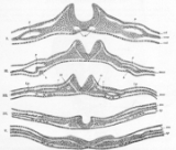
Folding
The process of the flat neural plate folding into the cylindrical neural tube is termed primary neurulation. As a result of the cellular shape changes, the neural plate forms the medial hinge point . The expanding epidermis puts pressure on the MHP and causes the neural plate to fold resulting in neural foldsNeural folds
In front of the primitive streak two longitudinal ridges, caused by a folding up of the ectoderm, make their appearance, one on either side of the middle line...
and the creation of the neural groove
Neural groove
The neural groove is a shallow median groove between the neural folds of an embryo. The neural folds are two longitudinal ridges that are caused by a folding up of the ectoderm in front of the primitive streak of the developing embryo...
. The neural folds form dorsolateral hinge points (DLHP) and pressure on this hinge causes the neural folds to meet and fuse at the midline. The fusion requires the regulation of cell adhesion molecules. The neural plate switches from E-cadherin expression to N-cadherin and N-CAM expression to recognize each other as the same tissue and close the tube. This change in expression stops the binding of the neural tube to the epidermis. Neural plate folding is a complicated step.
The notochord plays an integral role in the development of the neural tube. Prior to neurulation, during the migration of epiblastic endoderm cells towards the hypoblastic endoderm, the notochordal process opens into an arch termed the notochordal plate and attaches overlying neuroepithelium of the neural plate. The notochordal plate then serves as an anchor for the neural plate and pushes the two edges of the plate upwards while keeping the middle section anchored. Some of the notochodral cells become incorporated into the center section neural plate to later form the floor plate of the neural tube. The notochord plate separates and forms the solid notochord.
The folding of the neural tube to form an actual tube does not occur all at once. Instead, it begins approximately at the level of the fourth somite
Somite
A somite is a division of the body of an animal. In vertebrates this is mainly discernible in the embryo stage; in arthropods it is a characteristic of a hypothetical ancestor.- In vertebrates :...
at Carnegie stage
Carnegie stages
In embryology, Carnegie stages are a standardized system of 23 stages used to provide a unified developmental chronology of the vertebrate embryo....
9 (around Embryonic day 20 in human
Human
Humans are the only living species in the Homo genus...
s). The lateral edges of the neural plate touch in the midline and join together. This continues both cranially (toward the head) and caudal
Tail
The tail is the section at the rear end of an animal's body; in general, the term refers to a distinct, flexible appendage to the torso. It is the part of the body that corresponds roughly to the sacrum and coccyx in mammals, reptiles, and birds...
ly (toward the tail). The openings that are formed at the cranial and caudal regions are termed the cranial and caudal neuropores. In human
Human
Humans are the only living species in the Homo genus...
embryos, the cranial neuropore closes approximately on day 24 and the caudal neuropore on day 26 (Carnegie stages 11 and 13 respectively). Failure of the cranial (anterior) and caudal (posterior) neuropore closure results in conditions called anecephaly
Anencephaly
Anencephaly is a cephalic disorder that results from a neural tube defect that occurs when the cephalic end of the neural tube fails to close, usually between the 23rd and 26th day of pregnancy, resulting in the absence of a major portion of the brain, skull, and scalp...
and spina bifida
Spina bifida
Spina bifida is a developmental congenital disorder caused by the incomplete closing of the embryonic neural tube. Some vertebrae overlying the spinal cord are not fully formed and remain unfused and open. If the opening is large enough, this allows a portion of the spinal cord to protrude through...
, respectively. Additionally, failure of the neural tube to close throughout the length of the body results in a condition called craniorachischisis.
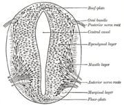
Patterning
Sonic hedgehog
Sonic hedgehog homolog is one of three proteins in the mammalian signaling pathway family called hedgehog, the others being desert hedgehog and Indian hedgehog . SHH is the best studied ligand of the hedgehog signaling pathway. It plays a key role in regulating vertebrate organogenesis, such as...
from the notochord induces its formation, the floor plate of the incipient neural tube also secretes SHH. After closure, the neural tube forms a basal plate
Basal plate
Basal plate may refer to:* Basal plate , the region of the neural tube ventral to the sulcus limitans* Basal plate , between this plate and the uterine muscular fibres are the stratum spongiosum and the boundary layer...
or floor plate
Floor plate
The floor plate is a structure integral to the developing nervous system of vertebrate organisms. Located on the ventral midline of the embryonic neural tube, the floor plate is a specialized glial structure that spans the anteroposterior axis from the midbrain to the tail regions...
and an alar plate
Alar plate
The alar plate is a neural structure in the embryonic nervous system, part of the dorsal side of neural tube, that involves the communication of general somatic and general visceral sensory impulses. The caudal part later becomes sensory axon part of the spinal cord.-External links:* *...
or roof plate in response to the combined effects of Shh and factors including BMP4 secreted by the roof plate. The basal plate forms most of the ventral portion of the nervous system, including the motor portion of the spinal cord and brain stem; the alar plate forms the dorsal portions, devoted mostly to sensory processing.
The dorsal epidermis expresses BMP4 and BMP7. The roof plate of the neural tube responds to those signals to express more BMP4 and other TGF-b signals to form a dorsal/ventral gradient among the neural tube. The notochord expresses Sonic Hedgehog (Shh). The floor plate responds to Shh by producing its own Shh and forming a gradient. These gradients allows for the differential expression of transcription factors.
Complexities of the model
In actuality, the folding of the neural tube is still not entirely understood and is still being studied. The simplistic model of the closure occurring in one step cranially and caudally does not explain the high frequency of neural tube defects. Proposed theories include closure of the neural tube occurs in regions, rather than entirely linearly.Secondary neurulation
In secondary neurulation, the neural ectoderm and some cells from the endoderm form the medullary cord. The medullary cord condenses, separates and then forms cavities. These cavities then merge to form a single tube. Secondary Neurulation occurs in the posterior section of most animals but it is better expressed in birds. Tubes from both primary and secondary neurulation eventually connect.Early brain development
The anterior segment of the neural tube forms the three main parts of the brain: the forebrain, midbrain, and the hindbrain. Formation of these structures begins with a swelling of the neural tube in a pattern specified by Hox genes. Ion pumps are used to increase the fluid pressure within the tube and create a bulge. A blockage between the brain and the spinal cord prevents the fluid accumulation from leaking out. These brain regions further divide into subregions. The hindbrain divides into different segments called rhombomeres. Neural crest cells form ganglia above each rhombomere. The neural tube becomes the germinal neuroepithelium and serves as a source of new neurons during brain development. The brain develops from the inside-out.Non-neural ectoderm tissue
Mesoderm surrounding the notochord at the sides will develop into the somites (future muscles, bones, and contributes to the formation of limbs of the vertebrate).Neural crest cells
Masses of tissue called the neural crestNeural crest
Neural crest cells are a transient, multipotent, migratory cell population unique to vertebrates that gives rise to a diverse cell lineage including melanocytes, craniofacial cartilage and bone, smooth muscle, peripheral and enteric neurons and glia....
that are located at the very edges of the lateral plates of the folding neural tube separate from the neural tube and migrate to become a variety of different but important cells.
Neural crest cells will migrate through the embryo and will give rise to several cell populations, including pigment cells and the cells of the peripheral nervous system.
Neural tube defects
Failure to complete the neurulation process will lead to an open neural tube, which is a common form of birth defect known as spina bifidaSpina bifida
Spina bifida is a developmental congenital disorder caused by the incomplete closing of the embryonic neural tube. Some vertebrae overlying the spinal cord are not fully formed and remain unfused and open. If the opening is large enough, this allows a portion of the spinal cord to protrude through...
. Spina bifida can lead to paralysis beneath the affected region of the spinal cord. Sufferers may require crutches or wheelchairs to move about, and may also suffer from lack of bladder and bowel control.
Neural tube defects are among the most common and disabling birth defects, occurring in roughly 1 in every 500 live births.