

Magnetic resonance neurography
Encyclopedia
Nerve
A peripheral nerve, or simply nerve, is an enclosed, cable-like bundle of peripheral axons . A nerve provides a common pathway for the electrochemical nerve impulses that are transmitted along each of the axons. Nerves are found only in the peripheral nervous system...
s in the body by optimizing selectivity for unique MRI water properties of nerves. It is a modification of magnetic resonance imaging
Magnetic resonance imaging
Magnetic resonance imaging , nuclear magnetic resonance imaging , or magnetic resonance tomography is a medical imaging technique used in radiology to visualize detailed internal structures...
. This technique yields a detailed image of a nerve from the resonance signal that arises from in the nerve itself rather than from surrounding tissues or from fat in the nerve lining. Because of the intraneural source of the image signal, the image provides a medically useful set of information about the internal state of the nerve such as the presence of irritation, nerve swelling (edema
Edema
Edema or oedema ; both words from the Greek , oídēma "swelling"), formerly known as dropsy or hydropsy, is an abnormal accumulation of fluid beneath the skin or in one or more cavities of the body that produces swelling...
), compression, pinch or injury. Standard magnetic resonance images can show the outline of some nerves in portions of their courses but do not show the intrinsic signal from nerve water. Magnetic resonance neurography is used to evaluate major nerve compressions such as those affecting the sciatic nerve
Sciatic nerve
The sciatic nerve is a large nerve fiber in humans and other animals. It begins in the lower back and runs through the buttock and down the lower limb...
(e.g. piriformis syndrome
Piriformis syndrome
Piriformis syndrome is a neuromuscular disorder that occurs when the sciatic nerve is compressed or otherwise irritated by the piriformis muscle causing pain, tingling and numbness in the buttocks and along the path of the sciatic nerve descending down the lower thigh and into the leg...
), the brachial plexus
Brachial plexus
The brachial plexus is a network of nerve fibers, running from the spine, formed by the ventral rami of the lower four cervical and first thoracic nerve roots...
nerves (e.g. thoracic outlet syndrome
Thoracic outlet syndrome
Thoracic outlet syndrome is a syndrome involving compression at the superior thoracic outlet involving compression of a neurovascular bundle passing between the anterior scalene and middle scalene...
), the pudendal nerve
Pudendal nerve
The pudendal nerve is a sensory and somatic nerve in the pelvic region which is a large branch of the sacral plexus that innervates the external genitalia of both sexes, as well as sphincters for the bladder and the rectum...
, or virtually any named nerve in the body. A related technique for imaging neural tracts in the brain and spinal cord is called magnetic resonance tractography
Tractography
In neuroscience, tractography is a procedure to demonstrate the neural tracts.It uses special techniques of magnetic resonance imaging , and computer-based image analysis.The results are presented in two- and three-dimensional images....
or diffusion tensor imaging.
History and physical basis
Magnetic resonance imaging (MRI) is based on differences in the physical properties of protonProton
The proton is a subatomic particle with the symbol or and a positive electric charge of 1 elementary charge. One or more protons are present in the nucleus of each atom, along with neutrons. The number of protons in each atom is its atomic number....
s in water molecules in different tissues in the body. The protons and the water molecules of which they are part have subtly different movement characteristics that relate to their biophysical
Biophysics
Biophysics is an interdisciplinary science that uses the methods of physical science to study biological systems. Studies included under the branches of biophysics span all levels of biological organization, from the molecular scale to whole organisms and ecosystems...
surroundings. Because of this, MRI is capable of differentiating one tissue from another; this provides "tissue contrast." From the time of the first clinical use of MRI in the mid 1970s until 1992, however, despite the active work of many thousands of researchers, there was no reliable method for visualizing nerve. In some parts of the body, nerves could be observed as areas of absent signal delineated by bright fat, or as bland grey structures that could not be reliably distinguished from other similar-appearing structures in cross sectional images.
In 1992, Aaron Filler and Franklyn Howe, working at St. George's Hospital Medical School
St George's Hospital
Founded in 1733, St George’s Hospital is one of the UK's largest teaching hospitals. It shares its main hospital site in Tooting, England with the St George's, University of London which trains NHS staff and carries out advanced medical research....
in London, succeeded in identifying the unique water properties of nerve water that would make it possible to generate tissue-specific nerve images. The result was an initial "pure" nerve image in which every other tissue was made to disappear leaving behind only the image of the nerves. The initial pure nerve image served as the basis of image processing techniques leading to discovery of a series of other MRI pulse sequence techniques that would make nerves imageable as well. Further, because they demonstrate water signal arising in the neural tissue itself, they can also reveal abnormalities that affect only the nerve and that do not affect surrounding tissues. More than three million patients seek medical attention every year for nerve-related disorders such as sciatica
Sciatica
Sciatica is a set of symptoms including pain that may be caused by general compression or irritation of one of five spinal nerve roots that give rise to each sciatic nerve, or by compression or irritation of the left or right or both sciatic nerves. The pain is felt in the lower back, buttock, or...
, carpal tunnel syndrome
Carpal tunnel syndrome
Carpal Tunnel Syndrome is an entrapment idiopathic median neuropathy, causing paresthesia, pain, and other symptoms in the distribution of the median nerve due to its compression at the wrist in the carpal tunnel. The pathophysiology is not completely understood but can be considered compression...
or various other nerve injuries
Nerve injury
Nerve injury is injury to nervous tissue. There is no single classification system that can describe all the many variations of nerve injury. Most systems attempt to correlate the degree of injury with symptoms, pathology and prognosis...
, yet before 1992, no radiologists were trained to image nerves, and most physicians believed it simply could not be done usefully
There are two main physical bases for the imaging discovery. Firstly, it was known at the time that water diffused preferentially along the long axis of neural tissue in the brain – a property called "anisotropic diffusion". Diffusion MRI
Diffusion MRI
Diffusion MRI is a magnetic resonance imaging method that produces in vivo images of biological tissues weighted with the local microstructural characteristics of water diffusion, which is capable of showing connections between brain regions...
had been developed to take advantage of this phenomenon to show contrast between white matter
White matter
White matter is one of the two components of the central nervous system and consists mostly of myelinated axons. White matter tissue of the freshly cut brain appears pinkish white to the naked eye because myelin is composed largely of lipid tissue veined with capillaries. Its white color is due to...
and grey matter
Grey matter
Grey matter is a major component of the central nervous system, consisting of neuronal cell bodies, neuropil , glial cells and capillaries. Grey matter contains neural cell bodies, in contrast to white matter, which does not and mostly contains myelinated axon tracts...
in the brain
Brain
The brain is the center of the nervous system in all vertebrate and most invertebrate animals—only a few primitive invertebrates such as sponges, jellyfish, sea squirts and starfishes do not have one. It is located in the head, usually close to primary sensory apparatus such as vision, hearing,...
. However, diffusion MRI proved ineffective for imaging of peripheral nerves for reasons that were not initially clear. Filler and Howe discovered that the problem was that the most of the image signal in nerve came from protons that were not involved in anisotropic diffusion. They developed a collection of methods to suppress the "isotropic signal" and this resulted in allowing the anisotropic signal to be unmasked. This was based on the discovery that Chemical Shift Selection could be used to suppress "short T2 water" in the nerve and that this mostly affected isotropic water.
The endoneurial fluid compartment in nerve can be unmasked by similar techniques resulting in a "T2" based neurography as well as the original diffusion based neurography technique. Endoneurial fluid increases when nerve is compressed, irritated or injured, leading to nerve image hyperintensity in an marnetic resonance neurography image. Subsequent research has further demonstrated the biophysical basis for the ability of MR Neurography to show nerve injury
Nerve injury
Nerve injury is injury to nervous tissue. There is no single classification system that can describe all the many variations of nerve injury. Most systems attempt to correlate the degree of injury with symptoms, pathology and prognosis...
and irritation.
Measurements of the T2 relaxation rate of nerve by Filler and Howe revealed that previous reports of a short relaxation time were wrong and that—once signal from lipid
Lipid
Lipids constitute a broad group of naturally occurring molecules that include fats, waxes, sterols, fat-soluble vitamins , monoglycerides, diglycerides, triglycerides, phospholipids, and others...
protons was suppressed—the primary image signal from nerve had long T2 relaxation rates best imaged with pulse sequence echo times in the range of 50 to 100 millisecond
Millisecond
A millisecond is a thousandth of a second.10 milliseconds are called a centisecond....
s. In addition, they later showed that T2-neurography differs from most other MR imaging in that the conspicuity or relative prominence of nerve is affected by the angle of voxel
Voxel
A voxel is a volume element, representing a value on a regular grid in three dimensional space. This is analogous to a pixel, which represents 2D image data in a bitmap...
orientation during the acquisition of the image. When acquisitions are done with echo times below 40 milliseconds, there can be "magic angle effects" that provide some spurious information, so MR Neurography is always done with echo times greater than 40 milliseconds. The need for long echo times also characterizes the type of inversion recovery fat suppression sequences used for neurography nerve imaging.
Within a few months of the initial findings on diffusion-based peripheral nerve imaging, the diffusion technique for peripheral nerve imaging was adapted to permit for visualization of neural tracts in the spinal cord
Spinal cord
The spinal cord is a long, thin, tubular bundle of nervous tissue and support cells that extends from the brain . The brain and spinal cord together make up the central nervous system...
and brain via Diffusion Tensor Imaging.
Clinical uses
The most significant impact of magnetic resonance neurography is on the evaluation of the large proximal nerve elements such as the brachial plexusBrachial plexus
The brachial plexus is a network of nerve fibers, running from the spine, formed by the ventral rami of the lower four cervical and first thoracic nerve roots...
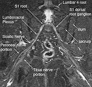
(the nerves between the cervical spine and the underarm that innervate shoulder, arm and hand), the lumbosacral plexus
Lumbosacral plexus
The anterior divisions of the lumbar nerves, sacral nerves, and coccygeal nerve form the lumbosacral plexus, the first lumbar nerve being frequently joined by a branch from the twelfth thoracic. For descriptive purposes this plexus is usually divided into three parts:* lumbar plexus* sacral plexus*...
(nerves between the lumbosacral spine and legs), the sciatic nerve
Sciatic nerve
The sciatic nerve is a large nerve fiber in humans and other animals. It begins in the lower back and runs through the buttock and down the lower limb...
in the pelvis, as well as other nerves such as the pudendal nerve
Pudendal nerve
The pudendal nerve is a sensory and somatic nerve in the pelvic region which is a large branch of the sacral plexus that innervates the external genitalia of both sexes, as well as sphincters for the bladder and the rectum...
that follow deep or complex courses.
Neurography has also been helpful for improving image diagnosis in spine disorders. It can help identify which spinal nerve is actually irritated as a supplement to routine spinal MRI. Standard spinal MRI only demonstrates the anatomy and numerous disk bulges
Spinal disc herniation
A spinal disc herniation , informally and misleadingly called a "slipped disc", is a medical condition affecting the spine due to trauma, lifting injuries, or idiopathic, in which a tear in the outer, fibrous ring of an intervertebral disc allows the soft, central portion A spinal disc herniation...
, bone spur
Bone spur
Osteophytes, commonly referred to as bone spurs, are bony projections that form along joint margins. They should not be confused with enthesophytes, which are bony projections which form at the attachment of a tendon or ligament.-Pathophysiology:...
s or stenoses
Stenosis
A stenosis is an abnormal narrowing in a blood vessel or other tubular organ or structure.It is also sometimes called a stricture ....
that may or may not actually cause nerve impingement symptoms.
Many nerves, such as the median
Median nerve
The median nerve is a nerve in humans and other animals. It is in the upper limb. It is one of the five main nerves originating from the brachial plexus....
and ulnar nerve
Ulnar nerve
In human anatomy, the ulnar nerve is a nerve which runs near the ulna bone. The ulnar collateral ligament of elbow joint is in relation with the ulnar nerve. The nerve is the largest unprotected nerve in the human body , so injury is common...
in the arm or the tibial nerve
Tibial nerve
The tibial nerve is a branch of the sciatic nerve. The tibial nerve passes through the popliteal fossa to pass below the arch of soleus.In the popliteal fossa the nerve gives off branches to gastrocnemius, popliteus, soleus and plantaris muscles, an articular branch to the knee joint, and a...
in the tarsal tunnel
Tarsal tunnel
The tarsal tunnel is found along the inner leg behind the medial malleolus.The tarsal tunnel is made up of bone on the inside and the flexor retinaculum on the outside.-Nerve distribution:...
, are just below the skin surface and can be tested for pathology with electromyography
Electromyography
Electromyography is a technique for evaluating and recording the electrical activity produced by skeletal muscles. EMG is performed using an instrument called an electromyograph, to produce a record called an electromyogram. An electromyograph detects the electrical potential generated by muscle...
, but this technique has always been difficult to apply for deep proximal nerves. Magnetic resonance neurography has greatly expanded the efficacy of nerve diagnosis by allowing uniform evaluation of virtually any nerve in the body.
There are numerous reports dealing with specialized uses of magnetic resonance neurography for nerve pathology such as cervical radiculopathy
Radiculopathy
Radiculopathy is not a specific condition, but rather a description of a problem in which one or more nerves are affected and do not work properly . The emphasis is on the nerve root...
, guidance for nerve blocks, demonstration of cysts in nerves, carpal tunnel syndrome
Carpal tunnel syndrome
Carpal Tunnel Syndrome is an entrapment idiopathic median neuropathy, causing paresthesia, pain, and other symptoms in the distribution of the median nerve due to its compression at the wrist in the carpal tunnel. The pathophysiology is not completely understood but can be considered compression...
, and obstetrical brachial plexus palsy
Erb's palsy
Erb's palsy is a paralysis of the arm caused by injury to the upper group of the arm's main nerves, specifically the upper trunk C5-C6 is severed. These form part of the brachial plexus, comprising the ventral rami of spinal nerves C5-C8, and T1. These injuries arise most commonly, but not...
. In addition several formal large scale outcome trials carried out with high quality "Class A" methodology have been published that have verified the clinical efficacy and validity of MR Neurography.
Use of magnetic resonance neurography is increasing in neurology and neurosurgery as the implications of its value in diagnosing various causes of sciatica becomes more widespread. There are 1.5 million lumbar MRI scans performed in the US each year for sciatica, leading to surgery for a herniated disk in about 300,000 patients per year. Of these, about 100,000 surgeries fail. Therefore there is successful treatment for sciatica in just 200,000 and failure of diagnosis or treatment in up to 1.3 million annually in the US alone. The success rate of the paradigm of lumbar MRI and disk resection for treatment of sciatica is therefore about 15%(Filler 2005). Neurography has been applied increasingly to evaluate the distal nerve roots, lumbo-sacral plexus and proximal sciatic nerve in the pelvis and thigh to find other causes of sciatica. It is increasingly important for brachial plexus imaging and for the diagnosis of thoracic outlet syndrome. Research and development in the clinical use of diagnostic neurography has taken place at Johns Hopkins
Johns Hopkins Hospital
The Johns Hopkins Hospital is the teaching hospital and biomedical research facility of Johns Hopkins University School of Medicine, located in Baltimore, Maryland . It was founded using money from a bequest by philanthropist Johns Hopkins...
, the Mayo Clinic
Mayo Clinic
Mayo Clinic is a not-for-profit medical practice and medical research group specializing in treating difficult patients . Patients are referred to Mayo Clinic from across the U.S. and the world, and it is known for innovative and effective treatments. Mayo Clinic is known for being at the top of...
, UCLA, UCSF, Harvard, the University of Washington in Seattle
University of Washington
University of Washington is a public research university, founded in 1861 in Seattle, Washington, United States. The UW is the largest university in the Northwest and the oldest public university on the West Coast. The university has three campuses, with its largest campus in the University...
, University of London
University of London
-20th century:Shortly after 6 Burlington Gardens was vacated, the University went through a period of rapid expansion. Bedford College, Royal Holloway and the London School of Economics all joined in 1900, Regent's Park College, which had affiliated in 1841 became an official divinity school of the...
, and Oxford University (see references below) as well as through the Neurography Institute. Recent patent litigation concerning MR Neurography has led some unlicensed centers to discontinue offering the technique. Courses have been offered for radiologists at the annual meetings of the Radiological Society of North America (RSNA
Radiological Society of North America
The Radiological Society of North America, Inc. is a professional membership society committed to excellence in patient care through education and research...
), and at the International Society for Magnetic Resonance in Medicine and for surgeons at the annual meetings of the American Association of Neurological Surgeons
American Association of Neurological Surgeons
The American Association of Neurological Surgeons is a professional body based in the United States with more than 8,000 members worldwide. The AANS is dedicated to advancing the specialty of neurological surgery in order to provide the highest quality of neurosurgical care to the public...
and the Congress of Neurological Surgeons. The use of imaging for diagnosis of nerve disorders represents a change from the way most physicians were trained to practice over the past several decades, as older routine tests fail to identify the diagnosis for nerve related disorders. The New England Journal of Medicine
New England Journal of Medicine
The New England Journal of Medicine is an English-language peer-reviewed medical journal published by the Massachusetts Medical Society. It describes itself as the oldest continuously published medical journal in the world.-History:...
in July 2009 published a report on whole body neurography using a diffusion based neurography technique. In 2010, RadioGraphics - a publication of the Radiological Society of North America that serves to provide continuing medical education to radiologists - published an article series taking the position that Neurography has an important role in the evaluation of entrapment neuropathies.
Magnetic resonance neurography does not pose any diagnostic disadvantage relative to standard magnetic resonance imaging because neurography studies typically include high resolution standard MRI image series for anatomical reference along with the neurographic sequences. However, the patient will generally have a slightly longer time in the scanner compared to a routine MRI scan. Magnetic resonance neurography can only be performed in 1.5 tesla and 3 tesla cylindrical type scanners and can't really be done effectively in lower power "open" MR scanners - this can pose significant challenges for claustrophobic patients. Although it has been in use for fifteen years and is the subject of more than 150 research publications, most insurance companies still classify this test as experimental and may decline reimbursement, resulting in the need to file appeals. Patients in some plans obtain standard insurance coverage for this widely used procedure.