

Active zone
Encyclopedia
The active zone is a term first used by Couteaux and Pecot-Dechavassinein in 1970 and is defined in the neuron as the site of neurotransmitter
release. Neurons contain structures called synapses that allow for the communication from one neuron to another. These synapses like the one diagrammed on the right contain a structure (top of picture) called a presynaptic bouton that stores vesicles
containing neurotransmitter and releases the contents of those vesicles upon the arrival of an action potential
. This neurotransmitter released by the presynaptic neuron then travels to the postsynaptic neuron (neuron on bottom) and activates receptors on the membrane of this neuron. The active zone is a region in the presynaptic bouton that mediates neurotransmitter release and is composed of the presynaptic membrane and a dense collection of proteins called the cytomatrix at the active zone (CAZ). The CAZ is identified in a electron microscope as a darkened (electron dense) area close to the presynaptic membrane. The proteins within the CAZ tether synaptic vesicles to the presynaptic membrane and mediate synaptic vesicle fusion to allow for neurotransmitter release. The machinery within the CAZ ensures that synaptic vesicles will be reliably and rapidly released upon the arrival of an action potential.
As an action potential
propagates down an axon it reaches the axon terminal called the presynaptic bouton. In the presynaptic bouton, the action potential activates calcium channels
(VDCCs) that cause a local influx of calcium. The increase in calcium is detected by proteins in the active zone and forces vesicles containing neurotransmitter to fuse with the membrane. This fusion of the vesicles with the membrane releases the contents of the vesicle into the synaptic cleft (space between the presynaptic bouton and the postsynaptic membrane). In the synapse the contents of the vesicles are neurotransmitters. These neurotransmitters diffuse across the cleft and bind to ligand gated ion channels and G-protein coupled receptors on the postsynaptic membrane. The binding of neurotransmitters to the postsynaptic receptors then induces a change in the postsynaptic neuron. The process of releasing neurotransmitter and binding to the postsynaptic receptor to cause a change in the postsynaptic neuron is called neurotransmission.
There are at least five major scaffold proteins that are enriched in the active zone; UNC13/Munc13, RIMs (Rab3-interacting molecule), Bassoon, Piccolo/aczonin, ELKS, and liprins-α. These scaffold proteins are thought to be the constituents of the dense pyramid like structures of the active zone and are thought to bring the synaptic vesicles into close proximity to the presynaptic membrane and the calcium channels. The protein ELKS binds to the cell adhesion
protein, β-neurexin
, and other proteins within the complex such as Piccolo and Bassoon. β-neurexin then binds to cell adhesion molecule, neuroligin
located on the postsynaptic membrane. Neuroligin then interacts with proteins that bind to postsynaptic receptors. Protein interactions like that seen between Piccolo/ELKS/β-neurexin/neuroligin ensures that machinery that mediates vesicle fusion is in close proximity to calcium channels and that vesicle fusion is adjacent to postsynaptic receptors. This close proximity vesicle fusion and postsynaptic receptors ensures that there is little delay between the activation of the postsynaptic receptors and the release of neurotransmitters.
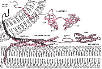
The release of neurotransmitter is accomplished by the fusion of neurotransmitter vesicles to the presynaptic membrane. Although the details of this mechanism are still being studied there is a consensus on some details of the process. Synaptic vesicle fusion with the presynaptic membrane is known to require a local increase of calcium and the formation of highly stable SNARE
complexes. One prevailing model of synaptic vesicle fusion is that SNARE complex formation is catalyzed by the proteins of the active zone such as Munc18, Munc13, and RIM. The formation of this complex is thought to "prime" the vesicle to be ready for vesicle fusion and release of neurotransmitter(see below: releasable pool). After the vesicle is primed then complexin
binds to the SNARE complex this is called "superprimed." The vesicles that are superprimed are within the readily releasable pool(see below) and are ready to be rapidly released. The arrival of an action potential opens voltage gated calcium channels near the SNARE/complexin complex. Calcium then binds to changes the conformation of synaptotagmin
. This change in conformation of allows synaptotagmin to then dislodge complexin, bind to the SNARE complex, and bind to the target membrane. When synaptotagmin binds to both the SNARE complex and the membrane this induces a mechanical force on the membrane so that it causes the vesicle membrane and presynaptic membrane to fuse. This fusion opens a membrane pore that releases the neurotransmitter. The pore increases in size until the entire vesicle membrane is indistinguishable from the presynaptic membrane.
is pinched off to form the synaptic vesicle and this vesicle is transported to the synaptic terminal. At the terminal (2) the vesicle is filled with neurotransmitter. (3) The vesicle is transported to the active zone and docked in close proximity to the plasma membrane. (4) During an action potential the vesicle is fuses with the membrane, releases the neurotransmitter and allows the membrane proteins previously on the vesicle to diffuse to the peri-active zone. (5) In the peri-active zone the membrane proteins are sequestered and are endocytosed
forming a clathrin
coated vesicle. (6) The vesicle is then filled with neurotransmitter and is then transported back to the active zone.
The endocytosis mechanism is slower than the exocytosis mechanism. This means that in intense activity the vesicle in the terminal can become depleted and no longer available to be released. To help prevent the depletion of synaptic vesicles the increase in calcium during intense activity can activate calcineurin
which dephosphorylate
proteins involved in clathrin-mediated endocytosis.
proteins. These vesicles are ready to release by a single action potential and are replenished by vesicles from the reserve pool. The releasable pool is sometimes subdivided into the readily releasable pool and the releasable pool.
. Calcineurin dephosphorylates a protein, synapsin
, that mediates the clustering of the reserve pool vesicles. Dephosphorylation of synapsin mobilize vesicles in the reserve pool and allows the vesicles to migrate to the active zone and replenish the readily releasable pool.
recruit proteins that mediate endocytotis such as dynamin, clathrin and endophilin. In Drosophilia the intersectin homolog, Dap160, is located in the periactive zone of the neuromuscular junction and mutant Dap160 deplete synaptic vesicles during high frequency stimulation.
after triggering an action potential in the presynaptic neuron. Measuring neurotransmitter release this way can be problematic because the effect of the postsynaptic neuron to the same amount of released neurotransmitter can change over time. Another way is to measure vesicle fusion with the presynaptic membrane directly using a patch pipette
. A cell membrane can be thought of as a capacitor
in that positive and negative ions are stored on both sides of the membrane. The larger the area of membrane the more ions that are necessary to hold the membrane at a certain potential. In electrophysiology this means that a current injection into the terminal will take less time to charge a membrane to a given potential before vesicle fusion than it will after vesicle fusion. The time course to charge the membrane to a potential and the resistance of the membrane is measured and with these values the capacitance of the membrane can be calculated by the equation Tau/Resistance=Capacitance. With this technique researchers can measure synaptic vesicle release directly by measuring increases in the membrane capacitance of the presynaptic terminal.
Neurotransmitter
Neurotransmitters are endogenous chemicals that transmit signals from a neuron to a target cell across a synapse. Neurotransmitters are packaged into synaptic vesicles clustered beneath the membrane on the presynaptic side of a synapse, and are released into the synaptic cleft, where they bind to...
release. Neurons contain structures called synapses that allow for the communication from one neuron to another. These synapses like the one diagrammed on the right contain a structure (top of picture) called a presynaptic bouton that stores vesicles
Vesicle (biology)
A vesicle is a bubble of liquid within another liquid, a supramolecular assembly made up of many different molecules. More technically, a vesicle is a small membrane-enclosed sack that can store or transport substances. Vesicles can form naturally because of the properties of lipid membranes , or...
containing neurotransmitter and releases the contents of those vesicles upon the arrival of an action potential
Action potential
In physiology, an action potential is a short-lasting event in which the electrical membrane potential of a cell rapidly rises and falls, following a consistent trajectory. Action potentials occur in several types of animal cells, called excitable cells, which include neurons, muscle cells, and...
. This neurotransmitter released by the presynaptic neuron then travels to the postsynaptic neuron (neuron on bottom) and activates receptors on the membrane of this neuron. The active zone is a region in the presynaptic bouton that mediates neurotransmitter release and is composed of the presynaptic membrane and a dense collection of proteins called the cytomatrix at the active zone (CAZ). The CAZ is identified in a electron microscope as a darkened (electron dense) area close to the presynaptic membrane. The proteins within the CAZ tether synaptic vesicles to the presynaptic membrane and mediate synaptic vesicle fusion to allow for neurotransmitter release. The machinery within the CAZ ensures that synaptic vesicles will be reliably and rapidly released upon the arrival of an action potential.
Function
The function of the active zone is to ensure that neurotransmitters can be reliably released in a specific location of a neuron and only released when the neuron fires an action potential.As an action potential
Action potential
In physiology, an action potential is a short-lasting event in which the electrical membrane potential of a cell rapidly rises and falls, following a consistent trajectory. Action potentials occur in several types of animal cells, called excitable cells, which include neurons, muscle cells, and...
propagates down an axon it reaches the axon terminal called the presynaptic bouton. In the presynaptic bouton, the action potential activates calcium channels
Voltage-dependent calcium channel
Voltage-dependent calcium channels are a group of voltage-gated ion channels found in excitable cells with a permeability to the ion Ca2+...
(VDCCs) that cause a local influx of calcium. The increase in calcium is detected by proteins in the active zone and forces vesicles containing neurotransmitter to fuse with the membrane. This fusion of the vesicles with the membrane releases the contents of the vesicle into the synaptic cleft (space between the presynaptic bouton and the postsynaptic membrane). In the synapse the contents of the vesicles are neurotransmitters. These neurotransmitters diffuse across the cleft and bind to ligand gated ion channels and G-protein coupled receptors on the postsynaptic membrane. The binding of neurotransmitters to the postsynaptic receptors then induces a change in the postsynaptic neuron. The process of releasing neurotransmitter and binding to the postsynaptic receptor to cause a change in the postsynaptic neuron is called neurotransmission.
Structure
The active zone is present in all chemical synapses examined so far and is present in all animal species. The active zones examined so far have at least two features in common, they all have protein dense material that project from the membrane and tethers synaptic vesicles close to the membrane and they have long filamentous projections originating at the membrane and terminating at vesicles slightly farther from the presynaptic membrane. The protein dense projections vary in size and shape depending on the type of synapse examined. One striking example of the dense projection is the ribbon synapse (see below) which contains a "ribbon" of protein dense material that is surrounded by a halo of synaptic vesicles and extends perpendicular to the presynaptic membrane and can be as long as 500 nm! The glutamate synapse contains smaller pyramid like structures that extend about 50 nm from the membrane. The neuromuscular synapse contains two rows of vesicles with a long proteinaceous band between them that is connected to regularly spaced horizontal ribs extending perpendicular to the band and parallel with the membrane. These ribs are then connected to the vesicles which are each positioned above a peg in the membrane (presumably a calcium channel). Previous research indicated that the active zone of glutamatergic neurons contained a highly regular array of pyramid shaped protein dense material and indicated that these pyramids were connected by filaments. This structure resembled a geometric lattice where vesicles were guided into holes of the lattic. This attractive model has come into question by recent experiments. Recent data shows that the glutamatergic active zone does contain the dense protein material projections but these projections were not in a regular array and contained long filaments projecting about 80 nm into the cytoplasm.There are at least five major scaffold proteins that are enriched in the active zone; UNC13/Munc13, RIMs (Rab3-interacting molecule), Bassoon, Piccolo/aczonin, ELKS, and liprins-α. These scaffold proteins are thought to be the constituents of the dense pyramid like structures of the active zone and are thought to bring the synaptic vesicles into close proximity to the presynaptic membrane and the calcium channels. The protein ELKS binds to the cell adhesion
Cell adhesion
Cellular adhesion is the binding of a cell to a surface, extracellular matrix or another cell using cell adhesion molecules such as selectins, integrins, and cadherins. Correct cellular adhesion is essential in maintaining multicellular structure...
protein, β-neurexin
NRXN1
Neurexin-1-alpha is a protein that in humans is encoded by the NRXN1 gene.-Genomics:The gene is found in a single copy on the short arm of chromosome 2 . The gene is 1,112,187 bases in length, is located on the Crick strand and encodes a protein of 1,477 amino acids .-Further reading:...
, and other proteins within the complex such as Piccolo and Bassoon. β-neurexin then binds to cell adhesion molecule, neuroligin
Neuroligin
300px|thumb|right|alt = Colored dice with checkered background|Tertiary structure of Neuroligin 4.Neuroligin , a type I membrane protein, is a protein on the postsynaptic membrane that mediates synapse formation between neurons. Neuroligins mediate signaling across the synapse and affect the...
located on the postsynaptic membrane. Neuroligin then interacts with proteins that bind to postsynaptic receptors. Protein interactions like that seen between Piccolo/ELKS/β-neurexin/neuroligin ensures that machinery that mediates vesicle fusion is in close proximity to calcium channels and that vesicle fusion is adjacent to postsynaptic receptors. This close proximity vesicle fusion and postsynaptic receptors ensures that there is little delay between the activation of the postsynaptic receptors and the release of neurotransmitters.
Neurotransmitter release mechanism
The release of neurotransmitter is accomplished by the fusion of neurotransmitter vesicles to the presynaptic membrane. Although the details of this mechanism are still being studied there is a consensus on some details of the process. Synaptic vesicle fusion with the presynaptic membrane is known to require a local increase of calcium and the formation of highly stable SNARE
Snare
Snare may refer to:* Snare trap, a kind of trap used for capturing animals* Snare drum* SNARE , a family of proteins involved in vesicle fusion* The Snares, a group of islands approximately 200 kilometres south of New Zealand...
complexes. One prevailing model of synaptic vesicle fusion is that SNARE complex formation is catalyzed by the proteins of the active zone such as Munc18, Munc13, and RIM. The formation of this complex is thought to "prime" the vesicle to be ready for vesicle fusion and release of neurotransmitter(see below: releasable pool). After the vesicle is primed then complexin
Complexin
Complexin is a cytoplasmic nerve tissue protein which binds to the SNARE protein complex with a high affinity. The transport vesicle protein synaptotagmin, among others, in the presence of Ca2+, displaces complexin allowing the SNARE protein complex to bind the transport vesicle to the...
binds to the SNARE complex this is called "superprimed." The vesicles that are superprimed are within the readily releasable pool(see below) and are ready to be rapidly released. The arrival of an action potential opens voltage gated calcium channels near the SNARE/complexin complex. Calcium then binds to changes the conformation of synaptotagmin
Synaptotagmin
Synaptotagmins constitute a family of membrane-trafficking proteins that are characterized by an N-terminal transmembrane region , a variable linker, and two C-terminal C2 domains - C2A and C2B. There are 15 members in the mammalian synaptotagmin family...
. This change in conformation of allows synaptotagmin to then dislodge complexin, bind to the SNARE complex, and bind to the target membrane. When synaptotagmin binds to both the SNARE complex and the membrane this induces a mechanical force on the membrane so that it causes the vesicle membrane and presynaptic membrane to fuse. This fusion opens a membrane pore that releases the neurotransmitter. The pore increases in size until the entire vesicle membrane is indistinguishable from the presynaptic membrane.
Synaptic vesicle cycle
The presynaptic bouton has an efficiently orchestrated process to release fuse vesicles to the presynaptic membrane to release neurotransmitter and regenerate neurotransmitter vesicles. This process called the synaptic vesicle cycle maintains the number of vesicles in the presynaptic bouton and allows the synaptic terminal to be an autonomous unit. The cycle begins with (1) a region of the golgi apparatusGolgi apparatus
The Golgi apparatus is an organelle found in most eukaryotic cells. It was identified in 1898 by the Italian physician Camillo Golgi, after whom the Golgi apparatus is named....
is pinched off to form the synaptic vesicle and this vesicle is transported to the synaptic terminal. At the terminal (2) the vesicle is filled with neurotransmitter. (3) The vesicle is transported to the active zone and docked in close proximity to the plasma membrane. (4) During an action potential the vesicle is fuses with the membrane, releases the neurotransmitter and allows the membrane proteins previously on the vesicle to diffuse to the peri-active zone. (5) In the peri-active zone the membrane proteins are sequestered and are endocytosed
Endocytosis
Endocytosis is a process by which cells absorb molecules by engulfing them. It is used by all cells of the body because most substances important to them are large polar molecules that cannot pass through the hydrophobic plasma or cell membrane...
forming a clathrin
Clathrin
Clathrin is a protein that plays a major role in the formation of coated vesicles. Clathrin was first isolated and named by Barbara Pearse in 1975. It forms a triskelion shape composed of three clathrin heavy chains and three light chains. When the triskelia interact they form a polyhedral lattice...
coated vesicle. (6) The vesicle is then filled with neurotransmitter and is then transported back to the active zone.
The endocytosis mechanism is slower than the exocytosis mechanism. This means that in intense activity the vesicle in the terminal can become depleted and no longer available to be released. To help prevent the depletion of synaptic vesicles the increase in calcium during intense activity can activate calcineurin
Calcineurin
Calcineurin is a protein phosphatase also known as protein phosphatase 3, PPP3CA, and calcium-dependent serine-threonine phosphatase, and formerly known as protein phosphatase 2B . It activates the T cells of the immune system and can be blocked by drugs...
which dephosphorylate
Dephosphorylation
Dephosphorylation is the essential process of removing phosphate groups from an organic compound by hydrolysis. Its opposite is phosphorylation...
proteins involved in clathrin-mediated endocytosis.
Vesicle pools
The synapse contains at least two clusters of synaptic vesicles, the readily releasable pool and the reserve pool. The readily releasable pool is located within the active zone and connected directly to the presynaptic membrane while the reserve pool is clustered by cytoskeletal and is not directly connected to the active zone.Releasable pool
The releasable pool is located in the active zone and is bound directly to the presynpatic membrane. It is stabilized by proteins within the active zone and bound to the presynaptic membrane by SNARESNARE (protein)
SNARE proteins are a large protein superfamily consisting of more than 60 members in yeast and mammalian cells....
proteins. These vesicles are ready to release by a single action potential and are replenished by vesicles from the reserve pool. The releasable pool is sometimes subdivided into the readily releasable pool and the releasable pool.
Reserve Pool
The reserve pool is not directly connected to the active zone. The increase in presynaptic calcium concentration activates the calcium sensitive phosphatase, calcineurinCalcineurin
Calcineurin is a protein phosphatase also known as protein phosphatase 3, PPP3CA, and calcium-dependent serine-threonine phosphatase, and formerly known as protein phosphatase 2B . It activates the T cells of the immune system and can be blocked by drugs...
. Calcineurin dephosphorylates a protein, synapsin
Synapsin
The synapsins are a family of proteins that have long been implicated in the regulation of neurotransmitter release at synapses. Specifically, they are thought to be involved in regulating the number of synaptic vesicles available for release via exocytosis at any one time.Current studies suggest...
, that mediates the clustering of the reserve pool vesicles. Dephosphorylation of synapsin mobilize vesicles in the reserve pool and allows the vesicles to migrate to the active zone and replenish the readily releasable pool.
Periactive zone
The periactive zone surrounds the active zone and is the site of endocytosis of the presynaptic terminal. In the periactive zone, scaffolding proteins such as intersectin 1ITSN1
Intersectin-1 is a protein that, in humans, is encoded by the ITSN1 gene.-Interactions:ITSN1 has been shown to interact with SNAP-25, SOS1, SCAMP1 and CDC42.-Further reading:...
recruit proteins that mediate endocytotis such as dynamin, clathrin and endophilin. In Drosophilia the intersectin homolog, Dap160, is located in the periactive zone of the neuromuscular junction and mutant Dap160 deplete synaptic vesicles during high frequency stimulation.
Ribbon Synapse Active Zone
The ribbon synapse is a special type of synapse found in sensory neurons such as photoreceptor cells, retinal bipolar cells, and hair cells. Ribbon synapses contain a dense protein structure that tethers an array of vesicles perpendicular to the presynaptic membrane. In an electron micrograph it appears as a ribbon like structure perpendicular to the membrane. Unlike the 'traditional' synapse, ribbon synapses can maintain a graded release of vesicles. In other words the more depolarized a neuron the higher the rate of vesicle fusion. The Ribbon synapse active zone is separated into two regions, the archiform density and the ribbon. The archiform density is the site of vesicle fusion and the ribbon stores the releasable pool of vesicles. The ribbon structure is composed primarily of the protein RIBEYE, about 64-69% of the ribbon volume, and is tethered to the archiform density by scaffolding proteins such as Bassoon.Proteins of the Active Zone
| Protein | Structure/Function |
| Structural Proteins | |
| Piccolo PCLO Protein piccolo is a protein that in humans is encoded by the PCLO gene.-Further reading:... |
|
| Bassoon | |
| RIMs RIMS1 Regulating synaptic membrane exocytosis protein 1 is a protein that in humans is encoded by the RIMS1 gene.-Interactions:RIMS1 has been shown to interact with UNC13B, RAB3A, ERC2 and YWHAH.-Further reading:... |
|
| ELKS (ERCs or CAST) ERC2 (gene) ERC protein 2 is a protein that in humans is encoded by the ERC2 gene.-Interactions:ERC2 has been shown to interact with PPFIA4, RIMS1 and liprin-alpha-1.-Further reading:... |
|
| CASK CASK Peripheral plasma membrane protein CASK is a protein that in humans is encoded by the CASK gene. This gene is also known by several other names: CMG 2 , calcium/calmodulin-dependent serine protein kinase 3 and membrane-associated guanylate kinase 2.-Genomics:This gene is located on the short arm of... |
|
| Mint | |
| Liprin-alpha-1 | |
| Docking and Priming | |
| Munc-13 UNC13B Protein unc-13 homolog B is a protein that in humans is encoded by the UNC13B gene.-Interactions:UNC13B has been shown to interact with STX1B, STX1A, RIMS1, SPTBN2 and DOC2A.-Further reading:... |
|
| Munc-18 Munc-18 Munc-18 protein is the mammalian homologue of the unc-18 protein and is a member of the Sec1/Munc18-like protein family... |
|
| SNAREs SNARE (protein) SNARE proteins are a large protein superfamily consisting of more than 60 members in yeast and mammalian cells.... |
|
| SNAP25 | |
| VAMP2 VAMP2 Vesicle-associated membrane protein 2 is a protein that in humans is encoded by the VAMP2 gene.-Interactions:VAMP2 has been shown to interact with STX4, SNAP-25, SNAP23, STX1A and RABAC1.-Further reading:... |
|
| syntaxin Syntaxin Syntaxins are a family of membrane integrated Q-SNARE proteins participating in exocytosis.- Domains :Syntaxins possess a single C-terminal transmembrane domain, a SNARE domain , and an N-terminal regulatory domain .... |
Located on the synaptic membrane and binds to SNAP-25 and synaptobrevin to mediate vesicle fusion. |
| Cytoskeletal Proteins | |
| Actin Actin Actin is a globular, roughly 42-kDa moonlighting protein found in all eukaryotic cells where it may be present at concentrations of over 100 μM. It is also one of the most highly-conserved proteins, differing by no more than 20% in species as diverse as algae and humans... |
|
| Tubulin Tubulin Tubulin is one of several members of a small family of globular proteins. The most common members of the tubulin family are α-tubulin and β-tubulin, the proteins that make up microtubules. Each has a molecular weight of approximately 55 kiloDaltons. Microtubules are assembled from dimers of α- and... |
|
| myosin Myosin Myosins comprise a family of ATP-dependent motor proteins and are best known for their role in muscle contraction and their involvement in a wide range of other eukaryotic motility processes. They are responsible for actin-based motility. The term was originally used to describe a group of similar... |
|
| spectrin Spectrin Spectrin is a cytoskeletal protein that lines the intracellular side of the plasma membrane of many cell types in pentagonal or hexagonal arrangements, forming a scaffolding and playing an important role in maintenance of plasma membrane integrity and cytoskeletal structure... |
|
| β-catenin | |
| Calcium Channel | |
| Voltage-dependent calcium channel Voltage-dependent calcium channel Voltage-dependent calcium channels are a group of voltage-gated ion channels found in excitable cells with a permeability to the ion Ca2+... (VDCC) |
Allows the rapid influx of calcium during an action potential. |
Measuring Neurotransmitter Release
Neurotransmitter release can be measured by determining the amplitude of the postsynaptic potentialPostsynaptic potential
Postsynaptic potentials are changes in the membrane potential of the postsynaptic terminal of a chemical synapse. Postsynaptic potentials are graded potentials, and should not be confused with action potentials although their function is to initiate or inhibit action potentials...
after triggering an action potential in the presynaptic neuron. Measuring neurotransmitter release this way can be problematic because the effect of the postsynaptic neuron to the same amount of released neurotransmitter can change over time. Another way is to measure vesicle fusion with the presynaptic membrane directly using a patch pipette
Patch clamp
The patch clamp technique is a laboratory technique in electrophysiology that allows the study of single or multiple ion channels in cells. The technique can be applied to a wide variety of cells, but is especially useful in the study of excitable cells such as neurons, cardiomyocytes, muscle...
. A cell membrane can be thought of as a capacitor
Capacitor
A capacitor is a passive two-terminal electrical component used to store energy in an electric field. The forms of practical capacitors vary widely, but all contain at least two electrical conductors separated by a dielectric ; for example, one common construction consists of metal foils separated...
in that positive and negative ions are stored on both sides of the membrane. The larger the area of membrane the more ions that are necessary to hold the membrane at a certain potential. In electrophysiology this means that a current injection into the terminal will take less time to charge a membrane to a given potential before vesicle fusion than it will after vesicle fusion. The time course to charge the membrane to a potential and the resistance of the membrane is measured and with these values the capacitance of the membrane can be calculated by the equation Tau/Resistance=Capacitance. With this technique researchers can measure synaptic vesicle release directly by measuring increases in the membrane capacitance of the presynaptic terminal.
See also
- Chemical synapseChemical synapseChemical synapses are specialized junctions through which neurons signal to each other and to non-neuronal cells such as those in muscles or glands. Chemical synapses allow neurons to form circuits within the central nervous system. They are crucial to the biological computations that underlie...
- NeurotransmitterNeurotransmitterNeurotransmitters are endogenous chemicals that transmit signals from a neuron to a target cell across a synapse. Neurotransmitters are packaged into synaptic vesicles clustered beneath the membrane on the presynaptic side of a synapse, and are released into the synaptic cleft, where they bind to...
- Neurotransmitter vesicle
- Vesicle fusionVesicle fusionVesicle fusion is the merging of a vesicle with other vesicles or a part of a cell membrane. In the latter case, it is the end stage of secretion from secretory vesicles, where their contents are expelled from the cell through exocytosis at the porosome...
- ExocytosisExocytosisExocytosis , also known as 'The peni-cytosis', is the durable process by which a cell directs the contents of secretory vesicles out of the cell membrane...
- Paired Pulse FacilitationNeural facilitationNeural facilitation, also known as paired pulse facilitation, is a concept in neuroscience where an increase in the postsynaptic potential is evoked by a second impulse....