
Sliding filament mechanism
Encyclopedia
- Ratchet mechanism redirects here (it is the name given in some textbooks to the sliding filament mechanism). For the mechanical device, see Ratchet (device)Ratchet (device)A ratchet is a device that allows continuous linear or rotary motion in only one direction while preventing motion in the opposite direction. Because most socket wrenches today use ratcheting handles, the term "ratchet" alone is often used to refer to a ratcheting wrench, and the terms "ratchet"...
The sliding filament theory describes a process used by muscle
Muscle
Muscle is a contractile tissue of animals and is derived from the mesodermal layer of embryonic germ cells. Muscle cells contain contractile filaments that move past each other and change the size of the cell. They are classified as skeletal, cardiac, or smooth muscles. Their function is to...
s to contract
Muscle contraction
Muscle fiber generates tension through the action of actin and myosin cross-bridge cycling. While under tension, the muscle may lengthen, shorten, or remain the same...
. It was independently developed by Andrew F. Huxley and Rolf Niedergerke and by Hugh Huxley
Hugh Huxley
Hugh Esmor Huxley FRS is a British biologist. He is professor of biology at Brandeis University, in Waltham, Massachusetts, United States....
and Jean Hanson in 1954.
Process of movement
Myosin
Myosins comprise a family of ATP-dependent motor proteins and are best known for their role in muscle contraction and their involvement in a wide range of other eukaryotic motility processes. They are responsible for actin-based motility. The term was originally used to describe a group of similar...
is a molecular motor that acts like an active ratchet
Ratchet (device)
A ratchet is a device that allows continuous linear or rotary motion in only one direction while preventing motion in the opposite direction. Because most socket wrenches today use ratcheting handles, the term "ratchet" alone is often used to refer to a ratcheting wrench, and the terms "ratchet"...
. Chains of actin
Actin
Actin is a globular, roughly 42-kDa moonlighting protein found in all eukaryotic cells where it may be present at concentrations of over 100 μM. It is also one of the most highly-conserved proteins, differing by no more than 20% in species as diverse as algae and humans...
proteins form high tensile passive 'thin' filaments that transmit the force generated by myosin to the ends of the muscle. Myosin also forms 'thick' filaments. Each myosin 'paddles' along an actin filament repeatedly binding, ratcheting and letting go, sliding the thick filament over the thin filament.
Calcium ions are released. This calcium bonds to troponin, allowing the myosin head to bind with the binding site.
- MyosinMyosinMyosins comprise a family of ATP-dependent motor proteins and are best known for their role in muscle contraction and their involvement in a wide range of other eukaryotic motility processes. They are responsible for actin-based motility. The term was originally used to describe a group of similar...
heads bind to the passive actinActinActin is a globular, roughly 42-kDa moonlighting protein found in all eukaryotic cells where it may be present at concentrations of over 100 μM. It is also one of the most highly-conserved proteins, differing by no more than 20% in species as diverse as algae and humans...
filaments at the myosin binding siteBinding siteIn biochemistry, a binding site is a region on a protein, DNA, or RNA to which specific other molecules and ions—in this context collectively called ligands—form a chemical bond...
s. - Upon strong binding, myosin and actin undergo an isomerization (myosin rotates at the myosin-actin interface) extending an extensible region in the neck of the myosin head.
- Shortening occurs when the extensible region pulls the filaments across each other (like the shortening of a spring). Myosin remains attached to the actin.
- The binding of ATPAdenosine triphosphateAdenosine-5'-triphosphate is a multifunctional nucleoside triphosphate used in cells as a coenzyme. It is often called the "molecular unit of currency" of intracellular energy transfer. ATP transports chemical energy within cells for metabolism...
allows myosin to detach from actin. While detached, ATP hydrolysis occurs "recharging" the myosin head. If the actin binding sites are still available, myosin can bind actin again. - The collective bending of numerous myosin heads (all in the same direction), combine to move the actin filament relative to the myosin filament. This results in muscle contraction.
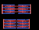
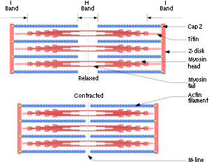
All muscle cells are composed of a number of actin and myosin filaments in series. The basic unit of organisation of these contractile proteins in striated muscle cells (i.e., the cells that compose cardiac and skeletal muscle, but not in smooth muscle tissue) is called the sarcomere
Sarcomere
A sarcomere is the basic unit of a muscle. Muscles are composed of tubular muscle cells . Muscle cells are composed of tubular myofibrils. Myofibrils are composed of repeating sections of sarcomeres, which appear under the microscope as dark and light bands...
. It consists of a central bidirectional thick filament flanked by two actin filaments, orientated in opposite directions. When each end of the myosin thick filament ratchets along the actin filament with which it overlaps, the two actin filaments are drawn closer together. Thus, the ends of the sarcomere
Sarcomere
A sarcomere is the basic unit of a muscle. Muscles are composed of tubular muscle cells . Muscle cells are composed of tubular myofibrils. Myofibrils are composed of repeating sections of sarcomeres, which appear under the microscope as dark and light bands...
are drawn in and the sarcomere shortens. Sarcomeres are connected together by so-called 'Z lines', which anchor the ends of actin filaments in such a way that the filaments on each side of the Z line point in opposite directions (with reversed polarity). By this means, sarcomeres are arranged in series. When a muscle fiber contracts, all sarcomeres contract simultaneously so that force is transmitted to the fiber ends.
Physiologically, this contraction is not uniform across the sarcomere; the central position of the thick filaments becomes unstable and can shift during contraction. However the actions of elastic proteins such as Titin
Titin
Titin , also known as connectin, is a protein that in humans is encoded by the TTN gene. Titin is a giant protein that functions as a molecular spring which is responsible for the passive elasticity of muscle. It is composed of 244 individually folded protein domains connected by unstructured...
are hypothesised to maintain uniform tension across the sarcomere and pull the thick filament into a central position.
Pre-process of movement
If the process of movement were to continue constantly, all muscles would constantly be contracted. Therefore, the body needs a way to control the ability of myosin to bind to the actin. This is accomplished by the introduction of calcium into the cytoplasmCytoplasm
The cytoplasm is a small gel-like substance residing between the cell membrane holding all the cell's internal sub-structures , except for the nucleus. All the contents of the cells of prokaryote organisms are contained within the cytoplasm...
of the muscle cell.
- When the muscle does not need to contract (is in a resting state), thin strands of a protein called tropomyosinTropomyosinTropomyosin is an actin-binding protein that regulates actin mechanics. It is important, among other things, for muscle contraction. Tropomyosin, along with the troponin complex, associate with actin in muscle fibers and regulate muscle contraction by regulating the binding of myosin...
are wrapped around the actin filaments, blocking the myosin binding sites. This inhibits the myosin from binding to actin, and therefore causes a chain of events leading to muscle relaxation. - Molecules called troponinTroponin400px|thumb|right|alt = Colored dice with checkered background|Ribbon representation of the human cardiac troponin core complex in the calcium-saturated form...
are attached to the tropomyosin. - When calcium is introduced into the muscle cell (fiber), calcium ions bind to troponin molecules.
- Calcium binding changes the shape of troponin, causing tropomyosin to be moved deeper into the groove of the actin dimer, therefore causing the myosin binding sites on the actin to be exposed.
- Myosin binds to the now-exposed binding sites, and muscles contract via the sliding-filament mechanism.
Nerve impulses affect the way in which calcium bonds to the troponin.