
Sarcomere
Encyclopedia
A sarcomere is the basic unit of a muscle
. Muscles are composed of tubular muscle cells (myocytes or myofibers). Muscle cells are composed of tubular myofibrils. Myofibrils are composed of repeating sections of sarcomeres, which appear under the microscope as dark and light bands. Sarcomeres are composed of long, fibrous proteins that slide past each other when the muscles contract and relax.
Two of the important proteins are myosin
, which forms the thick filament, and actin
, which forms the thin filament. Myosin has a long, fibrous tail and a globular head, which binds to actin. The myosin head also binds to ATP
, which is the source of energy for muscle movement. Myosin can only bind to actin when the binding sites on actin are exposed by calcium ions.
Actin molecules are bound to the Z line, which forms the borders of the sarcomere. Other bands appear when the sarcomere is relaxed.
A muscle cell from a biceps
may contain 100,000 sarcomeres. The myofibrils of smooth muscle
cells are not arranged into sarcomeres.
 The sarcomeres are what give skeletal and cardiac muscle
s their striated appearance.
The relationship between the proteins and the regions of the sarcomere are as follows:
The protein tropomyosin
covers the myosin binding sites of the actin molecules in the muscle cell. To allow the muscle cell to contract, tropomyosin must be moved to uncover the binding sites on the actin. Calcium ions bind with troponin-C molecules (which are dispersed throughout the tropomyosin protein) and alter the structure of the tropomyosin, forcing it to reveal the cross bridge binding site on the actin. The concentration of calcium within muscle cells is controlled by the sarcoplasmic reticulum, a unique form of endoplasmic reticulum. Muscle contraction ends when calcium ions are pumped back into the sarcoplasmic reticulum, allowing the contractile apparatus and thus muscle cell to relax.
During stimulation of the muscle cell, the motor neuron releases the neurotransmitter acetylcholine
, which travels across the neuromuscular junction (the synapse between the terminal bouton of the neuron and the muscle cell). Acetylcholine
binds to a post-synaptic nicotinic acetylcholine receptor
. A change in the receptor conformation allows an influx of sodium ions and initiation of a post-synaptic action potential. The action potential
then travels along T (transverse) tubules until it reaches the sarcoplasmic reticulum; the action potential from the motor neuron changes the permeability of the sarcoplasmic reticulum, allowing the flow of calcium ions into the sarcomere. The outflow of calcium allows the myosin heads access to the actin cross bridge binding sites, permitting muscle contraction.
molecule in a low-energy configuration and is unable to access the cross bridge binding sites on the actin. However, the myosin head can hydrolyze ATP into ADP and an inorganic phosphate ion. A portion of the energy released in this reaction changes the shape of the myosin head and promotes it to a high-energy configuration. Through the process of binding to the actin, the myosin head releases ADP and an inorganic phosphate ion, changing its configuration back to one of low energy. The myosin remains attached to actin in a state known as rigor, until a new ATP binds the myosin head. This binding of ATP to myosin releases the actin by cross-bridge dissociation. The ATP-associated myosin is ready for another cycle, beginning with hydrolysis of the ATP.
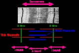
The A-band is visible as dark transverse lines across myofibers; the I-band is visible as lightly staining transverse lines, and the Z-line is visible as dark lines separating sarcomeres at the light-microscope level.
is creatine phosphate, which is used to provide ADP with a phosphate group for ATP synthesis in vertebrates.
s display a very limited range of sarcomere lengths, with roughly the same optimal length (length at peak length-tension) in all muscles of an individual as well as between species. Arthropods, however, show tremendous variation (over seven-fold) in sarcomere length, both between species and between muscles in a single individual. The reasons for the lack of substantial sarcomere variability in vertebrates is not fully known.
Muscle
Muscle is a contractile tissue of animals and is derived from the mesodermal layer of embryonic germ cells. Muscle cells contain contractile filaments that move past each other and change the size of the cell. They are classified as skeletal, cardiac, or smooth muscles. Their function is to...
. Muscles are composed of tubular muscle cells (myocytes or myofibers). Muscle cells are composed of tubular myofibrils. Myofibrils are composed of repeating sections of sarcomeres, which appear under the microscope as dark and light bands. Sarcomeres are composed of long, fibrous proteins that slide past each other when the muscles contract and relax.
Two of the important proteins are myosin
Myosin
Myosins comprise a family of ATP-dependent motor proteins and are best known for their role in muscle contraction and their involvement in a wide range of other eukaryotic motility processes. They are responsible for actin-based motility. The term was originally used to describe a group of similar...
, which forms the thick filament, and actin
Actin
Actin is a globular, roughly 42-kDa moonlighting protein found in all eukaryotic cells where it may be present at concentrations of over 100 μM. It is also one of the most highly-conserved proteins, differing by no more than 20% in species as diverse as algae and humans...
, which forms the thin filament. Myosin has a long, fibrous tail and a globular head, which binds to actin. The myosin head also binds to ATP
Adenosine triphosphate
Adenosine-5'-triphosphate is a multifunctional nucleoside triphosphate used in cells as a coenzyme. It is often called the "molecular unit of currency" of intracellular energy transfer. ATP transports chemical energy within cells for metabolism...
, which is the source of energy for muscle movement. Myosin can only bind to actin when the binding sites on actin are exposed by calcium ions.
Actin molecules are bound to the Z line, which forms the borders of the sarcomere. Other bands appear when the sarcomere is relaxed.
A muscle cell from a biceps
Biceps
Biceps may refer to:*Biceps brachii muscle, a muscle located on the inside of the upper arm*Biceps femoris muscle, one of the hamstring muscles of the back of each thigh*Biceps , a point in a metrical pattern...
may contain 100,000 sarcomeres. The myofibrils of smooth muscle
Smooth muscle
Smooth muscle is an involuntary non-striated muscle. It is divided into two sub-groups; the single-unit and multiunit smooth muscle. Within single-unit smooth muscle tissues, the autonomic nervous system innervates a single cell within a sheet or bundle and the action potential is propagated by...
cells are not arranged into sarcomeres.
Bands
Cardiac muscle
Cardiac muscle is a type of involuntary striated muscle found in the walls and histologic foundation of the heart, specifically the myocardium. Cardiac muscle is one of three major types of muscle, the others being skeletal and smooth muscle...
s their striated appearance.
- A sarcomere is defined as the segment between two neighbouring Z-lines (or Z-discs, or Z bodies). In electron micrographs of cross-striated muscle, the Z-line (from the GermanGerman languageGerman is a West Germanic language, related to and classified alongside English and Dutch. With an estimated 90 – 98 million native speakers, German is one of the world's major languages and is the most widely-spoken first language in the European Union....
"Zwischenscheibe", the disc in between the I bands) appears as a series of dark lines. - Surrounding the Z-line is the region of the I-band (for isotropic).
- Following the I-band is the A-band (for anisotropic). Named for their properties under a polarizing microscopeMicroscopeA microscope is an instrument used to see objects that are too small for the naked eye. The science of investigating small objects using such an instrument is called microscopy...
. - Within the A-band is a paler region called the H-band (from the German "heller", brighter). Named for their properties under a polarization microscopeMicroscopeA microscope is an instrument used to see objects that are too small for the naked eye. The science of investigating small objects using such an instrument is called microscopy...
. - Finally, inside the H-zone is a thin M-line (from the German "Mittelscheibe", the disc in the middle of the sarcomere) formed of cross-connecting elements of the cytoskeleton.
The relationship between the proteins and the regions of the sarcomere are as follows:
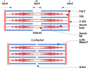
- Actin filaments are the major component of the I-band and extend into the A-band.
- Myosin filaments are bipolar and extend throughout the A-band. They are crosslinked at the centre by the M-band.
- The giant protein titinTitinTitin , also known as connectin, is a protein that in humans is encoded by the TTN gene. Titin is a giant protein that functions as a molecular spring which is responsible for the passive elasticity of muscle. It is composed of 244 individually folded protein domains connected by unstructured...
(connectin) extends from the Z-line of the sarcomere, where it binds to the thick filament (myosin)system, to the M-band, where it is thought to interact with the thick filaments. Titin (and its splice isoforms) is the biggest single highly elasticated protein found in nature. It provides binding sites for numerous proteins and is thought to play an important role as sarcomeric ruler and as blueprint for the assembly of the sarcomere. - Another giant protein, NebulinNebulinNebulin is an actin-binding protein which is localized to the I-band of the sarcomeres in skeletal muscle. It is a very large protein and binds as many as 200 actin monomers. Because its length is proportional to thin filament length, it is believed that nebulin acts as a thin filament "ruler"...
, is hypothesised to extend along the thin filaments and the entire I-Band. Similar to Titin, it is thought to act as a molecular ruler along for thin filament assembly. - Several proteins important for the stability of the sarcomeric structure are found in the Z-line as well as in the M-band of the sarcomere.
- Actin filaments and titin molecules are cross-linked in the Z-disc via the Z-line protein alpha-Actinin.
- The M-band proteins myomesinMyomesinMyomesin is an end line protein that is part of the M line. It is a protein found in the M-band of muscle sarcomeres in association with M-protein. It is found in both slow and fast muscle fibers while M-protein is only found in fast fibers...
as well as C-protein crosslink the thick filament system (myosins) and the M-band part of titin (the elastic filaments). - The interaction between actin and myosin filaments in the A-band of the sarcomere is responsible for the muscle contractionMuscle contractionMuscle fiber generates tension through the action of actin and myosin cross-bridge cycling. While under tension, the muscle may lengthen, shorten, or remain the same...
(sliding filament model).
Contraction
Upon muscle contraction, the A-bands do not change their length (1.85 micrometer in mammalian skeletal muscle), whereas the I-bands and the H-zone shorten. This causes the Z lines to come closer together.The protein tropomyosin
Tropomyosin
Tropomyosin is an actin-binding protein that regulates actin mechanics. It is important, among other things, for muscle contraction. Tropomyosin, along with the troponin complex, associate with actin in muscle fibers and regulate muscle contraction by regulating the binding of myosin...
covers the myosin binding sites of the actin molecules in the muscle cell. To allow the muscle cell to contract, tropomyosin must be moved to uncover the binding sites on the actin. Calcium ions bind with troponin-C molecules (which are dispersed throughout the tropomyosin protein) and alter the structure of the tropomyosin, forcing it to reveal the cross bridge binding site on the actin. The concentration of calcium within muscle cells is controlled by the sarcoplasmic reticulum, a unique form of endoplasmic reticulum. Muscle contraction ends when calcium ions are pumped back into the sarcoplasmic reticulum, allowing the contractile apparatus and thus muscle cell to relax.
During stimulation of the muscle cell, the motor neuron releases the neurotransmitter acetylcholine
Acetylcholine
The chemical compound acetylcholine is a neurotransmitter in both the peripheral nervous system and central nervous system in many organisms including humans...
, which travels across the neuromuscular junction (the synapse between the terminal bouton of the neuron and the muscle cell). Acetylcholine
Acetylcholine
The chemical compound acetylcholine is a neurotransmitter in both the peripheral nervous system and central nervous system in many organisms including humans...
binds to a post-synaptic nicotinic acetylcholine receptor
Nicotinic acetylcholine receptor
Nicotinic acetylcholine receptors, or nAChRs, are cholinergic receptors that form ligand-gated ion channels in the plasma membranes of certain neurons and on the postsynaptic side of the neuromuscular junction...
. A change in the receptor conformation allows an influx of sodium ions and initiation of a post-synaptic action potential. The action potential
Action potential
In physiology, an action potential is a short-lasting event in which the electrical membrane potential of a cell rapidly rises and falls, following a consistent trajectory. Action potentials occur in several types of animal cells, called excitable cells, which include neurons, muscle cells, and...
then travels along T (transverse) tubules until it reaches the sarcoplasmic reticulum; the action potential from the motor neuron changes the permeability of the sarcoplasmic reticulum, allowing the flow of calcium ions into the sarcomere. The outflow of calcium allows the myosin heads access to the actin cross bridge binding sites, permitting muscle contraction.
Rest
At rest, the myosin head is bound to an ATPAdenosine triphosphate
Adenosine-5'-triphosphate is a multifunctional nucleoside triphosphate used in cells as a coenzyme. It is often called the "molecular unit of currency" of intracellular energy transfer. ATP transports chemical energy within cells for metabolism...
molecule in a low-energy configuration and is unable to access the cross bridge binding sites on the actin. However, the myosin head can hydrolyze ATP into ADP and an inorganic phosphate ion. A portion of the energy released in this reaction changes the shape of the myosin head and promotes it to a high-energy configuration. Through the process of binding to the actin, the myosin head releases ADP and an inorganic phosphate ion, changing its configuration back to one of low energy. The myosin remains attached to actin in a state known as rigor, until a new ATP binds the myosin head. This binding of ATP to myosin releases the actin by cross-bridge dissociation. The ATP-associated myosin is ready for another cycle, beginning with hydrolysis of the ATP.
The A-band is visible as dark transverse lines across myofibers; the I-band is visible as lightly staining transverse lines, and the Z-line is visible as dark lines separating sarcomeres at the light-microscope level.
Storage
Most muscle cells only store enough ATP for a small number of muscle contractions. While muscle cells also store glycogen, most of the energy required for contraction is derived from phosphagens. One such phosphagenPhosphagen
The phosphagens are energy storage compounds, also known as high-energy phosphate compounds, are chiefly found in muscular tissue in animals. They allow a high-energy phosphate pool to be maintained in a concentration range, which, if it all were ATP, would create problems due to the ATP consuming...
is creatine phosphate, which is used to provide ADP with a phosphate group for ATP synthesis in vertebrates.
Comparative sarcomere structure
The structure of the sarcomere affects its function in several ways. The overlap of actin & myosin gives rise to the length-tension curve, which shows how sarcomere force output decreases if the muscle is stretched so that fewer cross-bridges can form or compressed until actin filaments interfere with each other. Length of the actin and myosin filaments (taken together as sarcomere length) affects force and velocity - longer sarcomeres have more cross-bridges and thus more force, but have a reduced range of shortening. VertebrateVertebrate
Vertebrates are animals that are members of the subphylum Vertebrata . Vertebrates are the largest group of chordates, with currently about 58,000 species described. Vertebrates include the jawless fishes, bony fishes, sharks and rays, amphibians, reptiles, mammals, and birds...
s display a very limited range of sarcomere lengths, with roughly the same optimal length (length at peak length-tension) in all muscles of an individual as well as between species. Arthropods, however, show tremendous variation (over seven-fold) in sarcomere length, both between species and between muscles in a single individual. The reasons for the lack of substantial sarcomere variability in vertebrates is not fully known.