
Mitral stenosis
Encyclopedia
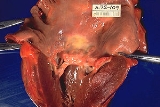
Mitral stenosis is a valvular heart disease
characterized by the narrowing of the orifice of the mitral valve
of the heart
.
Symptoms increase with exercise and pregnancy
 Almost all cases of mitral stenosis are due to disease in the heart secondary to rheumatic fever
and the consequent rheumatic heart disease. Uncommon causes of mitral stenosis are calcification of the mitral valve leaflets, and as a form of congenital heart disease. However, there are primary causes of mitral stenosis that emanate from a cleft mitral valve
.
Other causes include Bacterial endocarditis where the vegetations may favor increase risk of stenosis. It is the most common valvular heart disease in pregnancy
 The normal area of the mitral valve orifice is about 4 to 6 cm2. In normal cardiac physiology, the mitral valve
opens during left ventricular
diastole
, to allow blood
to flow from the left atrium
to the left ventricle
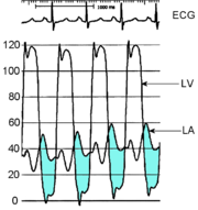
. A normal mitral valve will not impede the flow of blood from the left atrium to the left ventricle during (ventricular) diastole, and the pressures in the left atrium and the left ventricle during ventricular diastole will be equal. The result is that the left ventricle gets filled with blood during early ventricular diastole, with only a small portion of extra blood contributed by contraction of the left atrium (the "atrial kick") during late ventricular diastole.
When the mitral valve area goes below 2 cm2, the valve causes an impediment to the flow of blood into the left ventricle, creating a pressure gradient across the mitral valve. This gradient may be increased by increases in the heart rate
or cardiac output
. As the gradient across the mitral valve increases, the amount of time necessary to fill the left ventricle with blood increases. Eventually, the left ventricle requires the atrial kick to fill with blood. As the heart rate increases, the amount of time that the ventricle is in diastole and can fill up with blood (called the diastolic filling period) decreases. When the heart rate goes above a certain point, the diastolic filling period is insufficient to fill the ventricle with blood and pressure builds up in the left atrium, leading to pulmonary congestion.
When the mitral valve area goes less than 1 cm2, there will be an increase in the left atrial pressures (required to push blood through the stenotic valve). Since the normal left ventricular diastolic pressures is about 5 mmHg, a pressure gradient across the mitral valve of 20 mmHg due to severe mitral stenosis will cause a left atrial pressure of about 25 mmHg. This left atrial pressure is transmitted to the pulmonary vasculature and causes pulmonary hypertension
. Pulmonary capillary
pressures in this level cause an imbalance between the hydrostatic pressure and the oncotic pressure
, leading to extravasation of fluid from the vascular tree and pooling of fluid in the lungs (congestive heart failure
causing pulmonary edema
).
The constant pressure overload of the left atrium will cause the left atrium to increase in size. As the left atrium increases in size, it becomes more prone to develop atrial fibrillation
. When atrial fibrillation develops, the atrial kick is lost (since it is due to the normal atrial contraction).
In individuals with severe mitral stenosis, the left ventricular filling is dependent on the atrial kick. The loss of the atrial kick due to atrial fibrillation can cause a precipitous decrease in cardiac output and sudden congestive heart failure.
Patients with mitral stenosis prompts a series of hemodynamic changes that frequently cause deterioration of the patient's clinical status. A reduction in cardiac output, associated with acceleration of heart rate and shortening of the diastolic time, frequently leads to congestive heart failure. In addition, when AF sets in, systemic embolization becomes a real danger.
Mitral stenosis typically progresses slowly (over decades) from the initial signs of mitral stenosis to NYHA functional class
II symptoms to the development of atrial fibrillation to the development of NYHA functional class III or IV symptoms. Once an individual develops NYHA class III or IV symptoms, the progression of the disease accelerates and the patient's condition deteriorates.
of an individual with mitral stenosis, the first heart sound is unusually loud and may be palpable (tapping apex beat
) because of increased force in closing the mitral valve. The first heart sound is made by the mitral and tricuspid heart valves closing. These are normally synchronous, and the sounds are termed M1 and T1 respectively. M1 becomes louder in mitral stenosis. It may be the most prominent sign.
If pulmonary hypertension
secondary to mitral stenosis is severe, the P2 (pulmonic) component of the second heart sound (S2) will become loud.
An opening snap which is a high pitched additional sound may be heard after the A2 (aortic) component of the second heart sound (S2), which correlates to the forceful opening of the mitral valve. The mitral valve opens when the pressure in the left atrium is greater than the pressure in the left ventricle. This happens in ventricular diastole
(after closure of the aortic valve
), when the pressure in the ventricle precipitously drops. In individuals with mitral stenosis, the pressure in the left atrium correlates with the severity of the mitral stenosis. As the severity of the mitral stenosis increases, the pressure in the left atrium increases, and the mitral valve opens earlier in ventricular diastole.
A mid-diastolic rumbling murmur
with presystolic accentuation will be heard after the opening snap. The murmur is best heard at the apical region and is not radiated. Since it is low-pitched it is heard best with the bell of the stethoscope. Its duration increases with worsening disease. Rolling the patient towards left, as well as isometric exercise will accentuate the murmur. A thrill might be present when palpating at the apical region of the precordium.
Advanced disease may present with signs of right-sided heart failure such as parasternal heave
, jugular venous distension, hepatomegaly
, ascites
and/or pulmonary hypertension
, the latter often presenting with a loud P2.
Almost all signs increase with exercise and pregnancy.
Other peripheral signs include:
Medical sign
s of atrial fibrillation
include:
heart rate is about 100-150/min.
irregularly irregular pulse with a pulse deficit>10.
varying first heart sound intensity.
opening snap is not heard sometimes.
absent a waves in the neck veins.
presystolic accentuation of diastolic murmur disappears.
embolic manifestations may appear.
In most cases, the diagnosis of mitral stenosis is most easily made by echocardiography
, which shows left atrial enlargement, thick and calcified mitral valve with narrow and "fish-mouth"-shaped orifice and signs of right ventricular failure in advanced disease. It can also show decreased opening of the mitral valve leaflets, and increased blood flow velocity during diastole
. The trans-mitral gradient as measured by Doppler echocardiography is the gold standard
in the evaluation of the severity of mitral stenosis.
, which is a marker for the severity of mitral stenosis. This method of evaluating mitral stenosis tends to over-estimate the degree of mitral stenosis, however, because of the time lag in the pressure tracings seen on the right heart catheterization and the slow Y descent seen on the wedge tracings. If a trans-septal puncture is made during right heart catheterization, however, the pressure gradient can accurately quantify the severity of mitral stenosis.
may also assist in diagnosis, showing left atrial enlargement
.
Electrocardiography may show P mitrale, that is, broad, notched P waves in several or many leads with a prominent late negative component to the P wave in lead V1, and may also be seen in mitral regurgitation
, and, potentially, any cause of overload of the left atrium. Thus, P-sinistrocardiale may be a more appropriate term.
(the most common cause) is an asymptomatic latent phase following the initial episode of rheumatic fever. This latent period lasts an average of 16.3 ± 5.2 years. Once symptoms of mitral stenosis begin to develop, progression to severe disability takes 9.2 ± 4.3 years.
In individuals who were offered mitral valve surgery but refused, survival with medical therapy alone was 44 ± 6% at 5 years, and 32 ± 8% at 10 years after they were offered correction.
The treatment options for mitral stenosis include medical management, mitral valve replacement
by surgery, and percutaneous
mitral valvuloplasty by balloon catheter
.
The indication for invasive treatment with either a mitral valve replacement or valvuloplasty is NYHA functional class
III or IV symptoms.
Another option is balloon dilatation. To determine which patients would benefit from percutaneous balloon mitral valvuloplasty, a scoring system has been developed. Scoring is based on 4 echocardiographic criteria: leaflet mobility, leaflet thickening, subvalvar thickening, and calcification. Individuals with a score of ≥ 8 tended to have suboptimal results. Superb results with valvotomy are seen in individuals with a crisp opening snap, score < 8, and no calcium in the commissures.
Treatment also focuses on concomitant conditions often seen in mitral stenosis:
Under local anaesthetic, a catheter with a special balloon is passed from the right femoral vein
, up the inferior vena cava
and into the right atrium
. The interatrial septum
is punctured and the catheter
passed into the left atrium using a "trans-septal technique". The balloon is sub-divided into 3 segments and is dilated in 3 stages. 1st the distal portion (lying in the left ventricle) is inflated and pulled against the valve cusps. Second the proximal portion is dilated, in order to fix the centre segment at the valve orifice. Finally, the central section is inflated, this should take no longer than 30 seconds since full inflation obstructs the valve and causes congestion, leading to circulatory arrest and flash pulmonary edema
.
With careful patient pre-selection, percutaneous balloon mitral valvuloplasty (PBMV) is associated with good success rates and a low rate of complications. By far the most serious adverse event is the occurrence of acute severe mitral regurgitation. Severe mitral regurgitation usually results from a tear in one of the valve leaflets or the subvalvular apparatus. It can lead to pulmonary oedema and hemodynamic compromise, necessitating urgent surgical mitral valve replacement.
Other serious complications with PBMV usually relate to the technique of trans-septal puncture (TSP). The ideal site for TSP is the region of the fossa ovalis in the inter-atrial septum. Occasionally, however, the sharp needle used for TSP may inadvertently traumatize other cardiac structures, leading to cardiac tamponade or serious blood loss.
Although the immediate results of PBMV are often quite gratifying, the procedure does not provide permanent relief from mitral stenosis. Regular follow-up is mandatory, to detect restenosis. Long-term follow up data from patients undergoing PBMV indicates that upto 70-75% individuals can be free of restenosis 10 years following the procedure. The number falls to about 40% 15 years post-PBMV.
Valvular heart disease
Valvular heart disease is any disease process involving one or more of the valves of the heart . Valve problems may be congenital or acquired...
characterized by the narrowing of the orifice of the mitral valve
Mitral valve
The mitral valve is a dual-flap valve in the heart that lies between the left atrium and the left ventricle...
of the heart
Heart
The heart is a myogenic muscular organ found in all animals with a circulatory system , that is responsible for pumping blood throughout the blood vessels by repeated, rhythmic contractions...
.
Signs and symptoms
Symptoms of mitral stenosis include:- Heart failure symptoms, such as dyspnea on exertion, orthopnea and paroxysmal nocturnal dyspnea
- Palpitations
- Chest painChest painChest pain may be a symptom of a number of serious conditions and is generally considered a medical emergency. Even though it may be determined that the pain is non-cardiac in origin, this is often a diagnosis of exclusion made after ruling out more serious causes of the pain.-Differential...
- HemoptysisHemoptysisHemoptysis or haemoptysis is the expectoration of blood or of blood-stained sputum from the bronchi, larynx, trachea, or lungs Hemoptysis or haemoptysis is the expectoration (coughing up) of blood or of blood-stained sputum from the bronchi, larynx, trachea, or lungs Hemoptysis or haemoptysis ...
- Thromboembolism
- AscitesAscitesAscites is a gastroenterological term for an accumulation of fluid in the peritoneal cavity.The medical condition is also known as peritoneal cavity fluid, peritoneal fluid excess, hydroperitoneum or more archaically as abdominal dropsy. Although most commonly due to cirrhosis and severe liver...
and edemaEdemaEdema or oedema ; both words from the Greek , oídēma "swelling"), formerly known as dropsy or hydropsy, is an abnormal accumulation of fluid beneath the skin or in one or more cavities of the body that produces swelling...
(if right-sided heart failure develops)
Symptoms increase with exercise and pregnancy
Cause
Rheumatic fever
Rheumatic fever is an inflammatory disease that occurs following a Streptococcus pyogenes infection, such as strep throat or scarlet fever. Believed to be caused by antibody cross-reactivity that can involve the heart, joints, skin, and brain, the illness typically develops two to three weeks after...
and the consequent rheumatic heart disease. Uncommon causes of mitral stenosis are calcification of the mitral valve leaflets, and as a form of congenital heart disease. However, there are primary causes of mitral stenosis that emanate from a cleft mitral valve
Mitral valve
The mitral valve is a dual-flap valve in the heart that lies between the left atrium and the left ventricle...
.
Other causes include Bacterial endocarditis where the vegetations may favor increase risk of stenosis. It is the most common valvular heart disease in pregnancy
Pregnancy
Pregnancy refers to the fertilization and development of one or more offspring, known as a fetus or embryo, in a woman's uterus. In a pregnancy, there can be multiple gestations, as in the case of twins or triplets...
Pathophysiology
Mitral valve
The mitral valve is a dual-flap valve in the heart that lies between the left atrium and the left ventricle...
opens during left ventricular
Left ventricle
The left ventricle is one of four chambers in the human heart. It receives oxygenated blood from the left atrium via the mitral valve, and pumps it into the aorta via the aortic valve.-Shape:...
diastole
Diastole
Diastole is the period of time when the heart fills with blood after systole . Ventricular diastole is the period during which the ventricles are relaxing, while atrial diastole is the period during which the atria are relaxing...
, to allow blood
Blood
Blood is a specialized bodily fluid in animals that delivers necessary substances such as nutrients and oxygen to the cells and transports metabolic waste products away from those same cells....
to flow from the left atrium
Left atrium
The left atrium is one of the four chambers in the human heart. It receives oxygenated blood from the pulmonary veins, and pumps it into the left ventricle, via the mitral valve.-Foramen ovale:...
to the left ventricle
Left ventricle
The left ventricle is one of four chambers in the human heart. It receives oxygenated blood from the left atrium via the mitral valve, and pumps it into the aorta via the aortic valve.-Shape:...
. A normal mitral valve will not impede the flow of blood from the left atrium to the left ventricle during (ventricular) diastole, and the pressures in the left atrium and the left ventricle during ventricular diastole will be equal. The result is that the left ventricle gets filled with blood during early ventricular diastole, with only a small portion of extra blood contributed by contraction of the left atrium (the "atrial kick") during late ventricular diastole.
When the mitral valve area goes below 2 cm2, the valve causes an impediment to the flow of blood into the left ventricle, creating a pressure gradient across the mitral valve. This gradient may be increased by increases in the heart rate
Heart rate
Heart rate is the number of heartbeats per unit of time, typically expressed as beats per minute . Heart rate can vary as the body's need to absorb oxygen and excrete carbon dioxide changes, such as during exercise or sleep....
or cardiac output
Cardiac output
Cardiac output is the volume of blood being pumped by the heart, in particular by a left or right ventricle in the time interval of one minute. CO may be measured in many ways, for example dm3/min...
. As the gradient across the mitral valve increases, the amount of time necessary to fill the left ventricle with blood increases. Eventually, the left ventricle requires the atrial kick to fill with blood. As the heart rate increases, the amount of time that the ventricle is in diastole and can fill up with blood (called the diastolic filling period) decreases. When the heart rate goes above a certain point, the diastolic filling period is insufficient to fill the ventricle with blood and pressure builds up in the left atrium, leading to pulmonary congestion.
When the mitral valve area goes less than 1 cm2, there will be an increase in the left atrial pressures (required to push blood through the stenotic valve). Since the normal left ventricular diastolic pressures is about 5 mmHg, a pressure gradient across the mitral valve of 20 mmHg due to severe mitral stenosis will cause a left atrial pressure of about 25 mmHg. This left atrial pressure is transmitted to the pulmonary vasculature and causes pulmonary hypertension
Pulmonary hypertension
In medicine, pulmonary hypertension is an increase in blood pressure in the pulmonary artery, pulmonary vein, or pulmonary capillaries, together known as the lung vasculature, leading to shortness of breath, dizziness, fainting, and other symptoms, all of which are exacerbated by exertion...
. Pulmonary capillary
Capillary
Capillaries are the smallest of a body's blood vessels and are parts of the microcirculation. They are only 1 cell thick. These microvessels, measuring 5-10 μm in diameter, connect arterioles and venules, and enable the exchange of water, oxygen, carbon dioxide, and many other nutrient and waste...
pressures in this level cause an imbalance between the hydrostatic pressure and the oncotic pressure
Oncotic pressure
Oncotic pressure, or colloid osmotic pressure, is a form of osmotic pressure exerted by proteins in blood plasma that usually tends to pull water into the circulatory system.Throughout the body, dissolved compounds have an osmotic pressure...
, leading to extravasation of fluid from the vascular tree and pooling of fluid in the lungs (congestive heart failure
Congestive heart failure
Heart failure often called congestive heart failure is generally defined as the inability of the heart to supply sufficient blood flow to meet the needs of the body. Heart failure can cause a number of symptoms including shortness of breath, leg swelling, and exercise intolerance. The condition...
causing pulmonary edema
Pulmonary edema
Pulmonary edema , or oedema , is fluid accumulation in the air spaces and parenchyma of the lungs. It leads to impaired gas exchange and may cause respiratory failure...
).
The constant pressure overload of the left atrium will cause the left atrium to increase in size. As the left atrium increases in size, it becomes more prone to develop atrial fibrillation
Atrial fibrillation
Atrial fibrillation is the most common cardiac arrhythmia . It is a common cause of irregular heart beat, identified clinically by taking a pulse. Chaotic electrical activity in the two upper chambers of the heart result in the muscle fibrillating , instead of achieving coordinated contraction...
. When atrial fibrillation develops, the atrial kick is lost (since it is due to the normal atrial contraction).
In individuals with severe mitral stenosis, the left ventricular filling is dependent on the atrial kick. The loss of the atrial kick due to atrial fibrillation can cause a precipitous decrease in cardiac output and sudden congestive heart failure.
Patients with mitral stenosis prompts a series of hemodynamic changes that frequently cause deterioration of the patient's clinical status. A reduction in cardiac output, associated with acceleration of heart rate and shortening of the diastolic time, frequently leads to congestive heart failure. In addition, when AF sets in, systemic embolization becomes a real danger.
Mitral stenosis typically progresses slowly (over decades) from the initial signs of mitral stenosis to NYHA functional class
New York Heart Association Functional Classification
The New York Heart Association Functional Classification provides a simple way of classifying the extent of heart failure. It places patients in one of four categories based on how much they are limited during physical activity; the limitations/symptoms are in regards to normal breathing and...
II symptoms to the development of atrial fibrillation to the development of NYHA functional class III or IV symptoms. Once an individual develops NYHA class III or IV symptoms, the progression of the disease accelerates and the patient's condition deteriorates.
Physical examination
Upon auscultationHeart sounds
Heart sounds, or heartbeats, are the noises generated by the beating heart and the resultant flow of blood through it...
of an individual with mitral stenosis, the first heart sound is unusually loud and may be palpable (tapping apex beat
Apex beat
The apex beat, also called the point of maximum impulse , is the furthermost point outwards and downwards from the sternum at which the cardiac impulse can be felt...
) because of increased force in closing the mitral valve. The first heart sound is made by the mitral and tricuspid heart valves closing. These are normally synchronous, and the sounds are termed M1 and T1 respectively. M1 becomes louder in mitral stenosis. It may be the most prominent sign.
If pulmonary hypertension
Pulmonary hypertension
In medicine, pulmonary hypertension is an increase in blood pressure in the pulmonary artery, pulmonary vein, or pulmonary capillaries, together known as the lung vasculature, leading to shortness of breath, dizziness, fainting, and other symptoms, all of which are exacerbated by exertion...
secondary to mitral stenosis is severe, the P2 (pulmonic) component of the second heart sound (S2) will become loud.
An opening snap which is a high pitched additional sound may be heard after the A2 (aortic) component of the second heart sound (S2), which correlates to the forceful opening of the mitral valve. The mitral valve opens when the pressure in the left atrium is greater than the pressure in the left ventricle. This happens in ventricular diastole
Diastole
Diastole is the period of time when the heart fills with blood after systole . Ventricular diastole is the period during which the ventricles are relaxing, while atrial diastole is the period during which the atria are relaxing...
(after closure of the aortic valve
Aortic valve
The aortic valve is one of the valves of the heart. It is normally tricuspid , although in 1% of the population it is found to be congenitally bicuspid . It lies between the left ventricle and the aorta....
), when the pressure in the ventricle precipitously drops. In individuals with mitral stenosis, the pressure in the left atrium correlates with the severity of the mitral stenosis. As the severity of the mitral stenosis increases, the pressure in the left atrium increases, and the mitral valve opens earlier in ventricular diastole.
A mid-diastolic rumbling murmur
Heart murmur
Murmurs are extra heart sounds that are produced as a result of turbulent blood flow that is sufficient to produce audible noise. Most murmurs can only be heard with the assistance of a stethoscope ....
with presystolic accentuation will be heard after the opening snap. The murmur is best heard at the apical region and is not radiated. Since it is low-pitched it is heard best with the bell of the stethoscope. Its duration increases with worsening disease. Rolling the patient towards left, as well as isometric exercise will accentuate the murmur. A thrill might be present when palpating at the apical region of the precordium.
Advanced disease may present with signs of right-sided heart failure such as parasternal heave
Parasternal heave
A parasternal heave is a praecordial impulse that may be palpable in patients with cardiac or respiratory disease. A parasternal impulse may be felt when the heel of the hand is rested just to the left of the sternum with the fingers lifted slightly off the chest. Normally no impulse or a slight...
, jugular venous distension, hepatomegaly
Hepatomegaly
Hepatomegaly is the condition of having an enlarged liver. It is a nonspecific medical sign having many causes, which can broadly be broken down into infection, direct toxicity, hepatic tumours, or metabolic disorder. Often, hepatomegaly will present as an abdominal mass...
, ascites
Ascites
Ascites is a gastroenterological term for an accumulation of fluid in the peritoneal cavity.The medical condition is also known as peritoneal cavity fluid, peritoneal fluid excess, hydroperitoneum or more archaically as abdominal dropsy. Although most commonly due to cirrhosis and severe liver...
and/or pulmonary hypertension
Pulmonary hypertension
In medicine, pulmonary hypertension is an increase in blood pressure in the pulmonary artery, pulmonary vein, or pulmonary capillaries, together known as the lung vasculature, leading to shortness of breath, dizziness, fainting, and other symptoms, all of which are exacerbated by exertion...
, the latter often presenting with a loud P2.
Almost all signs increase with exercise and pregnancy.
Other peripheral signs include:
- Malar flush - pulmonary hypertension is prominent in patients with mitral stenosis
- Atrial fibrillationAtrial fibrillationAtrial fibrillation is the most common cardiac arrhythmia . It is a common cause of irregular heart beat, identified clinically by taking a pulse. Chaotic electrical activity in the two upper chambers of the heart result in the muscle fibrillating , instead of achieving coordinated contraction...
- irregular pulse and loss of 'a' wave in jugular venous pressure - Left parasternal heaveParasternal heaveA parasternal heave is a praecordial impulse that may be palpable in patients with cardiac or respiratory disease. A parasternal impulse may be felt when the heel of the hand is rested just to the left of the sternum with the fingers lifted slightly off the chest. Normally no impulse or a slight...
- presence of right ventricular hypertrophy due to pulmonary hypertension - Tapping apex beat which is not displaced
Medical sign
Medical sign
A medical sign is an objective indication of some medical fact or characteristic that may be detected by a physician during a physical examination of a patient....
s of atrial fibrillation
Atrial fibrillation
Atrial fibrillation is the most common cardiac arrhythmia . It is a common cause of irregular heart beat, identified clinically by taking a pulse. Chaotic electrical activity in the two upper chambers of the heart result in the muscle fibrillating , instead of achieving coordinated contraction...
include:
heart rate is about 100-150/min.
irregularly irregular pulse with a pulse deficit>10.
varying first heart sound intensity.
opening snap is not heard sometimes.
absent a waves in the neck veins.
presystolic accentuation of diastolic murmur disappears.
embolic manifestations may appear.
Echocardiography
| Degree of mitral stenosis | Mean gradient | Mitral valve area |
|---|---|---|
| Mild mitral stenosis | <5 mmHg | >1.5 cm2 |
| Moderate mitral stenosis | 5 - 10 mmHg | 1.0 - 1.5 cm2 |
| Severe mitral stenosis | > 10 mmHg | < 1.0 cm2 |
In most cases, the diagnosis of mitral stenosis is most easily made by echocardiography
Echocardiography
An echocardiogram, often referred to in the medical community as a cardiac ECHO or simply an ECHO, is a sonogram of the heart . Also known as a cardiac ultrasound, it uses standard ultrasound techniques to image two-dimensional slices of the heart...
, which shows left atrial enlargement, thick and calcified mitral valve with narrow and "fish-mouth"-shaped orifice and signs of right ventricular failure in advanced disease. It can also show decreased opening of the mitral valve leaflets, and increased blood flow velocity during diastole
Diastole
Diastole is the period of time when the heart fills with blood after systole . Ventricular diastole is the period during which the ventricles are relaxing, while atrial diastole is the period during which the atria are relaxing...
. The trans-mitral gradient as measured by Doppler echocardiography is the gold standard
Gold standard (test)
In medicine and statistics, gold standard test refers to a diagnostic test or benchmark that is the best available under reasonable conditions. It does not have to be necessarily the best possible test for the condition in absolute terms...
in the evaluation of the severity of mitral stenosis.
Cardiac chamber catheterization
Another method of measuring the severity of mitral stenosis is the simultaneous left and right heart chamber catheterization. The right heart catheterization (commonly known as Swan-Ganz catheterization) gives the physician the mean pulmonary capillary wedge pressure, which is a reflection of the left atrial pressure. The left heart catheterization, on the other hand, gives the pressure in the left ventricle. By simultaneously taking these pressures, it is possible to determine the gradient between the left atrium and left ventricle during ventricular diastoleDiastole
Diastole is the period of time when the heart fills with blood after systole . Ventricular diastole is the period during which the ventricles are relaxing, while atrial diastole is the period during which the atria are relaxing...
, which is a marker for the severity of mitral stenosis. This method of evaluating mitral stenosis tends to over-estimate the degree of mitral stenosis, however, because of the time lag in the pressure tracings seen on the right heart catheterization and the slow Y descent seen on the wedge tracings. If a trans-septal puncture is made during right heart catheterization, however, the pressure gradient can accurately quantify the severity of mitral stenosis.
Other assisting diagnostic techniques
Chest X-rayChest X-ray
In medicine, a chest radiograph, commonly called a chest X-ray , is a projection radiograph of the chest used to diagnose conditions affecting the chest, its contents, and nearby structures...
may also assist in diagnosis, showing left atrial enlargement
Left atrial enlargement
Left atrial enlargement or left atrial dilation is a form of cardiomegaly.In the general population, obesity appears to be the most important risk factor for LAE. Also, a study found that LAE can occur as a consequence of atrial fibrillation , although another study found that AF by itself does...
.
Electrocardiography may show P mitrale, that is, broad, notched P waves in several or many leads with a prominent late negative component to the P wave in lead V1, and may also be seen in mitral regurgitation
Mitral regurgitation
Mitral regurgitation , mitral insufficiency or mitral incompetence is a disorder of the heart in which the mitral valve does not close properly when the heart pumps out blood. It is the abnormal leaking of blood from the left ventricle, through the mitral valve, and into the left atrium, when...
, and, potentially, any cause of overload of the left atrium. Thus, P-sinistrocardiale may be a more appropriate term.
Natural history
The natural history of mitral stenosis secondary to rheumatic feverRheumatic fever
Rheumatic fever is an inflammatory disease that occurs following a Streptococcus pyogenes infection, such as strep throat or scarlet fever. Believed to be caused by antibody cross-reactivity that can involve the heart, joints, skin, and brain, the illness typically develops two to three weeks after...
(the most common cause) is an asymptomatic latent phase following the initial episode of rheumatic fever. This latent period lasts an average of 16.3 ± 5.2 years. Once symptoms of mitral stenosis begin to develop, progression to severe disability takes 9.2 ± 4.3 years.
In individuals who were offered mitral valve surgery but refused, survival with medical therapy alone was 44 ± 6% at 5 years, and 32 ± 8% at 10 years after they were offered correction.
Treatment
Treatment is not necessary in asymptomatic patients.The treatment options for mitral stenosis include medical management, mitral valve replacement
Mitral valve replacement
Mitral valve replacement is a cardiac surgery procedure in which a patient’s mitral valve is replaced by a different valve. Mitral valve replacement is typically performed robotically or manually, when the valve becomes too tight for blood to flow into the left ventricle, or too loose in which...
by surgery, and percutaneous
Percutaneous
In surgery, percutaneous pertains to any medical procedure where access to inner organs or other tissue is done via needle-puncture of the skin, rather than by using an "open" approach where inner organs or tissue are exposed .The percutaneous approach is commonly used in vascular procedures...
mitral valvuloplasty by balloon catheter
Balloon catheter
A balloon catheter is a type of "soft" catheter with an inflatable "balloon" at its tip which is used during a catheterization procedure to enlarge a narrow opening or passage within the body...
.
The indication for invasive treatment with either a mitral valve replacement or valvuloplasty is NYHA functional class
New York Heart Association Functional Classification
The New York Heart Association Functional Classification provides a simple way of classifying the extent of heart failure. It places patients in one of four categories based on how much they are limited during physical activity; the limitations/symptoms are in regards to normal breathing and...
III or IV symptoms.
Another option is balloon dilatation. To determine which patients would benefit from percutaneous balloon mitral valvuloplasty, a scoring system has been developed. Scoring is based on 4 echocardiographic criteria: leaflet mobility, leaflet thickening, subvalvar thickening, and calcification. Individuals with a score of ≥ 8 tended to have suboptimal results. Superb results with valvotomy are seen in individuals with a crisp opening snap, score < 8, and no calcium in the commissures.
Treatment also focuses on concomitant conditions often seen in mitral stenosis:
- Any angina is treated with short-acting nitrovasodilatorNitrovasodilatorA nitrovasodilator is an agent which causes vasodilation by donation of nitric oxide.Examples include nitroprusside and nitroglycerine.-References:...
s, beta-blockers and/or calcium blockers - Any hypertensionHypertensionHypertension or high blood pressure is a cardiac chronic medical condition in which the systemic arterial blood pressure is elevated. What that means is that the heart is having to work harder than it should to pump the blood around the body. Blood pressure involves two measurements, systolic and...
is treated aggressively, but caution must be taken in administering beta-blockers - Any heart failure is treated with digoxinDigoxinDigoxin INN , also known as digitalis, is a purified cardiac glycoside and extracted from the foxglove plant, Digitalis lanata. Its corresponding aglycone is digoxigenin, and its acetyl derivative is acetyldigoxin...
, diureticDiureticA diuretic provides a means of forced diuresis which elevates the rate of urination. There are several categories of diuretics. All diuretics increase the excretion of water from bodies, although each class does so in a distinct way.- Medical uses :...
s, nitrovasodilatorNitrovasodilatorA nitrovasodilator is an agent which causes vasodilation by donation of nitric oxide.Examples include nitroprusside and nitroglycerine.-References:...
s and, if not contraindicated, cautious inpatient administration of ACE inhibitorACE inhibitorACE inhibitors or angiotensin-converting enzyme inhibitors are a group of drugs used primarily for the treatment of hypertension and congestive heart failure...
s
Mitral valvuloplasty
Mitral valvuloplasty is a minimally invasive therapeutic procedure to correct an uncomplicated mitral stenosis by dilating the valve using a balloon.Under local anaesthetic, a catheter with a special balloon is passed from the right femoral vein
Femoral vein
In the human body, the femoral vein is a blood vessel that accompanies the femoral artery in the femoral sheath. It begins at the adductor canal and is a continuation of the popliteal vein...
, up the inferior vena cava
Inferior vena cava
The inferior vena cava , also known as the posterior vena cava, is the large vein that carries de-oxygenated blood from the lower half of the body into the right atrium of the heart....
and into the right atrium
Right atrium
The right atrium is one of four chambers in the hearts of mammals and archosaurs...
. The interatrial septum
Interatrial septum
The interatrial septum is the wall of tissue that separates the right and left atria of the heart.-Development:The interatrial septum forms during the first and second months of fetal development. Formation of the septum occurs in several stages...
is punctured and the catheter
Catheter
In medicine, a catheter is a tube that can be inserted into a body cavity, duct, or vessel. Catheters thereby allow drainage, administration of fluids or gases, or access by surgical instruments. The process of inserting a catheter is catheterization...
passed into the left atrium using a "trans-septal technique". The balloon is sub-divided into 3 segments and is dilated in 3 stages. 1st the distal portion (lying in the left ventricle) is inflated and pulled against the valve cusps. Second the proximal portion is dilated, in order to fix the centre segment at the valve orifice. Finally, the central section is inflated, this should take no longer than 30 seconds since full inflation obstructs the valve and causes congestion, leading to circulatory arrest and flash pulmonary edema
Pulmonary edema
Pulmonary edema , or oedema , is fluid accumulation in the air spaces and parenchyma of the lungs. It leads to impaired gas exchange and may cause respiratory failure...
.
With careful patient pre-selection, percutaneous balloon mitral valvuloplasty (PBMV) is associated with good success rates and a low rate of complications. By far the most serious adverse event is the occurrence of acute severe mitral regurgitation. Severe mitral regurgitation usually results from a tear in one of the valve leaflets or the subvalvular apparatus. It can lead to pulmonary oedema and hemodynamic compromise, necessitating urgent surgical mitral valve replacement.
Other serious complications with PBMV usually relate to the technique of trans-septal puncture (TSP). The ideal site for TSP is the region of the fossa ovalis in the inter-atrial septum. Occasionally, however, the sharp needle used for TSP may inadvertently traumatize other cardiac structures, leading to cardiac tamponade or serious blood loss.
Although the immediate results of PBMV are often quite gratifying, the procedure does not provide permanent relief from mitral stenosis. Regular follow-up is mandatory, to detect restenosis. Long-term follow up data from patients undergoing PBMV indicates that upto 70-75% individuals can be free of restenosis 10 years following the procedure. The number falls to about 40% 15 years post-PBMV.