
Gonioscopy
Encyclopedia
Gonioscopy describes the use of a goniolens (also known as a gonioscope) in conjunction with a slit lamp
or operating microscope to gain a view of the iridocorneal angle, or the anatomical angle formed between the eye's cornea
and iris
. The importance of this process is in diagnosing and monitoring various eye conditions associated with glaucoma
.
from the ocular tissue.
The mechanism for this process varies with each type of goniolens. Three examples of goniolenses are the:
There are many other goniolenses available for use, including modified versions the aforementioned, which prove valuable for surgical use (goniotomy).

Slit lamp
The slit lamp is an instrument consisting of a high-intensity light source that can be focused to shine a thin sheet of light into the eye. It is used in conjunction with a biomicroscope...
or operating microscope to gain a view of the iridocorneal angle, or the anatomical angle formed between the eye's cornea
Cornea
The cornea is the transparent front part of the eye that covers the iris, pupil, and anterior chamber. Together with the lens, the cornea refracts light, with the cornea accounting for approximately two-thirds of the eye's total optical power. In humans, the refractive power of the cornea is...
and iris
Iris (anatomy)
The iris is a thin, circular structure in the eye, responsible for controlling the diameter and size of the pupils and thus the amount of light reaching the retina. "Eye color" is the color of the iris, which can be green, blue, or brown. In some cases it can be hazel , grey, violet, or even pink...
. The importance of this process is in diagnosing and monitoring various eye conditions associated with glaucoma
Glaucoma
Glaucoma is an eye disorder in which the optic nerve suffers damage, permanently damaging vision in the affected eye and progressing to complete blindness if untreated. It is often, but not always, associated with increased pressure of the fluid in the eye...
.
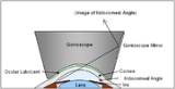
The goniolens or gonioscope
The goniolens allows the clinician - usually an ophthalmologist or optometrist - to view the irideocorneal angle through a mirror or prism, without which the angle is masked by total internal reflectionTotal internal reflection
Total internal reflection is an optical phenomenon that happens when a ray of light strikes a medium boundary at an angle larger than a particular critical angle with respect to the normal to the surface. If the refractive index is lower on the other side of the boundary and the incident angle is...
from the ocular tissue.
The mechanism for this process varies with each type of goniolens. Three examples of goniolenses are the:
- Koeppe direct goniolens: this transparent device is placed directly on the corneaCorneaThe cornea is the transparent front part of the eye that covers the iris, pupil, and anterior chamber. Together with the lens, the cornea refracts light, with the cornea accounting for approximately two-thirds of the eye's total optical power. In humans, the refractive power of the cornea is...
along with lubricating fluid, to avoid damaging its surface. The steeper curvature of this goniolens' exterior surface optically eliminates the total internal reflectionTotal internal reflectionTotal internal reflection is an optical phenomenon that happens when a ray of light strikes a medium boundary at an angle larger than a particular critical angle with respect to the normal to the surface. If the refractive index is lower on the other side of the boundary and the incident angle is...
problem and allows a view of the iridocorneal angle. Unfortunately it requires the patient to be lying down, and so it cannot be so easily used with an ordinary slit lampSlit lampThe slit lamp is an instrument consisting of a high-intensity light source that can be focused to shine a thin sheet of light into the eye. It is used in conjunction with a biomicroscope...
in an optometric environment. In an ophthalmological setting, an operating microscope is one available option. - Goldmann indirect goniolens: this truncated-cone like device utilises mirrors to reflect the light from the iridocorneal angle into the direction of the observer (as shown by the schematic diagram). In practice the image comes out roughly orthogonal to the back surface (nearer the practitioner), making observation and magnification with a slit lampSlit lampThe slit lamp is an instrument consisting of a high-intensity light source that can be focused to shine a thin sheet of light into the eye. It is used in conjunction with a biomicroscope...
easy and reliable. The small, curved front surface does not rest on the cornea, but instead vaults over it, with lubricating fluid filling the gap. The border of the front surface rests on the scleraScleraThe sclera , also known as the white or white of the eye, is the opaque , fibrous, protective, outer layer of the eye containing collagen and elastic fiber. In the development of the embryo, the sclera is derived from the neural crest...
. While the view obtained is smaller than that of the Koeppe goniolens, it can be used with the patient sitting upright, and other mirrors within the device can be used to obtain views of other parts of the eye, such as the retina and the ora serrata. - Zeiss indirect goniolens: this instrument uses a similar method to the Goldmann, but employs prisms in the place of mirrors. Its four symmetrical prisms allow visualisation of the iridocorneal angle in four quadrants of the eye simultaneously, and works well with a slit lampSlit lampThe slit lamp is an instrument consisting of a high-intensity light source that can be focused to shine a thin sheet of light into the eye. It is used in conjunction with a biomicroscope...
. Most importantly, the size and shape of the instrument - a smaller front surface that rests on the cornea without requiring lubricating fluid, only the patient's tear film - allows for indentation gonioscopy, which can be used for further diagnosis.
There are many other goniolenses available for use, including modified versions the aforementioned, which prove valuable for surgical use (goniotomy).
The gonioscopy process
Although the details vary based on the type of goniolens used, in general the gonioscopy process involves:- briefly explaining the procedure to the patient
- cleaning and sterilising the front (curved) surface of the goniolens
- applying lubricating fluid to the front surface if appropriate
- anaesthetising the patient's corneaCorneaThe cornea is the transparent front part of the eye that covers the iris, pupil, and anterior chamber. Together with the lens, the cornea refracts light, with the cornea accounting for approximately two-thirds of the eye's total optical power. In humans, the refractive power of the cornea is...
with topicalTopicalIn medicine, a topical medication is applied to body surfaces such as the skin or mucous membranes such as the vagina, anus, throat, eyes and ears.Many topical medications are epicutaneous, meaning that they are applied directly to the skin...
anaesthetic - preparing the slit lampSlit lampThe slit lamp is an instrument consisting of a high-intensity light source that can be focused to shine a thin sheet of light into the eye. It is used in conjunction with a biomicroscope...
for viewing through the goniolens - gently moving the patient's eyelids away from the cornea
- slowly applying the goniolens to the ocular surface, forming suction
- fine-tuning the slit lamp to optimise the view
- interpreting the gonioscopic image
- swivelling the goniolens to view each section of the iridocorneal angle
- when satisfied, very carefully breaking suction via the eyelids
- cleaning the instruments and irrigating the patient's eyes with [saline] if desired
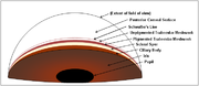
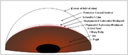
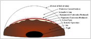
Interpreting the gonioscopic image
The typical view through most goniolenses is illustrated by these idealistic schematic diagrams. In reality the clinical picture can vary both within and between individual patients. This view of the iridocorneal angle provides information in several ways:- Iridocorneal angle width: The width of the iridocorneal angle is one factor affecting the drainage of aqueous humourAqueous humourThe aqueous humour is a clear, gelatinous fluid similar to plasma, but containing low-protein concentrations. It is secreted from the ciliary epithelium, a structure supporting the lens. It is located in the space between the lens and the cornea...
from the eye's anterior chamberAnterior chamberThe anterior chamber is the fluid-filled space inside the eye between the iris and the cornea's innermost surface, the endothelium. Aqueous humor is the fluid that fills the anterior chamber. Hyphema and glaucoma are two main pathologies in this area. In hyphema, blood fills the anterior chamber...
. A wide angle allows sufficient drainage of humour through the trabecular meshworkTrabecular meshworkThe trabecular meshwork is an area of tissue in the eye located around the base of the cornea, near the ciliary body, and is responsible for draining the aqueous humor from the eye via the anterior chamber .The tissue is spongy and lined by trabeculocytes; it allows fluid to drain into a set of...
(unless obstructed), whereas a narrow angle may impede the drainage system and leave the patient susceptible to acute angle-closure glaucomaGlaucomaGlaucoma is an eye disorder in which the optic nerve suffers damage, permanently damaging vision in the affected eye and progressing to complete blindness if untreated. It is often, but not always, associated with increased pressure of the fluid in the eye...
. Gonioscopy indicates the angular width of the iridocorneal angle by the number of ocular structures visible above the rim of the iris. Generally the more structures visible, the wider the angle. However, not all structures may be easily discriminated, especially the faint Schwalbe's lineSchwalbe's lineSchwalbe's line is the anatomical line found on the interior surface of the eye's cornea, and delineates the outer limit of the corneal endothelium layer. Specifically, it represents the termination of Descemet's membrane. In many cases it can be seen via gonioscopy....
at the top of the stack. Further information is obtained if a very narrow slit lamp beam may be shone upon the angle, as the angle width is generally proportional to the separation of the corneal beam and iris beam when they meet in the angle. - Anterior synechiae: Anterior synechiaeSynechiaA synechia is an eye condition where the iris adheres to either the cornea or lens . Synechiae can be caused by ocular trauma, iritis or iridocyclitis and may lead to certain types of glaucoma...
are simply stands of the irisIris (anatomy)The iris is a thin, circular structure in the eye, responsible for controlling the diameter and size of the pupils and thus the amount of light reaching the retina. "Eye color" is the color of the iris, which can be green, blue, or brown. In some cases it can be hazel , grey, violet, or even pink...
attaching to the iridocorneal angle or surrounding tissue. This may be exacerbated by ocular inflammationInflammationInflammation is part of the complex biological response of vascular tissues to harmful stimuli, such as pathogens, damaged cells, or irritants. Inflammation is a protective attempt by the organism to remove the injurious stimuli and to initiate the healing process...
, which can render the angle 'sticky' with inflammatory cells and substances, or by structural defects in the iris which lead to strands floating free into the anterior chamberAnterior chamberThe anterior chamber is the fluid-filled space inside the eye between the iris and the cornea's innermost surface, the endothelium. Aqueous humor is the fluid that fills the anterior chamber. Hyphema and glaucoma are two main pathologies in this area. In hyphema, blood fills the anterior chamber...
, as may occur with iris atrophy and congenital iris defects. Gonioscopy allows a direct view of these synechiae, and is thus especially helpful for the more subtle cases. - Indentation gonioscopy: An extension of the above two concepts, indentation gonioscopy involves the applied pressure of the goniolens against the eye, acutely raising the intraocular pressureIntraocular pressureIntraocular pressure is the fluid pressure inside the eye. Tonometry is the method eye care professionals use to determine this. IOP is an important aspect in the evaluation of patients at risk from glaucoma...
in the anterior chamberAnterior chamberThe anterior chamber is the fluid-filled space inside the eye between the iris and the cornea's innermost surface, the endothelium. Aqueous humor is the fluid that fills the anterior chamber. Hyphema and glaucoma are two main pathologies in this area. In hyphema, blood fills the anterior chamber...
and subsequently opening up the iridocorneal angle mechanically, allowing a greater understanding of the nature of the anterior synechiae. In the absence of synechiae, indentation gonioscopy may reveal the area where the cornea and iris are truly anatomically attached, as compared to where they are simply apposed against each other.