

Extraocular muscles
Encyclopedia
The extraocular muscles are the six muscle
s that control the movements of the (human) eye (there are four in bovines
). The actions of the extraocular muscles depend on the position of the eye
at the time of muscle contraction.
Note that intorsion and extorsion are not included in the following table; their actions are accounted for via summation of other actions.> Medial (towards nose)
Lateral (towards temple)
>-
| Elevation, abduction:
Inferior oblique
Elevation, adduction:
Superior rectus
>-
| Adduction:
Medial rectus
Abduction:
Lateral rectus
>-
| Depression, abduction:
Superior oblique
Depression, adduction:
Inferior rectus
In an eye examination
, the inability of the patient to move the eye in the specified direction can indicate a problem with the associated muscle, and the nerve associated with that muscle.
Coordination of Movement Between Both Eyes
Intermediate directions are controlled by simultaneous actions of multiple muscles. When one shifts the gaze horizontally, one eye will move laterally (toward the side) and the other will move medially (toward the midline). This may be neurally coordinated by the central nervous system, to make the eyes move together and almost involuntarily. This is a key factor in the study of squint, namely, the inability of the eyes to be directed to one point.
There are two main kinds of movement: conjugate movement (the eyes move in the same direction) and disjunctive (opposite directions). The former is typical when shifting gaze right or left, the latter is convergence of the two eyes on a near object. Disjunction can be performed voluntarily, but is usually triggered by the nearness of the target object. A "see-saw" movement, namely, one eye looking up and the other down, is possible, but not voluntarily; this effect is brought on by putting a prism in front of one eye, so the relevant image is apparently displaced. To avoid double vision from non-corresponding points, the eye with the prism must move up or down, following the image passing through the prism. Likewise conjugate torsion (rolling) on the anteroposterior axis (from the front to the back) can occur naturally, such as when one tips one's head to one shoulder; the torsion, in the opposite direction, keeps the image vertical.
The muscles show little inertia - a shutdown of one muscle is not due to checking of the antagonist, so the motion is not ballistic.
.
Four of these then course forward through the orbit and insert onto the globe on its anterior half (i.e., in front of the eye's equator). These muscles are named after their straight paths, and are called the four rectus muscles, or four recti.
(Note that lateral and medial are relative to the subject, with lateral toward the side and medial toward the midline, thus the medial rectus is the muscle closest to the nose).
Another way to remember which nerves innervate which muscles is to understand the meaning behind all of the Latin words.
Muscle
Muscle is a contractile tissue of animals and is derived from the mesodermal layer of embryonic germ cells. Muscle cells contain contractile filaments that move past each other and change the size of the cell. They are classified as skeletal, cardiac, or smooth muscles. Their function is to...
s that control the movements of the (human) eye (there are four in bovines
Bovinae
The biological subfamily Bovinae includes a diverse group of 10 genera of medium to large sized ungulates, including domestic cattle, the bison, African buffalo, the water buffalo, the yak, and the four-horned and spiral-horned antelopes...
). The actions of the extraocular muscles depend on the position of the eye
Human eye
The human eye is an organ which reacts to light for several purposes. As a conscious sense organ, the eye allows vision. Rod and cone cells in the retina allow conscious light perception and vision including color differentiation and the perception of depth...
at the time of muscle contraction.
List of muscles
| Muscle | Innervation | Origin | Insertion | Primary function | Secondary function | Tertiary function | Inserted into the sclera distance |
|---|---|---|---|---|---|---|---|
| Superior rectus Superior rectus muscle The superior rectus muscle is a muscle in the orbit. It is one of the extraocular muscles. It is innervated by the superior division of the oculomotor nerve... |
Superior branch of oculomotor nerve Superior branch of oculomotor nerve The superior branch of the oculomotor nerve or the superior division, the smaller, passes medialward over the optic nerve.It supplies the Superior rectus and Levator palpebrae superioris.... |
Annulus of Zinn Annulus of Zinn The annulus of Zinn, also known as the annular tendon or common tendinous ring, is a ring of fibrous tissue surrounding the optic nerve at its entrance at the apex of the orbit. It is the origin for five of the six extraocular muscles... from tendinous ring |
eye Human eye The human eye is an organ which reacts to light for several purposes. As a conscious sense organ, the eye allows vision. Rod and cone cells in the retina allow conscious light perception and vision including color differentiation and the perception of depth... (anterior, superior surface) |
Elevation Elevation (kinesiology) Elevation, in kinesiology is the anatomical term of motion for movement in a superior direction.It is the opposite of depression.-Muscles:* elevation of the scapula at the shoulders Elevation, in kinesiology is the anatomical term of motion for movement in a superior direction.It is the opposite of... |
Intorsion | Adduction Adduction Adduction is a movement which brings a part of the anatomy closer to the middle sagittal plane of the body. It is opposed to abduction.-Upper limb:* of arm at shoulder ** Subscapularis** Teres major** Pectoralis major** Infraspinatus... |
7.9mm |
| Inferior rectus Inferior rectus muscle The inferior rectus muscle is a muscle in the orbit.-Actions:It depresses, adducts, and helps extort the eye.The inferior rectus muscle is the only muscle that is capable of depressing the pupil when it is in a fully abducted position.... |
Inferior branch of oculomotor nerve Inferior branch of oculomotor nerve The inferior branch of the oculomotor nerve or the inferior division, the larger, divides into three branches.* One passes beneath the optic nerve to the medial rectus.* Another, to the inferior rectus.... |
Annulus of Zinn Annulus of Zinn The annulus of Zinn, also known as the annular tendon or common tendinous ring, is a ring of fibrous tissue surrounding the optic nerve at its entrance at the apex of the orbit. It is the origin for five of the six extraocular muscles... from tendinous ring |
eye Human eye The human eye is an organ which reacts to light for several purposes. As a conscious sense organ, the eye allows vision. Rod and cone cells in the retina allow conscious light perception and vision including color differentiation and the perception of depth... (anterior, inferior surface) |
Depression Depression (kinesiology) Depression, in kinesiology, is the anatomical term of motion for movement in an inferior direction.It is the opposite of elevation.This term is often applied to the shoulders Depression, in kinesiology, is the anatomical term of motion for movement in an inferior direction.It is the opposite of... |
Extorsion | Adduction | 6.6mm |
| Lateral rectus Lateral rectus muscle The lateral rectus muscle is a muscle in the orbit. It is one of six extraocular muscles that control the movements of the eye and the only muscle innervated by the abducens nerve, cranial nerve VI.... |
Abducens nerve | Annulus of Zinn Annulus of Zinn The annulus of Zinn, also known as the annular tendon or common tendinous ring, is a ring of fibrous tissue surrounding the optic nerve at its entrance at the apex of the orbit. It is the origin for five of the six extraocular muscles... from tendinous ring |
eye Human eye The human eye is an organ which reacts to light for several purposes. As a conscious sense organ, the eye allows vision. Rod and cone cells in the retina allow conscious light perception and vision including color differentiation and the perception of depth... (anterior, lateral surface) |
Abduction Abduction (kinesiology) Abduction, in functional anatomy, is a movement which draws a limb away from the median plane of the body. It is thus opposed to adduction.-Upper limb:* of arm at shoulder ** Supraspinatus** Deltoid* of hand at wrist... |
7.0mm | ||
| Medial rectus Medial rectus muscle The medial rectus muscle is a muscle in the orbit.As with most of the muscles of the orbit, it is innervated by the inferior division of the oculomotor nerve .... |
Inferior branch of oculomotor nerve Inferior branch of oculomotor nerve The inferior branch of the oculomotor nerve or the inferior division, the larger, divides into three branches.* One passes beneath the optic nerve to the medial rectus.* Another, to the inferior rectus.... |
Annulus of Zinn Annulus of Zinn The annulus of Zinn, also known as the annular tendon or common tendinous ring, is a ring of fibrous tissue surrounding the optic nerve at its entrance at the apex of the orbit. It is the origin for five of the six extraocular muscles... from tendinous ring |
eye Human eye The human eye is an organ which reacts to light for several purposes. As a conscious sense organ, the eye allows vision. Rod and cone cells in the retina allow conscious light perception and vision including color differentiation and the perception of depth... (anterior, medial surface) |
Adduction | 5.8mm | ||
| Superior oblique Superior oblique muscle For the abdominal muscle see: Abdominal external oblique muscleThe superior oblique muscle, or obliquus oculi superior, is a fusiform muscle originating in the upper, medial side of the orbit which abducts, depresses and internally rotates the eye... |
Trochlear nerve Trochlear nerve The trochlear nerve is a motor nerve that innervates a single muscle: the superior oblique muscle of the eye.... |
Annulus of Zinn Annulus of Zinn The annulus of Zinn, also known as the annular tendon or common tendinous ring, is a ring of fibrous tissue surrounding the optic nerve at its entrance at the apex of the orbit. It is the origin for five of the six extraocular muscles... via the Trochlea of superior oblique Trochlea of superior oblique The Trochlea of superior oblique is a pulley structure in the eye. The tendon of the superior oblique muscle passes through it. Situated on the superior nasal aspect of the frontal bone, it is the only cartilage found in the normal orbit.... which forms a 'pulley system'. |
eye Human eye The human eye is an organ which reacts to light for several purposes. As a conscious sense organ, the eye allows vision. Rod and cone cells in the retina allow conscious light perception and vision including color differentiation and the perception of depth... (posterior, superior, lateral surface) |
Intorsion | Depression | Abduction | |
| Inferior oblique Inferior oblique muscle The Obliquus oculi inferior is a thin, narrow muscle placed near the anterior margin of the floor of the orbit.-Action:Its actions are lateral rotation, elevation and abduction of the eye.... |
Inferior branch of oculomotor nerve Inferior branch of oculomotor nerve The inferior branch of the oculomotor nerve or the inferior division, the larger, divides into three branches.* One passes beneath the optic nerve to the medial rectus.* Another, to the inferior rectus.... |
Maxillary bone | eye Human eye The human eye is an organ which reacts to light for several purposes. As a conscious sense organ, the eye allows vision. Rod and cone cells in the retina allow conscious light perception and vision including color differentiation and the perception of depth... (posterior, inferior, lateral surface) |
Extorsion | Elevation | Abduction |
Importance
Since only the fovea provides sharp distinct vision, the eye must move to follow a target. It must be precise and fast. This is seen in scenarios like reading, wherein the reader must shift gaze constantly, or following a small object like a golf ball, in which the extraocular muscles must lead the eye to follow the head movements. Although under voluntary control, most movement is done without thinking, such as those based on head or other body movement, or movement of objects in the area. Researchers still have some work in order to find the parallel nature of the environment-based (involuntary) and voluntary control.Innervation
The nuclei or bodies of these nerves are found in the brain stem. The nuclei of the abducens and oculomotor nerves are connected. This is important in coordinating motion of the lateral rectus in one eye and the medial action on the other. In one eye, in two antagonistic muscles, like the lateral and medial recti, contraction of one leads to inhibition of the other. Muscles shows small degrees of activity even when resting, keeping the muscles taut. This "tonic" activity is brought on by discharges of the motor nerve to the muscle.Actions
Note that intorsion and extorsion are not included in the following table; their actions are accounted for via summation of other actions.>
>-
| Elevation, abduction:
Inferior oblique
Superior rectus
>-
| Adduction:
Medial rectus
Lateral rectus
>-
| Depression, abduction:
Superior oblique
Inferior rectus
- These motions are only for eye examinations. Note that they are different from the intrinsic motor functions of each muscle. This is done in an exam to separate out the muscle being tested specifically.
In an eye examination
Eye examination
An eye examination is a battery of tests performed by an ophthalmologist, optometrist, or orthoptist assessing vision and ability to focus on and discern objects, as well as other tests and examinations pertaining to the eyes....
, the inability of the patient to move the eye in the specified direction can indicate a problem with the associated muscle, and the nerve associated with that muscle.
Coordination of Movement Between Both Eyes
Intermediate directions are controlled by simultaneous actions of multiple muscles. When one shifts the gaze horizontally, one eye will move laterally (toward the side) and the other will move medially (toward the midline). This may be neurally coordinated by the central nervous system, to make the eyes move together and almost involuntarily. This is a key factor in the study of squint, namely, the inability of the eyes to be directed to one point.
There are two main kinds of movement: conjugate movement (the eyes move in the same direction) and disjunctive (opposite directions). The former is typical when shifting gaze right or left, the latter is convergence of the two eyes on a near object. Disjunction can be performed voluntarily, but is usually triggered by the nearness of the target object. A "see-saw" movement, namely, one eye looking up and the other down, is possible, but not voluntarily; this effect is brought on by putting a prism in front of one eye, so the relevant image is apparently displaced. To avoid double vision from non-corresponding points, the eye with the prism must move up or down, following the image passing through the prism. Likewise conjugate torsion (rolling) on the anteroposterior axis (from the front to the back) can occur naturally, such as when one tips one's head to one shoulder; the torsion, in the opposite direction, keeps the image vertical.
The muscles show little inertia - a shutdown of one muscle is not due to checking of the antagonist, so the motion is not ballistic.
Paths
Five with paths from annulus of zinn
Five of the extraocular muscles have their origin in the back of the orbit in a fibrous ring called the annulus of ZinnAnnulus of Zinn
The annulus of Zinn, also known as the annular tendon or common tendinous ring, is a ring of fibrous tissue surrounding the optic nerve at its entrance at the apex of the orbit. It is the origin for five of the six extraocular muscles...
.
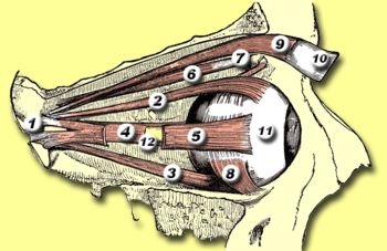
Four of these then course forward through the orbit and insert onto the globe on its anterior half (i.e., in front of the eye's equator). These muscles are named after their straight paths, and are called the four rectus muscles, or four recti.
- superior rectus - inserts on the globe at 2
- inferior rectus - inserts on the globe at 3
- medial rectus - inserts on the globe at 4
- lateral rectus - inserts on the globe at 5
(Note that lateral and medial are relative to the subject, with lateral toward the side and medial toward the midline, thus the medial rectus is the muscle closest to the nose).
Two with more complex paths
The other two extraocular muscles follow more complicated paths.- The superior oblique muscle originates at the back of the orbit (a little closer to the medial rectus, though medial to it, getting rounder as it courses forward to a rigid, cartilaginous pulley, called the trochlea, on the upper, nasal wall of the orbit. The muscle becomes tendinous about 10mm before it passes through the pulley, turning sharply across the orbit, and inserts on the lateral, posterior part of the globe. Thus, the superior oblique travels posteriorly for the last part of its path, going over the top of the eye. Due to its unique path, the superior oblique, when activated, pulls the eye downward and medially.
- The last muscle is the inferior oblique, which originates at the lower front of the nasal orbital wall, and passes under the LR to insert on the lateral, posterior part of the globe. Thus, the inferior oblique pulls the eye upward and laterally .
Rolling
The superior and inferior recti are not strictly vertical. The oblique pull of the obliques causes a rolling opposite each other. Although bearing mutual strict antagonism, the superior and inferior rectus team up with the inferior and superior oblique to move the eye up or down, respectively. The extent of rolling in the recti is less than the oblique, and opposite from it.Mnemonics
A good mnemonic to remember which muscles are innervated by what nerve is to paraphrase it as a molecular equation: LR6SO4R3. or (LR6SO4)3 i.e. "LR 6 SO 4 Whole 3."- Lateral Rectus - Cranial Nerve VI
- Superior Oblique - Cranial Nerve IV
- the Rest of the muscles - Cranial Nerve III.
Another way to remember which nerves innervate which muscles is to understand the meaning behind all of the Latin words.
- The fourth cranial nerve, the trochlear, is so named because the muscle it innervates, the superior oblique, runs through a little fascial pulley that changes its direction of pull (the trochlea of superior obliqueTrochlea of superior obliqueThe Trochlea of superior oblique is a pulley structure in the eye. The tendon of the superior oblique muscle passes through it. Situated on the superior nasal aspect of the frontal bone, it is the only cartilage found in the normal orbit....
). This pulley exists in the superiomedial corner of each orbit, and "trochl-" is Latin for "pulley." - The sixth cranial nerve, the abducens, is so named because it controls the lateral rectus, which abducts the eye (rotates it laterally) upon contraction.
- The third cranial nerve, the oculomotor, is so named because it is in charge of the movement (motor) of the eye (oculo-). It controls all of the other muscles.
See also
- Hering's law of equal innervationHering's law of equal innervationHering's law of equal innervation is used to explain the conjugacy of saccadic eye movement in stereoptic animals. The law proposes that conjugacy of saccades is due to innate connections in which the eye muscles responsible for each eye's movements are innervated equally...
- Sherrington's law of reciprocal innervationSherrington's law of reciprocal innervationSherrington's law of reciprocal innervation, also called Sherrington's law II explains how a muscle will relax when its opposite muscle is activated. René Descartes had hypothesized as much in 1626...