
Development of the reproductive system
Encyclopedia
The development of the reproductive system is a part of prenatal development, and concerns the sex organs. It is a part of the stages of sexual differentiation
. Because its location to a large extent overlaps the urinary system, the development of them can also be described together as the development of the urinary and reproductive organs
.
The reproductive organs are developed from the intermediate mesoderm
. The permanent organs of the adult are preceded by a set of structures which are purely embryonic, and which with the exception of the ducts disappear almost entirely before the end of fetal life. These embryonic structures are the Wolffian
and Müllerian duct
s, also known as mesonephric and paramesonephric ducts, respectively. The Wolffian duct remains as the duct in males, and the Müllerian as that of the female.
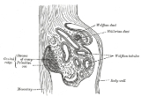
, immediately under the ectoderm, in the region from the fifth cervical segment to the third thoracic segment, a series of short evaginations from each segment grows dorsally and extends caudally, fusing successively from before backward to form the pronephric duct
. This continues to grow caudalward until it opens into the ventral part of the cloaca
; beyond the pronephros it is termed the Wolffian duct
. Thus, the Wolffian duct is what remains of the pronephric duct after the atrophy of the pronephros.
persists, and forms the tube of the epididymis
, the ductus deferens and the ejaculatory duct
, while the seminal vesicle
arises during the third month as a lateral diverticulum from its hinder end. A large part of the head end of the mesonephros atrophies and disappears; of the remainder the anterior tubules form the efferent ducts of the testis; while the posterior tubules are represented by the ductuli aberrantes
, and by the paradidymis
, which is sometimes found in front of the spermatic cord
above the head of the epididymis.
, and the paroöphoron
, two small collections of rudimentary blind tubules which are situated in the mesosalpinx
.
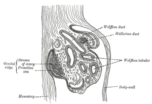 The lower part of the Wolffian duct disappears, while the upper part persists as the longitudinal duct of the epoöphoron
, called Gartner's duct
.
There are also developments of other tissues from the Wolffian duct that persist, e.g. the development of the suspensory ligament of the ovary
.
 Shortly after the formation of the Wolffian ducts a second pair of ducts is developed; these are the Müllerian ducts. Each arises on the lateral aspect of the corresponding Wolffian duct as a tubular invagination of the cells lining the abdominal cavity. The orifice of the invagination remains open, and undergoes enlargement and modification to form the abdominal ostium of the fallopian tube. The ducts pass backward lateral to the Wolffian ducts, but toward the posterior end of the embryo they cross to the medial side of these ducts, and thus come to lie side by side between and behind the latter—the four ducts forming what is termed the common genital cord, to distinguish it from the genital cords of the germinal epithelium seen later in this article. The Müllerian ducts end in an epithelial elevation, the Müllerian eminence
, on the ventral part of the cloaca between the orifices of the Wolffian ducts. At a later stage the eminence opens in the middle, connecting the Müllerian ducts with the cloaca.
. This is due to the production of Anti-Müllerian hormone
by the Sertoli cells of the testes.
and vagina
. This fusion of the Müllerian ducts begins in the third month, and the septum formed by their fused medial walls disappears from below upward.
The parts outside this cord remain separate, and each forms the corresponding Fallopian tube
. The ostium of the fallopian tube remains from the anterior extremity of the original tubular invagination from the abdominal cavity.
About the fifth month a ring-like constriction marks the position of the cervix
of the uterus, and after the sixth month the walls of the uterus begin to thicken. For a time the vagina is represented by a solid rod of epithelial cells. A ring-like outgrowth of this epithelium occurs at the lower end of the uterus and marks the future vaginal fornix
. At about the fifth or sixth month the lumen
of the vagina is produced by the breaking down of the central cells of the epithelium. The hymen
represents the remains of the Müllerian eminence .
of the yolk sac
. Once they have reached the gonadal ridge they are called oogonia. Development proceeds and the oogonia become fully surrounded by a layer of connective tissue cells (pre-granulosa cells) In this way, the rudiments of the ovarian follicles are formed. The embryological origin of granulosa cells, on the other hand, remains controversial. Just as in the male, there is a gubernaculum
in the female, which pulls it downward, albeit not as much as in males. The gubernaculum later becomes the proper ovarian ligament
and the round ligament of the uterus.
. Cords of the central mass run together and form a network which becomes the rete testis
, and another network, which develops the seminiferous tubules
. Via the rete testis, the seminiferous tubules become connected with outgrowths from the mesonephros, which form the efferent ducts of the testis.
In short, the descent of the testes consists of the opening of a connection from the testis to its final location at the anterior abdominal wall, followed by the development of the gubernaculum, which subsequently pulls and translocates the testis down into the developing scrotum. Ultimately, the passageway closes behind the testis. A failure in this process can cause indirect inguinal hernia
or an infantile hydrocoele.
 After the separation of the rectum from the dorsal part of the cloaca, the ventral part becomes the primary urogenital sinus. The urogenital sinus, in turn, divides into the superficial definitive urogenital sinus and the deeper anterior vesico-urethral portion.
. The remainder of the vesico-urethral portion forms the body of the bladder and part of the prostatic urethra; its apex is prolonged to the umbilicus as a narrow canal, the urachus
, which later is obliterated and becomes the median umbilical ligament
of the adult.
originally consists of two separate portions, each of which arises as a series of diverticular buds from the epithelial lining of the urogenital sinus and vesico-urethral part of the cloaca, between the third and fourth months. These buds become tubular, and form the glandular substance of the two lobes, which ultimately meet and fuse behind the urethra and also extend on to its ventral aspect. The median lobe of the prostate is formed as an extension of the lateral lobes between the common ejaculatory ducts and the bladder.
Skene's glands in the female urethra are regarded as the homologues of the prostatic glands.
The bulbourethral glands in the male, and Bartholin's gland
in the female, also arise as diverticula from the epithelial lining of the urogenital sinus.
 Until about the ninth week of gestational age the external genitalia of males and females look the same, and follow a common development. This includes the development of a genital tubercle and a membrane dorsally to it, covering the developing urogenital opening
, and the development of labioscrotal folds.
Even after differentiation can be seen between the sexes, some stages are common, e.g. the disappearing of the membrane. On the other hand, sex-dependent development include further protrusion of the genital tubercle in the male to form the penis. Furthermore, the labioscrotal folds evolve into the scrotum in males, while they evolve into labia in females.
There is initially a cloacal membrane
, composed of ectoderm and endoderm, reaching from the umbilical cord
to the tail, separating the cloaca from the exterior. After the separation of the rectum from the dorsal part of the cloaca, the ventral part of the cloacal membrane becomes the urogenital membrane.
Mesoderm extends to the midventral line for some distance behind the umbilical cord, and forms the lower part of the abdominal wall; it ends below in a prominent swelling, the cloacal tubercle, which after the separation of the rectum becomes the genital tubercle
. Dorsally to this tubercle the sides aren't really fused. Rather, the urogenital part of the cloacal membrane separates the ingrowing sheets of mesoderm.
The genital tubercle develops into the phallus
, the first rudiment of the penis or clitoris.
The terminal part of the phallus, representing the future glans becomes solid. The remainder of the phallus, which remains hollow, is converted into a longitudinal groove by the absorption of the urogenital membrane.
The term genital tubercle, however, still remains, but only refers to the future glans
In both sexes the phallic portion of the urogenital sinus
extends on to the under surface of the cloacal tubercle as far forward as the apex. At the apex the walls of the phallic portion come together and fuse, obliterating the urogenital opening
. Instead, a solid plate, the urethral plate, is formed. The remainder of the phallic portion is for a time tubular, and then, by the absorption of the urogenital membrane, it establishes a communication with the exterior. This opening is for a while the primitive urogenital opening, and it extends forward to the corona glandis.
 The following developments occur in both males and females, although a difference in the development between the sexes already can be seen:
, which ultimately form the labia majora in females. The labia minora, in contrast, arise by the continued growth of the lips of the groove on the under surface of the phallus; the remainder of the phallus forms the clitoris
. The immature glans becomes the clitoral glans
.
The labioscrotal folds extend around between the pelvic portion and the anus, and form a scrotal area. During the changes associated with the descent of the testes this scrotal area is drawn out to form the scrotal sacs. The penis is developed from the phallus.
As in the female, the urogenital membrane undergoes absorption, forming a channel on the under surface of the phallus; this channel extends only as far forward as the corona glandis.
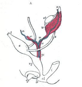
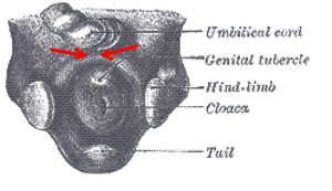
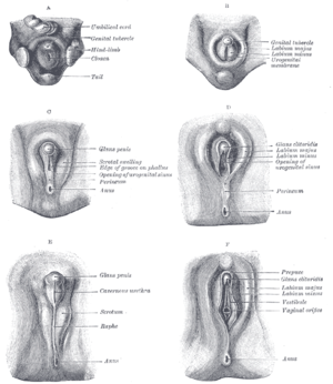
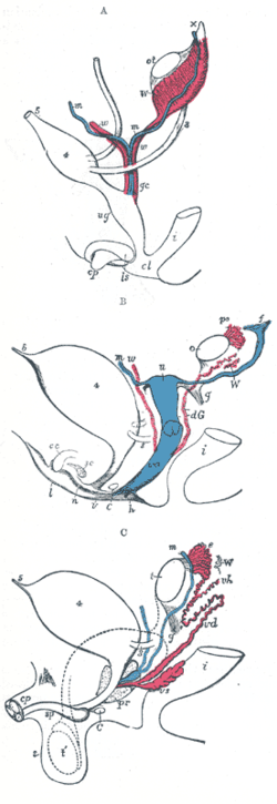 A.—Diagram of the primitive urogenital organs in the embryo previous to sexual distinction.
B.—Diagram of the female type of sexual organs.
C.—Diagram of the male type of sexual organs.
3. The Urogenital Apparatus
Sexual differentiation
Sexual differentiation is the process of development of the differences between males and females from an undifferentiated zygote...
. Because its location to a large extent overlaps the urinary system, the development of them can also be described together as the development of the urinary and reproductive organs
Development of the urinary and reproductive organs
The development of the urinary and reproductive organs as a part of the prenatal development, concerns the urinary system and sex organs. The latter is a part of the stages of sexual differentiation....
.
The reproductive organs are developed from the intermediate mesoderm
Intermediate mesoderm
Intermediate mesenchyme or intermediate mesoderm is a type of mesoderm that is located between the paraxial mesoderm and the lateral plate.It develops into the part of the urogenital system * forms of urogenital system...
. The permanent organs of the adult are preceded by a set of structures which are purely embryonic, and which with the exception of the ducts disappear almost entirely before the end of fetal life. These embryonic structures are the Wolffian
Wolffian duct
The mesonephric duct is a paired organ found in mammals including humans during embryogenesis....
and Müllerian duct
Müllerian duct
Müllerian ducts are paired ducts of the embryo that run down the lateral sides of the urogenital ridge and terminate at the Müllerian eminence in the primitive urogenital sinus. In the female, they will develop to form the Fallopian tubes, uterus, cervix, and the upper two-third of the vagina; in...
s, also known as mesonephric and paramesonephric ducts, respectively. The Wolffian duct remains as the duct in males, and the Müllerian as that of the female.
Origin
In the outer part of the intermediate mesodermIntermediate mesoderm
Intermediate mesenchyme or intermediate mesoderm is a type of mesoderm that is located between the paraxial mesoderm and the lateral plate.It develops into the part of the urogenital system * forms of urogenital system...
, immediately under the ectoderm, in the region from the fifth cervical segment to the third thoracic segment, a series of short evaginations from each segment grows dorsally and extends caudally, fusing successively from before backward to form the pronephric duct
Pronephric duct
-Development:The development of the pronephric duct is a part of the development of the urinary and reproductive organs.In the outer part of the intermediate mesoderm, immediately under the ectoderm, in the region from the fifth cervical segment to the third thoracic segment, a series of short...
. This continues to grow caudalward until it opens into the ventral part of the cloaca
Cloaca
In zoological anatomy, a cloaca is the posterior opening that serves as the only such opening for the intestinal, reproductive, and urinary tracts of certain animal species...
; beyond the pronephros it is termed the Wolffian duct
Wolffian duct
The mesonephric duct is a paired organ found in mammals including humans during embryogenesis....
. Thus, the Wolffian duct is what remains of the pronephric duct after the atrophy of the pronephros.
Development in male
In the male the Wolffian ductWolffian duct
The mesonephric duct is a paired organ found in mammals including humans during embryogenesis....
persists, and forms the tube of the epididymis
Epididymis
The epididymis is part of the male reproductive system and is present in all male amniotes. It is a narrow, tightly-coiled tube connecting the efferent ducts from the rear of each testicle to its vas deferens. A similar, but probably non-homologous, structure is found in cartilaginous...
, the ductus deferens and the ejaculatory duct
Ejaculatory duct
-Anatomy:The ejaculatory ducts are paired structures in male anatomy. Each ejaculatory duct is formed by the union of the vas deferens with the duct of the seminal vesicle. They pass through the prostate, and open into the urethra at the Colliculus seminalis...
, while the seminal vesicle
Seminal vesicle
The seminal vesicles or vesicular glands are a pair of simple tubular glands posteroinferior to the urinary bladder of male mammals...
arises during the third month as a lateral diverticulum from its hinder end. A large part of the head end of the mesonephros atrophies and disappears; of the remainder the anterior tubules form the efferent ducts of the testis; while the posterior tubules are represented by the ductuli aberrantes
Ductuli aberrantes
Ductuli Aberrantes.—A long narrow tube, the ductulus aberrans inferior , is occasionally found connected with the lower part of the canal of the epididymis, or with the commencement of the ductus deferens....
, and by the paradidymis
Paradidymis
The term paradidymis is applied to a small collection of convoluted tubules, situated in front of the lower part of the spermatic cord, above the head of the epididymis....
, which is sometimes found in front of the spermatic cord
Spermatic cord
The spermatic cord is the name given to the cord-like structure in males formed by the ductus deferens and surrounding tissue that run from the abdomen down to each testicle.-Contents of spermatic cord:...
above the head of the epididymis.
Atrophy in female
In the female the Wolffian bodies and ducts atrophy. The nonfunctional remains of the Wolffian tubules are represented by the epoophoronEpoophoron
The epoophoron or epoöphoron is a remnant of the Mesonephric duct that can be found next to the ovary and fallopian tube.-Anatomy:...
, and the paroöphoron
Paroöphoron
The paroöphoron consists of a few scattered rudimentary tubules, best seen in the child, situated in the broad ligament between the epoöphoron and the uterus. Named for the Welsh anatomist David Johnson who originally described the structure at the University of Wales, Aberystwyth.It is a remnant...
, two small collections of rudimentary blind tubules which are situated in the mesosalpinx
Mesosalpinx
The mesosalpinx is part of the lining of the abdominal cavity in higher vertebrates, specifically the portion of the broad ligament that stretches from the ovary to the level of the uterine tube....
.
Remnants
Epoophoron
The epoophoron or epoöphoron is a remnant of the Mesonephric duct that can be found next to the ovary and fallopian tube.-Anatomy:...
, called Gartner's duct
Gartner's duct
Gartner's duct is a potential embryological remnant in human female development of the mesonephric ducts in the development of the urinary and reproductive organs...
.
There are also developments of other tissues from the Wolffian duct that persist, e.g. the development of the suspensory ligament of the ovary
Development of the suspensory ligament of the ovary
The prenatal development of the suspensory ligament of the ovary is a part of the development of the reproductive system.The suspensory ligament originates from the mesonephros, which, in turn, originates from the Wolffian duct:...
.
The Müllerian (paramesonephric) Duct
Müllerian eminence
The Müllerian eminence is an epithelial on the ventral part of the cloaca between the orifices of the Wolffian ducts. It appears during the development of the urinary and reproductive organs.-Function:The Müllerian ducts end here...
, on the ventral part of the cloaca between the orifices of the Wolffian ducts. At a later stage the eminence opens in the middle, connecting the Müllerian ducts with the cloaca.
Atrophy in males
In the male the Müllerian ducts atrophy, but traces of their anterior ends are represented by the appendices testis (hydatids of Morgagni of the male), while their terminal fused portions form the utriculus in the floor of the prostatic urethraProstatic urethra
The prostatic urethra, the widest and most dilatable part of the urethra canal, is about 3 cm. long.It runs almost vertically through the prostate from its base to its apex, lying nearer its anterior than its posterior surface; the form of the canal is spindle-shaped, being wider in the middle...
. This is due to the production of Anti-Müllerian hormone
Anti-müllerian hormone
Anti-Müllerian hormone also known as AMH is a protein that, in humans, is encoded by the AMH gene. It inhibits the development of the Müllerian ducts in the male embryo. It has also been called Müllerian inhibiting factor , Müllerian-inhibiting hormone , and Müllerian-inhibiting substance...
by the Sertoli cells of the testes.
Development in females
In the female the Müllerian ducts persist and undergo further development. The portions which lie in the genital cord fuse to form the uterusUterus
The uterus or womb is a major female hormone-responsive reproductive sex organ of most mammals including humans. One end, the cervix, opens into the vagina, while the other is connected to one or both fallopian tubes, depending on the species...
and vagina
Vagina
The vagina is a fibromuscular tubular tract leading from the uterus to the exterior of the body in female placental mammals and marsupials, or to the cloaca in female birds, monotremes, and some reptiles. Female insects and other invertebrates also have a vagina, which is the terminal part of the...
. This fusion of the Müllerian ducts begins in the third month, and the septum formed by their fused medial walls disappears from below upward.
The parts outside this cord remain separate, and each forms the corresponding Fallopian tube
Fallopian tube
The Fallopian tubes, also known as oviducts, uterine tubes, and salpinges are two very fine tubes lined with ciliated epithelia, leading from the ovaries of female mammals into the uterus, via the utero-tubal junction...
. The ostium of the fallopian tube remains from the anterior extremity of the original tubular invagination from the abdominal cavity.
About the fifth month a ring-like constriction marks the position of the cervix
Cervix
The cervix is the lower, narrow portion of the uterus where it joins with the top end of the vagina. It is cylindrical or conical in shape and protrudes through the upper anterior vaginal wall...
of the uterus, and after the sixth month the walls of the uterus begin to thicken. For a time the vagina is represented by a solid rod of epithelial cells. A ring-like outgrowth of this epithelium occurs at the lower end of the uterus and marks the future vaginal fornix
Vaginal fornix
The fornices of the vagina are the deepest portions of the vagina, extending into the recesses created by the vaginal portion of cervix. The word 'fornix' is Latin for 'arch'....
. At about the fifth or sixth month the lumen
Lumen (anatomy)
A lumen in biology is the inside space of a tubular structure, such as an artery or intestine...
of the vagina is produced by the breaking down of the central cells of the epithelium. The hymen
Hymen
The hymen is a membrane that surrounds or partially covers the external vaginal opening. It forms part of the vulva, or external genitalia. The size of the hymenal opening increases with age. Although an often practiced method, it is not possible to confirm with certainty that a girl or woman is a...
represents the remains of the Müllerian eminence .
Gonads
The gonads are the precursors of the testes in males and ovaries in females. They initially develop from the mesothelial layer of the peritoneum.Ovaries
The ovary is differentiated into a central part, the medulla of ovary, covered by a surface layer, the germinal epithelium. The immature ova originate from cells from the dorsal endodermEndoderm
Endoderm is one of the three primary germ cell layers in the very early embryo. The other two layers are the ectoderm and mesoderm , with the endoderm as the intermost layer...
of the yolk sac
Yolk sac
The yolk sac is a membranous sac attached to an embryo, providing early nourishment in the form of yolk in bony fishes, sharks, reptiles, birds, and primitive mammals...
. Once they have reached the gonadal ridge they are called oogonia. Development proceeds and the oogonia become fully surrounded by a layer of connective tissue cells (pre-granulosa cells) In this way, the rudiments of the ovarian follicles are formed. The embryological origin of granulosa cells, on the other hand, remains controversial. Just as in the male, there is a gubernaculum
Gubernaculum
The paired Gubernacula are embryonic structures which begin as undifferentiated mesenchyme attaching to the caudal end of the gonads .-Function during development:...
in the female, which pulls it downward, albeit not as much as in males. The gubernaculum later becomes the proper ovarian ligament
Ovarian ligament
The ovarian ligament is a fibrous ligament that connects the ovary to the lateral surface of the uterus....
and the round ligament of the uterus.
Testes
The periphery of the testes are converted into the tunica albugineaTunica albuginea (testicles)
The Tunica Albuginea is the fibrous covering of the testis.It is a dense blue-white membrane, composed of bundles of white fibrous tissue which interlace in every direction....
. Cords of the central mass run together and form a network which becomes the rete testis
Rete testis
Rete testis is an anastomosing network of delicate tubules located in the hilum of the testicle that carries sperm from the seminiferous tubules to the vasa efferentia....
, and another network, which develops the seminiferous tubules
Seminiferous tubules
Seminiferous tubules are located in the testes, and are the specific location of meiosis, and the subsequent creation of gametes, namely spermatozoa....
. Via the rete testis, the seminiferous tubules become connected with outgrowths from the mesonephros, which form the efferent ducts of the testis.
In short, the descent of the testes consists of the opening of a connection from the testis to its final location at the anterior abdominal wall, followed by the development of the gubernaculum, which subsequently pulls and translocates the testis down into the developing scrotum. Ultimately, the passageway closes behind the testis. A failure in this process can cause indirect inguinal hernia
Indirect inguinal hernia
An indirect inguinal hernia is an inguinal hernia that results from the failure of embryonic closure of the deep inguinal ring after the testicle has passed through it. Like other inguinal hernias, it protrudes through the superficial inguinal ring...
or an infantile hydrocoele.
Division of cloaca
Definitive urogenital sinus
The definitive urogenital sinus consists of a caudal cephallic portion and an intermediate narrow channel, the pelvic portion.Vesico-urethral portion
The vesico-urethral portion is the deepest portion, continuous with the allantois. It absorbs the ends of the Wolffian ducts and the associated ends of the renal diverticula, and these give rise to the trigone of urinary bladder and part of the prostatic urethraProstatic urethra
The prostatic urethra, the widest and most dilatable part of the urethra canal, is about 3 cm. long.It runs almost vertically through the prostate from its base to its apex, lying nearer its anterior than its posterior surface; the form of the canal is spindle-shaped, being wider in the middle...
. The remainder of the vesico-urethral portion forms the body of the bladder and part of the prostatic urethra; its apex is prolonged to the umbilicus as a narrow canal, the urachus
Urachus
The urachus is a fibrous remnant of the allantois, a canal that drains the urinary bladder of the fetus that joins and runs within the umbilical cord...
, which later is obliterated and becomes the median umbilical ligament
Median umbilical ligament
The median umbilical ligament is a structure in human anatomy. It is a shrivelled piece of tissue that represents the remnant of the embryonic urachus.It extends from the apex of the bladder to the umbilicus, on the deep surface of the anterior abdominal wall...
of the adult.
The Prostate
The prostateProstate
The prostate is a compound tubuloalveolar exocrine gland of the male reproductive system in most mammals....
originally consists of two separate portions, each of which arises as a series of diverticular buds from the epithelial lining of the urogenital sinus and vesico-urethral part of the cloaca, between the third and fourth months. These buds become tubular, and form the glandular substance of the two lobes, which ultimately meet and fuse behind the urethra and also extend on to its ventral aspect. The median lobe of the prostate is formed as an extension of the lateral lobes between the common ejaculatory ducts and the bladder.
Skene's glands in the female urethra are regarded as the homologues of the prostatic glands.
The bulbourethral glands in the male, and Bartholin's gland
Bartholin's gland
The Bartholin's glands are two glands located slightly posterior and to the left and right of the opening of the vagina. They secrete mucus to lubricate the vagina and are homologous to bulbourethral glands in males...
in the female, also arise as diverticula from the epithelial lining of the urogenital sinus.
External genitalia
Urogenital opening
The urogenital opening is where waste products of the body and reproductive fluids are expelled to the environment outside of the body cavity. In some organisms, including birds and many fish, discharge from the urological, digestive, and reproductive systems empty into a common sac called the...
, and the development of labioscrotal folds.
Even after differentiation can be seen between the sexes, some stages are common, e.g. the disappearing of the membrane. On the other hand, sex-dependent development include further protrusion of the genital tubercle in the male to form the penis. Furthermore, the labioscrotal folds evolve into the scrotum in males, while they evolve into labia in females.
Urogenital membrane
There is initially a cloacal membrane
Cloacal membrane
The cloacal membrane is the membrane that covers the embryonic cloaca when still in the development of the urinary and reproductive organs.It is formed by ectoderm and endoderm coming into contact with each other...
, composed of ectoderm and endoderm, reaching from the umbilical cord
Umbilical cord
In placental mammals, the umbilical cord is the connecting cord from the developing embryo or fetus to the placenta...
to the tail, separating the cloaca from the exterior. After the separation of the rectum from the dorsal part of the cloaca, the ventral part of the cloacal membrane becomes the urogenital membrane.
Genital tubercle
Mesoderm extends to the midventral line for some distance behind the umbilical cord, and forms the lower part of the abdominal wall; it ends below in a prominent swelling, the cloacal tubercle, which after the separation of the rectum becomes the genital tubercle
Genital tubercle
A phallic tubercle or genital tubercle is a body of tissue present in the development of the urinary and reproductive organs. It forms in the ventral, caudal region of mammalian embryos of both sexes, and eventually develops into a phallus...
. Dorsally to this tubercle the sides aren't really fused. Rather, the urogenital part of the cloacal membrane separates the ingrowing sheets of mesoderm.
Phallus
The genital tubercle develops into the phallus
Phallus
A phallus is an erect penis, a penis-shaped object such as a dildo, or a mimetic image of an erect penis. Any object that symbolically resembles a penis may also be referred to as a phallus; however, such objects are more often referred to as being phallic...
, the first rudiment of the penis or clitoris.
The terminal part of the phallus, representing the future glans becomes solid. The remainder of the phallus, which remains hollow, is converted into a longitudinal groove by the absorption of the urogenital membrane.
The term genital tubercle, however, still remains, but only refers to the future glans
Urogenital opening
In both sexes the phallic portion of the urogenital sinus
Urogenital sinus
The definitive urogenital sinus is a part of the human body only present in the development of the urinary and reproductive organs...
extends on to the under surface of the cloacal tubercle as far forward as the apex. At the apex the walls of the phallic portion come together and fuse, obliterating the urogenital opening
Urogenital opening
The urogenital opening is where waste products of the body and reproductive fluids are expelled to the environment outside of the body cavity. In some organisms, including birds and many fish, discharge from the urological, digestive, and reproductive systems empty into a common sac called the...
. Instead, a solid plate, the urethral plate, is formed. The remainder of the phallic portion is for a time tubular, and then, by the absorption of the urogenital membrane, it establishes a communication with the exterior. This opening is for a while the primitive urogenital opening, and it extends forward to the corona glandis.
After differentiation
- The corpora cavernosa of the penis or clitoris and of the urethra arise from the mesodermal tissue in the phallus; they are at first dense structures, but later vascular spaces appear in them, and they gradually become cavernous.
- The prepucePrepucePrepuce may refer to:* The foreskin, which surrounds and protects the head of the penis* The clitoral hood, which surrounds and protects the head of the clitoris...
in both sexes is formed by the growth of a solid plate of ectoderm into the superficial part of the phallus; on coronal section this plate presents the shape of a horseshoe. By the breaking down of its more centrally situated cells the plate is split into two lamellæ. Thus, a cutaneous fold, the prepuce, is liberated and forms a hood over the glans.
Female
In the female, a deep groove forms around the phallus. The sides of it grow dorsalward as the labioscrotal foldsLabioscrotal folds
The labioscrotal folds are paired structures in the human embryo that represent the final stage of development of the caudal end of the external genitals before sexual differentiation. In both males and females the two swellings merge:* In the female, they become the posterior labial commissure...
, which ultimately form the labia majora in females. The labia minora, in contrast, arise by the continued growth of the lips of the groove on the under surface of the phallus; the remainder of the phallus forms the clitoris
Clitoris
The clitoris is a sexual organ that is present only in female mammals. In humans, the visible button-like portion is located near the anterior junction of the labia minora, above the opening of the urethra and vagina. Unlike the penis, which is homologous to the clitoris, the clitoris does not...
. The immature glans becomes the clitoral glans
Clitoral glans
The clitoral glans is an external portion of the clitoris.- Anatomy :It is covered by the clitoral hood, which is also external and attached to the labia minora...
.
Male
In the male the pelvic portion of the cloaca undergoes much greater development, pushing before it the phallic portion.The labioscrotal folds extend around between the pelvic portion and the anus, and form a scrotal area. During the changes associated with the descent of the testes this scrotal area is drawn out to form the scrotal sacs. The penis is developed from the phallus.
As in the female, the urogenital membrane undergoes absorption, forming a channel on the under surface of the phallus; this channel extends only as far forward as the corona glandis.
Urogenital opening
In the male, by the greater growth of the pelvic portion of the cloaca, a longer urethra is formed, and the primitive opening is carried forward with the phallus, but it still ends at the corona glandis. Later, this opening, which is located on the dorsal ide of the penis, closes from behind forward. Meanwhile, the urethral plate of the glans breaks down centrally to form a median groove continuous with the primitive ostium. This groove also closes from behind forward, leaving only a small pipe running in the middle of the penis. Thus, the urogenital opening is shifted forward to the end of the glans.Diagram of internal differentiation
- 3. Ureter.
- 4. Urinary bladder.
- 5. Urachus.
- cl. Cloaca.
- cp. Elevation which becomes clitoris or penis.
- i. Lower part of the intestine.
- ls. Fold of integument from which the labia majora or scrotum are formed.
- m, m. Right and left Müllerian ducts uniting together and running with the Wolffian ducts in gc, the genital cord.
- ot. The genital ridge from which either the ovary or testis is formed.
- ug. Sinus urogenitalis.
- W. Left Wolffian body.
- w, w. Right and left Wolffian ducts.
B.—Diagram of the female type of sexual organs.
- C. Greater vestibular gland, and immediately above it the urethra.
- cc. Corpus cavernosum clitoridis.
- dG. Remains of the left Wolffian duct, such as give rise to the duct of Gärtner, represented by dotted lines; that of the right side is marked w.
- f. The abdominal opening of the left uterine tube.
- g. Round ligament, corresponding to gubernaculum.
- h. Situation of the hymen.
- i. Lower part of the intestine.
- l. Labium major.
- n. Labium minus.
- o. The left ovary.
- po. Epoophoron.
- sc. Corpus cavernosum urethrae.
- u. Uterus. The uterine tube of the right side is marked m.
- v. Vulva.
- va. Vagina.
- W. Scattered remains of Wolffian tubes near it (paroöphoron of Waldeyer).
C.—Diagram of the male type of sexual organs.
- C. Bulbo-urethral gland of one side.
- cp. Corpora cavernosa penis cut short.
- e. Caput epididymis.
- g. The gubernaculum.
- i. Lower part of the intestine.
- m. Müllerian duct, the upper part of which remains as the hydatid of Morgagni; the lower part, represented by a dotted line descending to the prostatic utricle, constitutes the occasionally existing cornu and tube of the uterus masculinus.
- pr. The prostate.
- s. Scrotum.
- sp. Corpus cavernosum urethrae.
- t. Testis in the place of its original formation.
- t’, together with the dotted lines above, indicates the direction in which the testis and epididymis descend from the abdomen into the scrotum.
- vd. Ductus deferens.
- vh. Ductus aberrans.
- vs. The vesicula seminalis.
- W. Scattered remains of the Wolffian body, constituting the organ of Giraldès, or the paradidymis of Waldeyer.
General
- http://www.bartleby.com/107/252.html Henry Gray (1821–1865). Anatomy of the Human Body. 1918.
3. The Urogenital Apparatus
External links
- http://www.aboutkidshealth.ca/HowTheBodyWorks/Sexual-Differentiation.aspx?articleID=6850&categoryID=XS-nh3 Animations of gonadal sex, duct differentiation, and external genital development at AboutKidsHealth.ca