

Dental radiography
Encyclopedia
Dental radiographs, commonly referred to as X-ray
films, or informally, X-rays, are pictures of the teeth, bones, and surrounding soft tissues to screen for and help identify problems with the teeth, mouth, and jaw. X-ray pictures can show cavities, cancerous or benign masses, hidden dental structures (such as wisdom teeth), and bone loss that cannot be seen during a visual examination. Dental X-rays may also be done as follow-up after dental treatments.
A radiographic image is formed by a controlled burst of X-ray radiation which penetrates oral structures at different levels, depending on varying anatomical densities, before striking the film or sensor. Teeth appear lighter because less radiation penetrates them to reach the film. Dental caries
, infections and other changes in the bone density, and the periodontal ligament
, appear darker because X-rays readily penetrate these less dense structures. Dental restorations (fillings, crowns) may appear lighter or darker, depending on the density of the material.
The dosage of X-ray radiation received by a dental patient is typically small (around 0.005 mSv
), equivalent to a few days' worth of background environmental radiation exposure, or similar to the dose received during a cross-country airplane flight (concentrated into one short burst aimed at a small area). Incidental exposure is further reduced by the use of a lead shield, lead apron, sometimes with a lead thyroid collar. Technician exposure is reduced by stepping out of the room, or behind adequate shielding material, when the X-ray source is activated.
Once photographic film
has been exposed to X-ray radiation, it needs to be developed, traditionally using a process where the film is exposed to a series of chemicals in a dark room, as the films are sensitive to normal light. This can be a time-consuming process, and incorrect exposures or mistakes in the development process can necessitate retakes, exposing the patient to additional radiation. Digital x-rays, which replace the film with an electronic sensor, address some of these issues, and are becoming widely used in dentistry as the technology evolves. They may require less radiation and are processed much quicker than conventional radiographic films, often instantly viewable on a computer. However digital sensors are extremely costly and have historically had poor resolution
, though this is much improved in modern sensors.
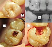 It is possible for both tooth decay
and periodontal disease
to be missed during a clinical exam, and radiographic evaluation of the dental and periodontal tissues is a critical segment of the comprehensive oral examination. The photographic montage at right depicts a situation in which extensive decay had been overlooked by a number of dentists prior to radiographic evaluation
teeth. The objective of this type of view is to capture the tip of the root on the film. This is often helpful in determining the cause of pain in a specific tooth, because it allows a dentist to visualize the tooth as well as the surrounding bone in their entirety. This view is often used to determine the need for endodontic therapy
as well as to visualize the successful progression of endodontic therapy once it is initiated.
It can be used in case of detection hyperdontia
(supernumerary teeth) & impacted teeth.
The name periapical is derived from the Greek peri, which means "around," and apical, which means "tip."
s, which are the demarcation lines on the teeth which separate tooth crown from tooth root. Routine bitewing radiographs are commonly used to examine for interdental caries and recurrent caries under existing restorations. When there is extensive bone loss, the films may be situated with their longer dimension in the vertical axis so as to better visualize their levels in relation to the teeth. Because bitewing views are taken from a more or less perpendicular angle to the buccal
surface of the teeth, they more accurately exhibit the bone levels than do periapical views. Bitewings of the anterior teeth are not routinely taken.
The name bitewing refers to a little tab of paper or plastic situated in the center of the X-ray film, which when bitten on, allows the film to hover so that it captures an even amount of maxilla
ry and mandibular information.
anatomy of either the floor of the mouth or the palate
. The occlusal film, which is about three to four times the size of the film used to take a periapical or bitewing, is inserted into the mouth so as to entirely separate the maxillary and mandibular teeth, and the film is exposed either from under the chin or angled down from the top of the nose. Sometimes, it is placed in the inside of the cheek to confirm the presence of a sialolith
in Stenson's duct, which carries saliva
from the parotid gland
. The occlusal view is not included in the standard full mouth series.
The Faculty of General Dental Practice
of the Royal College of Surgeons of England
publication Selection Criteria in Dental Radiography
holds that given current evidence
full mouth series are to be discouraged due to the large numbers of radiographs involved, many of which will not be necessary for the patient's treatment. An alternative approach using bitewing screening with selected periapical views is suggested as a method of minimising radiation dose to the patient while maximizing diagnostic yield.
A lateral cephalogram is used to evaluate dentofacial proportions and clarify the anatomic basis for a malocclusion, and an antero-posterior radiograph provides a face-forward view.
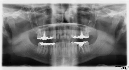 Panoramic films are extraoral films, in which the film is exposed while outside the patients' mouth, and they were developed by the United States Army
as a quick way to get an overall view of a soldiers' oral health. Exposing eighteen films per soldier was very time consuming, and it was felt that a single panoramic film could speed up the process of examining and assessing the dental health of the soldiers; soldiers with toothaches are not very effective. It was later discovered that while panoramic films can prove very useful in detecting and localizing mandibular fractures and other pathologic
entities of the mandible, they were not very good at assessing periodontal
bone loss or tooth decay.
Computed Tomography
There is increasing use of CT (computed tomography
) scans in dentistry, particularly to plan dental implants; there may be significant levels of radiation and potential risk. Specially designed CBCT (cone beam CT) scanners can be used instead, which produce adequate imaging with a tenfold reduction in radiation.
X-ray
X-radiation is a form of electromagnetic radiation. X-rays have a wavelength in the range of 0.01 to 10 nanometers, corresponding to frequencies in the range 30 petahertz to 30 exahertz and energies in the range 120 eV to 120 keV. They are shorter in wavelength than UV rays and longer than gamma...
films, or informally, X-rays, are pictures of the teeth, bones, and surrounding soft tissues to screen for and help identify problems with the teeth, mouth, and jaw. X-ray pictures can show cavities, cancerous or benign masses, hidden dental structures (such as wisdom teeth), and bone loss that cannot be seen during a visual examination. Dental X-rays may also be done as follow-up after dental treatments.
A radiographic image is formed by a controlled burst of X-ray radiation which penetrates oral structures at different levels, depending on varying anatomical densities, before striking the film or sensor. Teeth appear lighter because less radiation penetrates them to reach the film. Dental caries
Dental caries
Dental caries, also known as tooth decay or a cavity, is an irreversible infection usually bacterial in origin that causes demineralization of the hard tissues and destruction of the organic matter of the tooth, usually by production of acid by hydrolysis of the food debris accumulated on the...
, infections and other changes in the bone density, and the periodontal ligament
Periodontal ligament
The periodontal fiber or periodontal ligament, commonly abbreviated as the PDL, is a group of specialized connective tissue fibers that essentially attach a tooth to the alveolar bone within which it sits...
, appear darker because X-rays readily penetrate these less dense structures. Dental restorations (fillings, crowns) may appear lighter or darker, depending on the density of the material.
The dosage of X-ray radiation received by a dental patient is typically small (around 0.005 mSv
Sievert
The sievert is the International System of Units SI derived unit of dose equivalent radiation. It attempts to quantitatively evaluate the biological effects of ionizing radiation as opposed to just the absorbed dose of radiation energy, which is measured in gray...
), equivalent to a few days' worth of background environmental radiation exposure, or similar to the dose received during a cross-country airplane flight (concentrated into one short burst aimed at a small area). Incidental exposure is further reduced by the use of a lead shield, lead apron, sometimes with a lead thyroid collar. Technician exposure is reduced by stepping out of the room, or behind adequate shielding material, when the X-ray source is activated.
Once photographic film
Photographic film
Photographic film is a sheet of plastic coated with an emulsion containing light-sensitive silver halide salts with variable crystal sizes that determine the sensitivity, contrast and resolution of the film...
has been exposed to X-ray radiation, it needs to be developed, traditionally using a process where the film is exposed to a series of chemicals in a dark room, as the films are sensitive to normal light. This can be a time-consuming process, and incorrect exposures or mistakes in the development process can necessitate retakes, exposing the patient to additional radiation. Digital x-rays, which replace the film with an electronic sensor, address some of these issues, and are becoming widely used in dentistry as the technology evolves. They may require less radiation and are processed much quicker than conventional radiographic films, often instantly viewable on a computer. However digital sensors are extremely costly and have historically had poor resolution
Image resolution
Image resolution is an umbrella term that describes the detail an image holds. The term applies to raster digital images, film images, and other types of images. Higher resolution means more image detail....
, though this is much improved in modern sensors.
Dental caries
Dental caries, also known as tooth decay or a cavity, is an irreversible infection usually bacterial in origin that causes demineralization of the hard tissues and destruction of the organic matter of the tooth, usually by production of acid by hydrolysis of the food debris accumulated on the...
and periodontal disease
Periodontal disease
Periodontitis is a set of inflammatory diseases affecting the periodontium, i.e., the tissues that surround and support the teeth. Periodontitis involves progressive loss of the alveolar bone around the teeth, and if left untreated, can lead to the loosening and subsequent loss of teeth...
to be missed during a clinical exam, and radiographic evaluation of the dental and periodontal tissues is a critical segment of the comprehensive oral examination. The photographic montage at right depicts a situation in which extensive decay had been overlooked by a number of dentists prior to radiographic evaluation
Intraoral radiographic views
Placing the radiographic film or sensor inside the mouth produces an intraoral radiographic view.Periapical view
The periapical view is taken of both anterior and posteriorCommonly used terms of relationship and comparison in dentistry
There are numerous commonly used terms of relationship and comparison that refer to different aspects of teeth and are frequently utilized in articles about dentistry...
teeth. The objective of this type of view is to capture the tip of the root on the film. This is often helpful in determining the cause of pain in a specific tooth, because it allows a dentist to visualize the tooth as well as the surrounding bone in their entirety. This view is often used to determine the need for endodontic therapy
Endodontic therapy
Endodontic therapy is a sequence of treatment for the pulp of a tooth which results in the elimination of infection and protection of the decontaminated tooth from future microbial invasion...
as well as to visualize the successful progression of endodontic therapy once it is initiated.
It can be used in case of detection hyperdontia
Hyperdontia
Hyperdontia is the condition of having supernumerary teeth, or teeth which appear in addition to the regular number of teeth.-Types:Supernumerary teeth can be classified by shape and by position...
(supernumerary teeth) & impacted teeth.
The name periapical is derived from the Greek peri, which means "around," and apical, which means "tip."
Bitewing view
The bitewing view is taken to visualize the crowns of the posterior teeth and the height of the alveolar bone in relation to the cementoenamel junctionCementoenamel junction
The cementoenamel junction, frequently abbreviated as the CEJ, is an anatomical border identified on a tooth. It is the location where the enamel, which covers the anatomical crown of a tooth, and the cementum, which covers the anatomical root of a tooth, meet...
s, which are the demarcation lines on the teeth which separate tooth crown from tooth root. Routine bitewing radiographs are commonly used to examine for interdental caries and recurrent caries under existing restorations. When there is extensive bone loss, the films may be situated with their longer dimension in the vertical axis so as to better visualize their levels in relation to the teeth. Because bitewing views are taken from a more or less perpendicular angle to the buccal
Commonly used terms of relationship and comparison in dentistry
There are numerous commonly used terms of relationship and comparison that refer to different aspects of teeth and are frequently utilized in articles about dentistry...
surface of the teeth, they more accurately exhibit the bone levels than do periapical views. Bitewings of the anterior teeth are not routinely taken.
The name bitewing refers to a little tab of paper or plastic situated in the center of the X-ray film, which when bitten on, allows the film to hover so that it captures an even amount of maxilla
Maxilla
The maxilla is a fusion of two bones along the palatal fissure that form the upper jaw. This is similar to the mandible , which is also a fusion of two halves at the mental symphysis. Sometimes The maxilla (plural: maxillae) is a fusion of two bones along the palatal fissure that form the upper...
ry and mandibular information.
Occlusal view
The occlusal view is indicated when there is a desire to reveal the skeletal or pathologicPathology
Pathology is the precise study and diagnosis of disease. The word pathology is from Ancient Greek , pathos, "feeling, suffering"; and , -logia, "the study of". Pathologization, to pathologize, refers to the process of defining a condition or behavior as pathological, e.g. pathological gambling....
anatomy of either the floor of the mouth or the palate
Palate
The palate is the roof of the mouth in humans and other mammals. It separates the oral cavity from the nasal cavity. A similar structure is found in crocodilians, but, in most other tetrapods, the oral and nasal cavities are not truly separate. The palate is divided into two parts, the anterior...
. The occlusal film, which is about three to four times the size of the film used to take a periapical or bitewing, is inserted into the mouth so as to entirely separate the maxillary and mandibular teeth, and the film is exposed either from under the chin or angled down from the top of the nose. Sometimes, it is placed in the inside of the cheek to confirm the presence of a sialolith
Sialolithiasis
Sialolithiasis refers to the formation of stones in the salivary glands. Stones are most commonly found in the submandibular gland, where stones can obstruct Wharton's duct...
in Stenson's duct, which carries saliva
Saliva
Saliva , referred to in various contexts as spit, spittle, drivel, drool, or slobber, is the watery substance produced in the mouths of humans and most other animals. Saliva is a component of oral fluid. In mammals, saliva is produced in and secreted from the three pairs of major salivary glands,...
from the parotid gland
Parotid gland
The paired parotid glands are the largest of the salivary glands. They are each found wrapped around the mandibular ramus, and secrete saliva through Stensen's ducts into the oral cavity, to facilitate mastication and swallowing and to begin the digestion of starches.-Location:The parotid glands...
. The occlusal view is not included in the standard full mouth series.
Full mouth series
A full mouth series is a complete set of intraoral X-rays taken of a patients' teeth and adjacent hard tissue. This is often abbreviated as either FMS or FMX (or CMRS, meaning Complete Mouth Radiographic Series). The full mouth series is composed of 18 films:- four bitewings
- two molarMolar (tooth)Molars are the rearmost and most complicated kind of tooth in most mammals. In many mammals they grind food; hence the Latin name mola, "millstone"....
bitewings (left and right) - two premolarPremolarThe premolar teeth or bicuspids are transitional teeth located between the canine and molar teeth. In humans, there are two premolars per quadrant, making eight premolars total in the mouth. They have at least two cusps. Premolars can be considered as a 'transitional tooth' during chewing, or...
bitewings (left and right)
- two molar
- eight posterior periapicals
- two maxillary molar periapicals (left and right)
- two maxillary premolar periapicals (left and right)
- two mandibular molar periapicals (left and right)
- two mandibular premolar periapicals (left and right)
- six anterior periapicals
- two maxillary canine-lateral incisor periapicals (left and right)
- two mandibular canine-lateral incisor periapicals (left and right)
- two central incisor periapicals (maxillary and mandibular)
The Faculty of General Dental Practice
Faculty of General Dental Practice
-History:The FGDP was formed in 1992 as the academic home for general dental practitioners. It opened its membership up to Dental Care Professionals in 2005, and now supports the whole dental team. As of March 2011 there are approximately 4500 members of the FGDP...
of the Royal College of Surgeons of England
Royal College of Surgeons of England
The Royal College of Surgeons of England is an independent professional body and registered charity committed to promoting and advancing the highest standards of surgical care for patients, regulating surgery, including dentistry, in England and Wales...
publication Selection Criteria in Dental Radiography
Selection Criteria in Dental Radiography
This is a seminal publication of the Faculty of General Dental Practice of the Royal College of Surgeons of England.The publication marked the standardisation of dental radiography and now provides the dental profession with a set of evidence based criteria in order to minimse radiation exposure...
holds that given current evidence
Evidence-based medicine
Evidence-based medicine or evidence-based practice aims to apply the best available evidence gained from the scientific method to clinical decision making. It seeks to assess the strength of evidence of the risks and benefits of treatments and diagnostic tests...
full mouth series are to be discouraged due to the large numbers of radiographs involved, many of which will not be necessary for the patient's treatment. An alternative approach using bitewing screening with selected periapical views is suggested as a method of minimising radiation dose to the patient while maximizing diagnostic yield.
Extraoral radiographic views
Placing the radiographic film or sensor outside the mouth, on the opposite side of the head from the X-ray source, produces an extra-oral radiographic view.A lateral cephalogram is used to evaluate dentofacial proportions and clarify the anatomic basis for a malocclusion, and an antero-posterior radiograph provides a face-forward view.
Panoramic films
United States Army
The United States Army is the main branch of the United States Armed Forces responsible for land-based military operations. It is the largest and oldest established branch of the U.S. military, and is one of seven U.S. uniformed services...
as a quick way to get an overall view of a soldiers' oral health. Exposing eighteen films per soldier was very time consuming, and it was felt that a single panoramic film could speed up the process of examining and assessing the dental health of the soldiers; soldiers with toothaches are not very effective. It was later discovered that while panoramic films can prove very useful in detecting and localizing mandibular fractures and other pathologic
Pathology
Pathology is the precise study and diagnosis of disease. The word pathology is from Ancient Greek , pathos, "feeling, suffering"; and , -logia, "the study of". Pathologization, to pathologize, refers to the process of defining a condition or behavior as pathological, e.g. pathological gambling....
entities of the mandible, they were not very good at assessing periodontal
Periodontology
Periodontology or Periodontics is the specialty of dentistry that studies supporting structures of teeth, diseases, and conditions that affect them...
bone loss or tooth decay.
Computed TomographyComputed tomographyX-ray computed tomography or Computer tomography , is a medical imaging method employing tomography created by computer processing...
There is increasing use of CT (computed tomographyTomography
Tomography refers to imaging by sections or sectioning, through the use of any kind of penetrating wave. A device used in tomography is called a tomograph, while the image produced is a tomogram. The method is used in radiology, archaeology, biology, geophysics, oceanography, materials science,...
) scans in dentistry, particularly to plan dental implants; there may be significant levels of radiation and potential risk. Specially designed CBCT (cone beam CT) scanners can be used instead, which produce adequate imaging with a tenfold reduction in radiation.
See also
- OrthopantomogramOrthopantomogramAn Orthopantomogram or Dental Panoramic Radiograph , also known as an "orthopantogram" or "panorex", is a panoramic scanning dental X-ray of the upper and lower jaw. It shows a two-dimensional view of a half-circle from ear to ear. An OPT relies on tomography i.e...
- X-rayX-rayX-radiation is a form of electromagnetic radiation. X-rays have a wavelength in the range of 0.01 to 10 nanometers, corresponding to frequencies in the range 30 petahertz to 30 exahertz and energies in the range 120 eV to 120 keV. They are shorter in wavelength than UV rays and longer than gamma...
- RadiographyRadiographyRadiography is the use of X-rays to view a non-uniformly composed material such as the human body. By using the physical properties of the ray an image can be developed which displays areas of different density and composition....
- Digital radiography
- Selection Criteria in Dental RadiographySelection Criteria in Dental RadiographyThis is a seminal publication of the Faculty of General Dental Practice of the Royal College of Surgeons of England.The publication marked the standardisation of dental radiography and now provides the dental profession with a set of evidence based criteria in order to minimse radiation exposure...
External links
- Frequently Asked Questions about X-Rays - American Dental Association.
- RADIOGRAPHY WIKI A fledgling radiography specific wiki
- Discussions on Digital Radiography in Dentistry DDSGadget