

Cardiovascular Magnetic Resonance
Encyclopedia
Cardiovascular magnetic resonance imaging (CMR), sometimes known as cardiac MRI, is a medical imaging technology for the non-invasive assessment of the function and structure of the cardiovascular system. It is derived from and based on the same basic principles as magnetic resonance imaging
(MRI) but with optimisation for use in the cardiovascular system. These optimisations are principally in the use of ECG gating and rapid imaging techniques or sequences. By combining a variety of such techniques into protocols, key functional and morphological features of the cardiovascular system can be assessed.
(NMR) was first described in molecular beams (1938) and bulk matter (1946), work later acknowledged by the award of a joint Nobel prize
in 1952. Further investigation laid out the principles of relaxation times leading to nuclear spectroscopy
. In 1973, the first simple NMR image was published and the first medical imaging in 1977, entering the clinical arena in the early 1980s. In 1984, NMR medical imaging was renamed MRI. Initial attempts to image the heart were confounded by respiratory and cardiac motion, solved by using cardiac ECG gating, faster scan techniques and breath hold imaging. Increasingly sophisticated techniques were developed including cine imaging and techniques to characterise heart
muscle as normal or abnormal (fat infiltration, oedematous, iron loaded, acutely infarcted or fibrosed).
As MRI became more complex and application to cardiovascular imaging became more sophisticated, the Society for Cardiovascular Magnetic Resonance, http://www.scmr.org SCMR was set up (1996) with an academic journal, (JCMR) in 1999, which is going open source in 2008. In a move analogous to the development of ‘echocardiography
’ from cardiac ultrasound, the term ‘Cardiovascular Magnetic Resonance’ (CMR) was proposed and has gained acceptance as the name for the field.
MR, which are abundant in human tissue. By using magnetic fields and radiofrequency (RF) pulses, the patient's own 1H nuclei absorb and then emit energy, which can be measured and translated into images, without using ionising radiation.
to appear black. These are high resolution still images which in certain circumstances identify abnormal myocardium through differences in intrinsic contrast.
with CMR, but the image quality is limited. Instead most sequences use ECG
gating to acquire images at each stage of the cardiac cycle
over several heart beats. This technique forms the basis of functional assessment by CMR. Blood typically appears bright in these sequences due to the contrast properties of blood and its rapid flow. The technique can discriminate very well between blood and myocardium. The current technique typically used for this is called
balanced steady state free precession
(SSFP), implemented as TrueFISP, b-FFE or Fiesta, depending on scanner manufacturer.
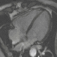
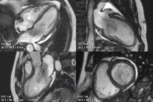
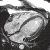
A 4 chamber view of the heart using SSFP cine imaging. Compare the image orientation (4 chamber) with the short axis view of the movie above
is best seen after giving a contrast agent, typically one containing gadolinium
bound to DTPA. With a special sequence, Inversion Recovery (IR) normal heart muscle appears dark, whilst areas of infarction appear bright white.
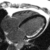
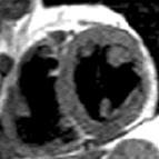
CMR in the 4 chamber view comparing the cine (left) with the late gadolinium image using inversion recovery (right). The subendocardial infarct is clearly seen. Fat around the heart also appears white.
, the heart muscle is starved of oxygen
by a coronary artery narrowing, especially during stress. This appears as a transient perfusion defect when a dose of contrast is given into a vein
. Knowing whether a perfusion defect is present and where it is helps guide intervention and treatment for coronary artery narrowings.
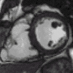
CMR perfusion. Contrast appears in the right ventricle then left ventricle before blushing into the muscle, which is normal (left) and abnormal (right, an inferior perfusion defect).
A good overview of the clinical indications for CMR can be found here and here
(cardiac ultrasound) cannot provide sufficient diagnostic information, 2) as an alternative to diagnostic cardiac catheterization which involve risks including x-ray radiation exposure, 3) to obtain diagnostic information for which CMR offers unique advantages such as blood flow measurement or identification of cardiac masses, and 4) when clinical assessment and other diagnostic tests are inconsistent. Examples of conditions in which CMR is often used include tetralogy of Fallot
, transposition of the great arteries, coarctation of the aorta, single ventricle heart disease, abnormalities of the pulmonary veins, atrial septal defect
, connective tissue diseases such as Marfan syndrome
, vascular rings, abnormal origins of the coronary arteries, and cardiac tumors.
Atrial septal defect with dilation of the right ventricle by CMR
Partial Anomalous Pulmonary Venous Drainage by CMR
CMR examinations in children typically last 15 to 60 minutes. In order to avoid blurry images the child must remain very still during the examination. In general, most children 7 years of age and older can cooperate sufficiently for a good quality examination. Providing an age-appropriate explanation of the procedure to the child in advance will increase the likelihood of a successful study. After proper safety screening, parents can be allowed into the MRI scanner room to help their child complete the examination. Some centers will allow children to listen to music or watch movies through a specialized MRI-compatible audiovisual system to reduce anxiety and improve cooperation. If the child cannot cooperate sufficiently, sedation with intravenous medications or general anesthesia may be necessary. In very young babies, it may be possible to perform the examination while they are in a natural sleep.
Enlarged right ventricle with poor function in a patient with repaired tetralogy of Fallot by CMR
and 3 tesla. The 3 tesla has advantages as it potentially doubles the amount of information acquired in the same. It offers particular advantages for perfusion
. The downside of 3 tesla is cost and potentially artefact degrading the pictures.
A resource for anyone thinking about CMR as a career can be found here
Magnetic resonance imaging
Magnetic resonance imaging , nuclear magnetic resonance imaging , or magnetic resonance tomography is a medical imaging technique used in radiology to visualize detailed internal structures...
(MRI) but with optimisation for use in the cardiovascular system. These optimisations are principally in the use of ECG gating and rapid imaging techniques or sequences. By combining a variety of such techniques into protocols, key functional and morphological features of the cardiovascular system can be assessed.
History and nomenclature
The phenomenon of nuclear magnetic resonanceNuclear magnetic resonance
Nuclear magnetic resonance is a physical phenomenon in which magnetic nuclei in a magnetic field absorb and re-emit electromagnetic radiation...
(NMR) was first described in molecular beams (1938) and bulk matter (1946), work later acknowledged by the award of a joint Nobel prize
Nobel Prize
The Nobel Prizes are annual international awards bestowed by Scandinavian committees in recognition of cultural and scientific advances. The will of the Swedish chemist Alfred Nobel, the inventor of dynamite, established the prizes in 1895...
in 1952. Further investigation laid out the principles of relaxation times leading to nuclear spectroscopy
Spectroscopy
Spectroscopy is the study of the interaction between matter and radiated energy. Historically, spectroscopy originated through the study of visible light dispersed according to its wavelength, e.g., by a prism. Later the concept was expanded greatly to comprise any interaction with radiative...
. In 1973, the first simple NMR image was published and the first medical imaging in 1977, entering the clinical arena in the early 1980s. In 1984, NMR medical imaging was renamed MRI. Initial attempts to image the heart were confounded by respiratory and cardiac motion, solved by using cardiac ECG gating, faster scan techniques and breath hold imaging. Increasingly sophisticated techniques were developed including cine imaging and techniques to characterise heart
Heart
The heart is a myogenic muscular organ found in all animals with a circulatory system , that is responsible for pumping blood throughout the blood vessels by repeated, rhythmic contractions...
muscle as normal or abnormal (fat infiltration, oedematous, iron loaded, acutely infarcted or fibrosed).
As MRI became more complex and application to cardiovascular imaging became more sophisticated, the Society for Cardiovascular Magnetic Resonance, http://www.scmr.org SCMR was set up (1996) with an academic journal, (JCMR) in 1999, which is going open source in 2008. In a move analogous to the development of ‘echocardiography
Echocardiography
An echocardiogram, often referred to in the medical community as a cardiac ECHO or simply an ECHO, is a sonogram of the heart . Also known as a cardiac ultrasound, it uses standard ultrasound techniques to image two-dimensional slices of the heart...
’ from cardiac ultrasound, the term ‘Cardiovascular Magnetic Resonance’ (CMR) was proposed and has gained acceptance as the name for the field.
Physics
CMR uses the same basic principles as other MRI techniques with the addition of ECG gating. Most CMR uses only 1H nucleiProton
The proton is a subatomic particle with the symbol or and a positive electric charge of 1 elementary charge. One or more protons are present in the nucleus of each atom, along with neutrons. The number of protons in each atom is its atomic number....
MR, which are abundant in human tissue. By using magnetic fields and radiofrequency (RF) pulses, the patient's own 1H nuclei absorb and then emit energy, which can be measured and translated into images, without using ionising radiation.
Techniques
CMR uses several different techniques within a single scan. The combination of these results in a comprehensive assessment of the heart and cardiovascular system. Examples are below:Visualising heart muscle scar or fat without using a contrast agent
Typically a sequence called spin-echo is used. This causes the bloodBlood
Blood is a specialized bodily fluid in animals that delivers necessary substances such as nutrients and oxygen to the cells and transports metabolic waste products away from those same cells....
to appear black. These are high resolution still images which in certain circumstances identify abnormal myocardium through differences in intrinsic contrast.
Heart function using cine imaging
Images of the heart may be acquired in real-timeReal-time MRI
Real-time magnetic resonance imaging refers to the continuous monitoring of moving objects in real time. Because MRIis based on time-consuming scanning of k-space, real-time MRI was possible only with low image quality or low temporal resolution...
with CMR, but the image quality is limited. Instead most sequences use ECG
Electrocardiogram
Electrocardiography is a transthoracic interpretation of the electrical activity of the heart over a period of time, as detected by electrodes attached to the outer surface of the skin and recorded by a device external to the body...
gating to acquire images at each stage of the cardiac cycle
Cardiac cycle
The cardiac cycle is a term referring to all or any of the events related to the flow or blood pressure that occurs from the beginning of one heartbeat to the beginning of the next. The frequency of the cardiac cycle is described by the heart rate. Each beat of the heart involves five major stages...
over several heart beats. This technique forms the basis of functional assessment by CMR. Blood typically appears bright in these sequences due to the contrast properties of blood and its rapid flow. The technique can discriminate very well between blood and myocardium. The current technique typically used for this is called
balanced steady state free precession
Steady-state free precession imaging
Steady-state free precession imaging is a magnetic resonance imaging technique which uses steady states of magnetizations. In general, SSFP MRI sequences are based on a gradient-echo MRI sequence with a short repetition time which in its generic form has been described as the FLASH MRI technique...
(SSFP), implemented as TrueFISP, b-FFE or Fiesta, depending on scanner manufacturer.
A 4 chamber view of the heart using SSFP cine imaging. Compare the image orientation (4 chamber) with the short axis view of the movie above
Infarct imaging using contrast
ScarScar
Scars are areas of fibrous tissue that replace normal skin after injury. A scar results from the biological process of wound repair in the skin and other tissues of the body. Thus, scarring is a natural part of the healing process. With the exception of very minor lesions, every wound results in...
is best seen after giving a contrast agent, typically one containing gadolinium
Gadolinium
Gadolinium is a chemical element with the symbol Gd and atomic number 64. It is a silvery-white, malleable and ductile rare-earth metal. It is found in nature only in combined form. Gadolinium was first detected spectroscopically in 1880 by de Marignac who separated its oxide and is credited with...
bound to DTPA. With a special sequence, Inversion Recovery (IR) normal heart muscle appears dark, whilst areas of infarction appear bright white.
CMR in the 4 chamber view comparing the cine (left) with the late gadolinium image using inversion recovery (right). The subendocardial infarct is clearly seen. Fat around the heart also appears white.
Perfusion
In anginaAngina
Angina pectoris, commonly known as angina, is chest pain due to ischemia of the heart muscle, generally due to obstruction or spasm of the coronary arteries . Coronary artery disease, the main cause of angina, is due to atherosclerosis of the cardiac arteries...
, the heart muscle is starved of oxygen
Oxygen
Oxygen is the element with atomic number 8 and represented by the symbol O. Its name derives from the Greek roots ὀξύς and -γενής , because at the time of naming, it was mistakenly thought that all acids required oxygen in their composition...
by a coronary artery narrowing, especially during stress. This appears as a transient perfusion defect when a dose of contrast is given into a vein
Vein
In the circulatory system, veins are blood vessels that carry blood towards the heart. Most veins carry deoxygenated blood from the tissues back to the heart; exceptions are the pulmonary and umbilical veins, both of which carry oxygenated blood to the heart...
. Knowing whether a perfusion defect is present and where it is helps guide intervention and treatment for coronary artery narrowings.
CMR perfusion. Contrast appears in the right ventricle then left ventricle before blushing into the muscle, which is normal (left) and abnormal (right, an inferior perfusion defect).
Uses
In the investigation of cardiovascular disease the physician has a wide variety of tools available. The key disadvantages of CMR are limited availability, expense, operator dependence and a lack of outcome data. The key advantages are image quality, non-invasiveness, accuracy, versatility and no ionising radiation.A good overview of the clinical indications for CMR can be found here and here
Children and congenital heart disease
Congenital heart defects are the most common type of major birth defect. Accurate diagnosis is essential for the development of appropriate treatment plans. CMR can provide comprehensive information about the nature of congenital hearts defects in a safe fashion without using x-rays or entering the body. It is rarely used as the first or sole diagnostic test for congenital heart disease. Rather, it is typically used in concert with other diagnostic techniques. In general, the clinical reasons for a CMR examination fall into one or more of the following categories: 1) when echocardiographyEchocardiography
An echocardiogram, often referred to in the medical community as a cardiac ECHO or simply an ECHO, is a sonogram of the heart . Also known as a cardiac ultrasound, it uses standard ultrasound techniques to image two-dimensional slices of the heart...
(cardiac ultrasound) cannot provide sufficient diagnostic information, 2) as an alternative to diagnostic cardiac catheterization which involve risks including x-ray radiation exposure, 3) to obtain diagnostic information for which CMR offers unique advantages such as blood flow measurement or identification of cardiac masses, and 4) when clinical assessment and other diagnostic tests are inconsistent. Examples of conditions in which CMR is often used include tetralogy of Fallot
Tetralogy of Fallot
Tetralogy of Fallot is a congenital heart defect which is classically understood to involve four anatomical abnormalities...
, transposition of the great arteries, coarctation of the aorta, single ventricle heart disease, abnormalities of the pulmonary veins, atrial septal defect
Atrial septal defect
Atrial septal defect is a form of congenital heart defect that enables blood flow between the left and right atria via the interatrial septum. The interatrial septum is the tissue that divides the right and left atria...
, connective tissue diseases such as Marfan syndrome
Marfan syndrome
Marfan syndrome is a genetic disorder of the connective tissue. People with Marfan's tend to be unusually tall, with long limbs and long, thin fingers....
, vascular rings, abnormal origins of the coronary arteries, and cardiac tumors.
Atrial septal defect with dilation of the right ventricle by CMR
Partial Anomalous Pulmonary Venous Drainage by CMR
CMR examinations in children typically last 15 to 60 minutes. In order to avoid blurry images the child must remain very still during the examination. In general, most children 7 years of age and older can cooperate sufficiently for a good quality examination. Providing an age-appropriate explanation of the procedure to the child in advance will increase the likelihood of a successful study. After proper safety screening, parents can be allowed into the MRI scanner room to help their child complete the examination. Some centers will allow children to listen to music or watch movies through a specialized MRI-compatible audiovisual system to reduce anxiety and improve cooperation. If the child cannot cooperate sufficiently, sedation with intravenous medications or general anesthesia may be necessary. In very young babies, it may be possible to perform the examination while they are in a natural sleep.
Enlarged right ventricle with poor function in a patient with repaired tetralogy of Fallot by CMR
Different magnet types
CMR scanners have to be modern. 'Open' magnet are not so good for cardiac as they do not cope with the beating heart very well. There are two magnet strengths mainly in use in CMR - 1.5 teslaTesla (unit)
The tesla is the SI derived unit of magnetic field B . One tesla is equal to one weber per square meter, and it was defined in 1960 in honour of the inventor, physicist, and electrical engineer Nikola Tesla...
and 3 tesla. The 3 tesla has advantages as it potentially doubles the amount of information acquired in the same. It offers particular advantages for perfusion
Perfusion
In physiology, perfusion is the process of nutritive delivery of arterial blood to a capillary bed in the biological tissue. The word is derived from the French verb "perfuser" meaning to "pour over or through."...
. The downside of 3 tesla is cost and potentially artefact degrading the pictures.
Training
Training is being increasingly protocolised and is now formal with stages of training and accreditation.A resource for anyone thinking about CMR as a career can be found here
External links
- The Society for Cardiovascular Magnetic Resonance
- The Journal for Cardiovascular Magnetic Resonance
- An Atlas of normal cardiac structure and function by CMR
- Having a CMR scan
- Cardiac MRI technical primer
- ReviseMRI.com
- Hull physics lecture series
- the basics of MRI
- MRI tutor
- Cardiac MRI technical primer
- A good description of the experience of receiving a CMR can be found here
- For online case examples, see here