

Susceptibility weighted imaging
Encyclopedia
Magnetic resonance imaging
Magnetic resonance imaging , nuclear magnetic resonance imaging , or magnetic resonance tomography is a medical imaging technique used in radiology to visualize detailed internal structures...
(MRI) different from traditional spin density, T1, or T2 imaging. SWI uses a fully flow compensated, long echo, gradient echo (GRE) scan to acquire images. This method exploits the susceptibility
Magnetic susceptibility
In electromagnetism, the magnetic susceptibility \chi_m is a dimensionless proportionality constant that indicates the degree of magnetization of a material in response to an applied magnetic field...
differences between tissues and uses the phase image to detect these differences. The magnitude and phase data are combined to produce an enhanced contrast magnitude image which is exquisitely sensitive to venous blood, hemorrhage and iron storage. The imaging of venous blood with SWI is a blood-oxygen-level dependent (BOLD) technique which is why it was (and is sometimes still) referred to as BOLD venography. Due to its sensitivity to venous blood SWI is commonly used in traumatic brain injuries
Traumatic brain injury
Traumatic brain injury , also known as intracranial injury, occurs when an external force traumatically injures the brain. TBI can be classified based on severity, mechanism , or other features...
(TBI) and for high resolution brain venographies but has many other clinical applications. SWI is offered as a clinical package by Siemens but can be run on any manufacturer’s machine at field strengths of 1.0 T, 1.5 T, 3.0 T and higher.
Acquisition and image processing
SWI uses a fully velocity compensated, three dimensional, RF spoiled, high-resolution, 3D gradient echo scan. Both the magnitude and phase images are saved, and the phase image is high pass (HP) filtered to removed unwanted artifacts. The magnitude image is then combined with the phase image to create an enhanced contrast magnitude image referred to as the susceptibility weighted (SW) image. It is also common to create minimum intensity projections (mIP) over 8 to 10 mm to better visualize vein connectivity. In this way four sets of images are generated, the original magnitude, HP filtered phase, susceptibility weighted, and mIPs over the susceptibility weighted images.Phase filtering
The values in the phase images are constrained from -π to π so if the value goes above π it wraps to -π, this is known as aliasing. Inhomogeneities in the magnetic field cause low frequency background gradients. This causes all the phase values to slowly increase across the image which creates phase wrapping and obscures the image. This type of artifact can be removed by phase unwrapping or by high pass filtering the original complex data to remove the low frequency variations in the phase image.Susceptibility weighted image creation

Traumatic brain injury (TBI)
Diffuse axonal injury
Diffuse axonal injury is one of the most common and devastating types of traumatic brain injury, meaning that damage occurs over a more widespread area than in focal brain injury. DAI, which refers to extensive lesions in white matter tracts, is one of the major causes of unconsciousness and...
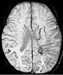
(DAI) in trauma patients is often difficult as the injuries tend to be relatively small in size and can be easily missed by low resolution scans. SWI is usually run at relatively high resolution (1 mm3) and is extremely sensitive to bleeding in the gray matter/white matter boundaries making it is possible to see very small lesions increasing the ability to detect more subtle injuries.
Stroke and hemorrhage
Diffusion weighted imagingDiffusion MRI
Diffusion MRI is a magnetic resonance imaging method that produces in vivo images of biological tissues weighted with the local microstructural characteristics of water diffusion, which is capable of showing connections between brain regions...
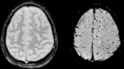
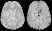
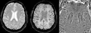
offers a powerful means to detect acute stroke. Although it is well known that gradient echo imaging can detect hemorrhage, it is best detected with SWI. In the example shown here, the gradient echo image shows the region of likely cytotoxic edema whereas the SW image shows the likely localization of the stroke and the vascular territory affected (data acquired at 1.5 T).
The bright region in the gradient echo weighted image shows the area affected in this acute stroke example. The arrows in the SWI image may show the tissue at risk that has been affected by the stroke (A, B, C) and the location of the stroke itself (D). The reason that we are able to see the affected vascular territory could be because there is a reduced level of oxygen saturation in this tissue, suggesting that the flow to this region of the brain could be reduced post stroke. Another possible explanation is that there is an increase in local venous blood volume. In either case, this image suggests that the tissue associated with this vascular territory could be tissue at risk. Future stroke research will involve comparisons of perfusion weighted imaging and SWI to learn more about local flow and oxygen saturation.
Sturge-Weber disease
Sturge-Weber syndrome
Sturge–Weber syndrome, sometimes referred to as encephalotrigeminal angiomatosis, is a rare congenital neurological and skin disorder. It is one of the phakomatoses and is often associated with port-wine stains of the face, glaucoma, seizures, mental retardation, and ipsilateral leptomeningeal...
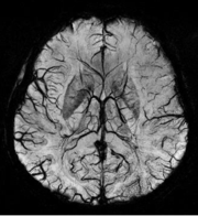
who did not display neurological symptoms is shown to the left. The initial conventional MR imaging methods did not demonstrate any abnormality. The abnormal venous vasculature in the left occipital lobe extending between the posterior horn of the ventricle and the cortical surface is clearly visible in the venogram. Due to the high resolution even collaterals can be resolved.
Tumors
Part of the characterization of tumors lies in understanding the angiographic behavior of lesions both from the perspective of angiogenesis and micro-hemorrhages. Aggressive tumors tend to have rapidly growing vasculature and many micro-hemorrhages. Hence, the ability to detect these changes in the tumor could lead to a better determination of the tumor status. The enhanced sensitivity of SWI to venous blood and blood products due to their differences in susceptibility compared to normal tissue leads to better contrast in detecting tumor boundaries and tumor hemorrhage.Multiple sclerosis
Multiple sclerosisMultiple sclerosis
Multiple sclerosis is an inflammatory disease in which the fatty myelin sheaths around the axons of the brain and spinal cord are damaged, leading to demyelination and scarring as well as a broad spectrum of signs and symptoms...
(MS) is usually studied with FLAIR
Fluid attenuated inversion recovery
Fluid attenuated inversion recovery is a pulse sequence used in magnetic resonance imaging which was invented by Dr. Graeme Bydder. FLAIR can be used with both three dimensional imaging or two dimensional imaging ....
and contrast enhanced T1 imaging. SWI adds to this by revealing the venous connectivity in some lesions and presents evidence of iron in some lesions. This key new information may help understand the physiology of MS.
Vascular dementia and cerebral amyloid angiopathy (CAA)
Pneumocephalus
Recent studies suggest that SWI might be suitable for monitorizing neurosurgical patients recovering from PneumocephalusPneumocephalus
Pneumocephalus is the presence of air or gas within the cranial cavity. It is usually associated with disruption of the skull: after head and facial trauma, tumors of the skull base, after neurosurgery or otorhinolaryngology, and rarely, spontaneously...
, as air can be easily detected with SWI.