

Gamma camera
Encyclopedia
Nuclear medicine
In nuclear medicine procedures, elemental radionuclides are combined with other elements to form chemical compounds, or else combined with existing pharmaceutical compounds, to form radiopharmaceuticals. These radiopharmaceuticals, once administered to the patient, can localize to specific organs...
to view and analyse images of the human body or the distribution of medically injected, inhaled, or ingested radionuclides emitting gamma ray
Gamma ray
Gamma radiation, also known as gamma rays or hyphenated as gamma-rays and denoted as γ, is electromagnetic radiation of high frequency . Gamma rays are usually naturally produced on Earth by decay of high energy states in atomic nuclei...
s.
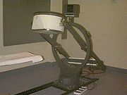
Construction
The system accumulates events, or counts, of gamma
Gamma ray
Gamma radiation, also known as gamma rays or hyphenated as gamma-rays and denoted as γ, is electromagnetic radiation of high frequency . Gamma rays are usually naturally produced on Earth by decay of high energy states in atomic nuclei...
photon
Photon
In physics, a photon is an elementary particle, the quantum of the electromagnetic interaction and the basic unit of light and all other forms of electromagnetic radiation. It is also the force carrier for the electromagnetic force...
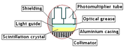
s that are absorbed by the crystal in the camera. Usually a large flat crystal of sodium iodide with thallium doping in a light-sealed housing is used. The highly efficient capture method of this combination for detecting gamma rays was discovered by noted physicist Robert Hofstadter
Robert Hofstadter
Robert Hofstadter was an American physicist. He was the joint winner of the 1961 Nobel Prize in Physics "for his pioneering studies of electron scattering in atomic nuclei and for his consequent discoveries concerning the structure of nucleons."-Biography :Born in New York City, he entered City...
in 1948 http://nobelprize.org/nobel_prizes/physics/laureates/1961/hofstadter-bio.html).
The crystal scintillate
Scintillation (physics)
Scintillation is a flash of light produced in a transparent material by an ionization event. See scintillator and scintillation counter for practical applications.-Overview:...
s in response to incident gamma radiation. When a gamma photon leaves the patient (who has been injected with a radioactive pharmaceutical), it knocks an electron loose from an iodine atom in the crystal, and a faint flash of light is produced when the dislocated electron again finds a minimal energy state. The initial phenomenon of the excited electron is similar to the photoelectric effect
Photoelectric effect
In the photoelectric effect, electrons are emitted from matter as a consequence of their absorption of energy from electromagnetic radiation of very short wavelength, such as visible or ultraviolet light. Electrons emitted in this manner may be referred to as photoelectrons...
and (particularly with gamma rays) the Compton effect. After the flash of light is produced, it is detected. Photomultiplier
Photomultiplier
Photomultiplier tubes , members of the class of vacuum tubes, and more specifically phototubes, are extremely sensitive detectors of light in the ultraviolet, visible, and near-infrared ranges of the electromagnetic spectrum...
tubes (PMTs) behind the crystal detect the fluorescent flashes (events) and a computer sums the counts. The computer reconstructs and displays a two dimensional image of the relative spatial count density on a monitor. This reconstructed image reflects the distribution and relative concentration of radioactive tracer elements present in the organs and tissues imaged.
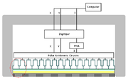
Signal processing
Hal AngerHal Anger
Hal Oscar Anger was an American electrical engineer and biophysicist at Donner Laboratory, University of California, Berkeley, known for his invention of the Anger camera....
developed the first gamma camera in 1957. His original design, frequently called the Anger camera, is still widely used today. The Anger camera uses sets of vacuum tube
Vacuum tube
In electronics, a vacuum tube, electron tube , or thermionic valve , reduced to simply "tube" or "valve" in everyday parlance, is a device that relies on the flow of electric current through a vacuum...
photomultipliers. Generally each tube has an exposed face of about 3 inches in diameter and the tubes are arranged in hexagon configurations, behind the absorbing crystal. The electronic circuit connecting the photodetectors is wired so as to reflect the relative coincidence of light fluorescence as sensed by the members of the hexagon detector array. All the PMTs simultaneously detect the (presumed) same flash of light to varying degrees, depending on their position from the actual individual event. Thus the spatial location of each single flash of fluorescence is reflected as a pattern of voltages within the interconnecting circuit array.
The location of the interaction between the gamma ray and the crystal can be determined by processing the voltage signals from the photomultipliers; in simple terms, the location can be found by weighting the position of each photomultiplier tube by the strength of its signal, and then calculating a mean position from the weighted positions. The total sum of the voltages from each photomultiplier is proportional to the energy of the gamma ray interaction, thus allowing discrimination between different isotopes or between scattered and direct photons.
Spatial resolution
In order to obtain spatial information about the gammaGamma
Gamma is the third letter of the Greek alphabet. In the system of Greek numerals it has a value of 3. It was derived from the Phoenician letter Gimel . Letters that arose from Gamma include the Roman C and G and the Cyrillic letters Ge Г and Ghe Ґ.-Greek:In Ancient Greek, gamma represented a...
emissions from an imaging subject (e.g. a person's heart muscle cells which have absorbed an intravenous injected radioactive, usually thallium-201 or technetium-99m
Technetium-99m
Technetium-99m is a metastable nuclear isomer of technetium-99, symbolized as 99mTc. The "m" indicates that this is a metastable nuclear isomer, i.e., that its half-life of 6 hours is considerably longer than most nuclear isomers that undergo gamma decay...
, medicinal imaging agent) a method of correlating the detected photons with their point of origin is required.
The conventional method is to place a collimator
Collimator
A collimator is a device that narrows a beam of particles or waves. To "narrow" can mean either to cause the directions of motion to become more aligned in a specific direction or to cause the spatial cross section of the beam to become smaller.- Optical collimators :In optics, a collimator may...
over the detection crystal/PMT array. The collimator consists of a thick sheet of lead
Lead
Lead is a main-group element in the carbon group with the symbol Pb and atomic number 82. Lead is a soft, malleable poor metal. It is also counted as one of the heavy metals. Metallic lead has a bluish-white color after being freshly cut, but it soon tarnishes to a dull grayish color when exposed...
, typically 1-3 inches thick, with thousands of adjacent holes through it. The individual holes limit photons which can be detected by the crystal to a cone; the point of the cone is at the midline center of any given hole and extends from the collimator surface outward. However, the collimator is also one of the sources of blurring within the image; lead does not totally attenuate incident gamma photons, there can be some crosstalk between holes.
Unlike a lens, as used in visible light cameras, the collimator attenuates most (>99%) of incident photons and thus greatly limits the sensitivity of the camera system. Large amounts of radiation must be present so as to provide enough exposure for the camera system to detect sufficient scintillation dots to form a picture.
Other methods of image localization (pinhole
Pinhole camera
A pinhole camera is a simple camera without a lens and with a single small aperture – effectively a light-proof box with a small hole in one side. Light from a scene passes through this single point and projects an inverted image on the opposite side of the box...
, rotating slat collimator with CZT
Cadmium zinc telluride
Cadmium zinc telluride, or CZT, is a compound of cadmium, zinc and tellurium or, more strictly speaking, an alloy of cadmium telluride and zinc telluride. A direct bandgap semiconductor, it is used in a variety of applications, including radiation detectors, photorefractive gratings,...
(Gagnon & Matthews) and others) have been proposed and tested; however, none have entered widespread routine clinical use.
The best current camera system designs can differentiate two separate point sources of gamma photons located a minimum of 1.8 cm apart, at 5 cm away from the camera face. Spatial resolution decreases rapidly at increasing distances from the camera face. This limits the spatial accuracy of the computer image: it is a fuzzy image made up of many dots of detected but not precisely located scintillation. This is a major limitation for heart muscle imaging systems; the thickest normal heart muscle in the left ventricle is about 1.2 cm and most of the left ventricle muscle is about 0.8 cm, always moving and much of it beyond 5 cm from the collimator face. To help compensate, better imaging systems limit scintillation counting to a portion of the heart contraction cycle, called gating, however this further limits system sensitivity.
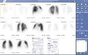
Imaging techniques using gamma cameras
ScintigraphyScintigraphy
Scintigraphy is a form of diagnostic test used in nuclear medicine, wherein radioisotopes are taken internally, and the emitted radiation is captured by external detectors to form two-dimensional images...
("scint") is the use of gamma cameras to capture emitted radiation from internal radioisotopes to create two-dimensional images.
SPECT (single photon emission computed tomography) imaging, as used in nuclear cardiac stress test
Cardiac stress test
Cardiac stress test is a test used in medicine and cardiology to measure the heart's ability to respond to external stress in a controlled clinical environment....
ing, is performed using gamma cameras, usually one, two or three detectors or heads, are slowly rotated around the patient's torso.
Multi-headed gamma cameras can also be used for Positron emission tomography
Positron emission tomography
Positron emission tomography is nuclear medicine imaging technique that produces a three-dimensional image or picture of functional processes in the body. The system detects pairs of gamma rays emitted indirectly by a positron-emitting radionuclide , which is introduced into the body on a...
scanning, provided that their hardware and software can be configured to detect 'coincidences' (near simultaneous events on 2 different heads). Gamma camera PET is markedly inferior to PET imaging with a purpose designed PET scanner, as the scintillator crystal has poor sensitivity for the high-energy annihilation photons, and the detector area is significantly smaller. However, given the low cost of a gamma camera and its additional flexibility compared to a dedicated PET scanner, this technique is useful where the expense and resource implications of a PET scanner cannot be justified.