

Sarfus
Encyclopedia
- an upright or inverted optical microscopeOptical microscopeThe optical microscope, often referred to as the "light microscope", is a type of microscope which uses visible light and a system of lenses to magnify images of small samples. Optical microscopes are the oldest design of microscope and were possibly designed in their present compound form in the...
in crossed polarization configuration and - specific supporting plates - called surfs - on which the sample to observe is deposited.
Sarfus visualization is based on the perfect control of the reflection properties of polarized light on a surf, which leads to increase the axial sensitivity of optical microscope
Optical microscope
The optical microscope, often referred to as the "light microscope", is a type of microscope which uses visible light and a system of lenses to magnify images of small samples. Optical microscopes are the oldest design of microscope and were possibly designed in their present compound form in the...
by a factor of around 100 without reducing its lateral resolution. Thus this new technique increases the sensitivity of standard optical microscope
Optical microscope
The optical microscope, often referred to as the "light microscope", is a type of microscope which uses visible light and a system of lenses to magnify images of small samples. Optical microscopes are the oldest design of microscope and were possibly designed in their present compound form in the...
to a point that it becomes possible to directly visualize thin films (down to 0.3 nanometer) and isolated nano-objects in real-time but also in air or in water.
Principles
A recent study on polarized light coherence leads to the development of new supports – the surfs – having contrast amplification properties for standard optical microscopy in cross polarizers mode. Made of optical layers on an opaque or transparent substrate, these supports do not modify the light polarization after reflection even if the numerical aperture of the incident source is important. This property is modified when a sample is present on a surf, a non-null light component is then detected after the analyzer rendering the sample visible.The performances of these supports are estimated from the measurement of the contrast (C) of the sample defines by: C = (I1-I0)/(I0+I1) where I0 and I1 represent the intensities reflected by the bare surf and by the analyzed sample on the surf, respectively. For a one nanometer-film thickness, the surfs display a contrast 200 times higher than on silicon wafer.
This high contrast increase allows the visualization with standard optical microscope of films with thicknesses down to 0.3 nm, as well as nano-objects (down to 2 nm diameter) and this, without any kind of sample labelling (neither fluorescence, nor radioactive marker). An illustration of the contrast enhance is given hereafter with the observation in optical microscopy between cross polarizers of a Langmuir-Blodgett structure on a silicon wafer and on a surf.
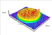
In addition to visualization, recent developments have allowed accessing to the thickness measurement of the analyzed sample. A colorimetric correspondence is carried out between a calibration standard made of nano-steps and the analyzed sample. Indeed, due to optical interference, a correlation exists between RGB (red, green, blue) parameters of the sample and its optical thickness. This leads to 3D-representation of the analyzed samples, the measurement of profile sections, roughness and other topological measurements.
Experimental setup
The experimental set-up is simple: the sample to be characterized is deposited by usual deposit techniques such as dip-coating, spin-coating, deposit pipette, evaporation… on a surf instead of the traditional microscope slide. The support is then placed on the microscope stage.Synergy with existing equipments
The sarfus technique can be integrated in existing analysis equipment (Atomic force microscopeAtomic force microscope
Atomic force microscopy or scanning force microscopy is a very high-resolution type of scanning probe microscopy, with demonstrated resolution on the order of fractions of a nanometer, more than 1000 times better than the optical diffraction limit...
(AFM), Raman spectroscopy
Raman spectroscopy
Raman spectroscopy is a spectroscopic technique used to study vibrational, rotational, and other low-frequency modes in a system.It relies on inelastic scattering, or Raman scattering, of monochromatic light, usually from a laser in the visible, near infrared, or near ultraviolet range...
, etc.) to add new functionalities, such as optical image, thickness measurement, kinetic study, and also for sample pre-localization to save time and consumables (AFM tips, etc.).
Applications
Life sciences
- Biological films
- BiochipBiochipThe development of biochips is a major thrust of the rapidly growing biotechnology industry, which encompasses a very diverse range ofresearch efforts including genomics, proteomics, and pharmaceuticals, among other activities...
- Phospholipids
- Soft lithographySoft lithography200px|right|thumb|Figure 1 - "Inking" a stamp. PDMS stamp with pattern is placed in Ethanol and ODT solution200px|right|thumb|Figure 2 - ODT from the solution settles down onto the PDMS stamp. Stamp now has ODT attached to it which acts as the ink....
- Cell adhesionCell adhesionCellular adhesion is the binding of a cell to a surface, extracellular matrix or another cell using cell adhesion molecules such as selectins, integrins, and cadherins. Correct cellular adhesion is essential in maintaining multicellular structure...
Thin films and surface treatment
- Polymers films
- Langmuir-Blodgett films
- Liquid crystals
- Plasma treatment
- Self-assembled monolayerSelf-assembled monolayerA self assembled monolayer is an organized layer of amphiphilic molecules in which one end of the molecule, the “head group” shows a specific, reversible affinity for a substrate...
s
Nanomaterials
- Carbon nanotubes
- Nanoparticles
- Nanowires
- GrapheneGrapheneGraphene is an allotrope of carbon, whose structure is one-atom-thick planar sheets of sp2-bonded carbon atoms that are densely packed in a honeycomb crystal lattice. The term graphene was coined as a combination of graphite and the suffix -ene by Hanns-Peter Boehm, who described single-layer...
- DNADNADeoxyribonucleic acid is a nucleic acid that contains the genetic instructions used in the development and functioning of all known living organisms . The DNA segments that carry this genetic information are called genes, but other DNA sequences have structural purposes, or are involved in...
molecules