

Ocular immune system
Encyclopedia
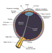
Human eye
The human eye is an organ which reacts to light for several purposes. As a conscious sense organ, the eye allows vision. Rod and cone cells in the retina allow conscious light perception and vision including color differentiation and the perception of depth...
from infection and regulates healing processes following injuries. The interior of the eye lacks lymph vessels but is highly vascularized, and many immune cells reside in the uvea
Uvea
The uvea , also called the uveal layer, uveal coat, uveal tract, or vascular tunic, is the pigmented middle of the three concentric layers that make up an eye. The name is possibly a reference to its reddish-blue or almost black colour, wrinkled appearance and grape-like size and shape when...
, including mostly macrophages, dendritic cells, and mast cells . These cells fight off intraocular infections, and intraocular inflammation can manifest as uveitis
Uveitis
Uveitis specifically refers to inflammation of the middle layer of the eye, termed the "uvea" but in common usage may refer to any inflammatory process involving the interior of the eye....
(including iritis
Iritis
Iritis is a form of anterior uveitis and refers to the inflammation of the iris of the eye.-Types:There are two main types of iritis: acute and chronic. They differ in numerous ways....
) or retinitis. The cornea of the eye is immunologically a very special tissue. Its constant exposure to the exterior world means that it is vulnerable to a wide range of microorganisms while its moist mucosal surface makes the cornea particularly susceptible to attack. At the same time, its lack of vasculature and relative immune separation from the rest of the body makes immune defense difficult. Lastly, the cornea is a multifunctional tissue. It provides a large part of the eye’s refractive power, meaning it has to maintain remarkable transparency, but must also serve as a barrier to keep pathogens from reaching the rest of the eye, similar to function of the dermis and epidermis in keeping underlying tissues protected. Immune reactions within the cornea come from surrounding vascularized tissues as well as innate immune responsive cells that reside within the cornea.
Immune difficulties for the cornea
The most important function of the cornea is to transmit and refract light so as to allow high-resolution images to be produced on the back of the retina. To do this, collagen within the cornea is highly ordered to be 30 nanometers in diameter and placed 60 nanometers apart so as to reduce light scatter. Furthermore, the tissue is not vascularized, and does not contain lymphoid cells or other defense mechanisms, apart from some dendritic cells (DC) . Both of these factors necessitate the small number of cells within the cornea. However, this necessitates keeping immune cells at a relative distance, effectively creating a time delay between exposures to a pathogen and mounting of an immune response . Therefore, many immune and protective responses within the cornea, such as moistening and nutrition, come from non-local sources, such as the conjunctiva.Immune responses of the Cornea
Innate immune responses defend against pathogens and toxin in a non-discriminatory manner. They provide an inherent barrier against corneal infection while also serving as a primary mode of defense that is present from birth. For instance, the orbit and the eyelid can guard against both traumatic events and exterior debris that may contain microorganisms. Other components of the ocular innate immune system include tears, epithelial cells, keratocytesCorneal keratocyte
Corneal keratocytes are specialized fibroblasts residing in the stroma. This corneal layer, representing about 85-90% of corneal thickness, is built up from highly regular collagenous lamellae and extracellular matrix components. Keratocytes play the major role in keeping it transparent, healing...
, corneal nerves, the complement system, and interferons.
Acquired immune responses are much more pathogen-specific than their innate immune counterparts. These pathways are cell-mediated and are understood to be controlled in part by Langerhans cells in the cornea. These Langerhans cells are antigen-presenting cells, which pick up pieces of invading pathogens and use them to elicit an immune response. Cell-mediated immune responses are usually slower acting and more efficient, but can cause damage to surrounding tissue, resulting in damage to the vision.
Mucosa-associated lymphoid tissue
Both innate and acquired responses are important in ocular defenses. One major pathway in which both are incorporated is the network of lymphoid cells that form the mucosa-associated lymphoid tissue (MALT). MALT is a major component in all mucosal organs, including the respiratory, genital, digestive, and ocular tracts. Regulated migrations of immune cells are known to occur between these mucosal organs. However, the role of MALT in human ocular defenses is not fully understood. However, it is known that the lacrimal glands and the conjunctiva contribute to ocular defenses via secretion of both immunoglobulins and lymphoid tissues. The latter is understood to be organized into clumps of lymphoid follicles as well as diffuse lymphoid tissues.In the follicular form of MALT, antigens are taken up by the follicles and presented to lymphocytes by antigen presenting cells. This leads to activation of B and T cells that carry out the immune reaction. Diffuse lymphoid tissues, on the other hand, is composed mainly of interspersed effector cells . Generally, both pathways lead to activation and migration of immune cells within the mucosal tissues, including the conjunctiva.
Conjunctival immune response
The conjunctivaConjunctiva
The conjunctiva covers the sclera and lines the inside of the eyelids. It is composed of rare stratified columnar epithelium.-Function:...
covers the sclera, or whites of the eyes, as well as the insides of the eyelids and provides nutrients to underlying and surrounding tissue. The conjunctiva is also one of the closest vascularized tissues to the cornea. As such, it provides a major source of immune components in the cornea
Cornea
The cornea is the transparent front part of the eye that covers the iris, pupil, and anterior chamber. Together with the lens, the cornea refracts light, with the cornea accounting for approximately two-thirds of the eye's total optical power. In humans, the refractive power of the cornea is...
. Not only does the conjunctiva produce IgA
IGA
Iga or IGA may stand for:-Given name:* a female given name of Polish origin. The name originates from the female given name Jadwiga and stands for gia,or gina in the USA....
, like the lacrimal gland
Lacrimal gland
The lacrimal glands are paired almond-shaped glands, one for each eye, that secrete the aqueous layer of the tear film. They are situated in the upper, outer portion of each orbit, in the lacrimal fossa of the orbit formed by the frontal bone. Inflammation of the lacrimal glands is called...
s, but it also contains macrophages, neutrophilic granulocytes, mast cells, lymphocytes, and other aspects of the general mucosal immune system . Like the rest of the MALT pathway, the conjunctiva has been found to possess lymphoid follicles, which develop at puberty and decline in old age, as well as diffuse lymphoid tissues. The conjunctiva also possess macrophages that play a part in modulating the T-cell immune response and mediating both the innate and acquired immune responses.
Lacrimal immune response
The tear film is composed of three layers: the lipid, aqueous, and mucin . These play a role in creating a smooth surface to facilitate refraction, lubricating the movement of the eyelid, passively transporting gases such as oxygen and carbon dioxide, and protecting the cornea. This last function is achieved through functions of various layers within the tear film. Tears bathe corneal epithelial cells in a moist environment, preventing them from drying out and weakening. However, the liquid layer of the tear film also contains antimicrobial properties resulting from the presence of lysozymeLysozyme
Lysozyme, also known as muramidase or N-acetylmuramide glycanhydrolase, are glycoside hydrolases, enzymes that damage bacterial cell walls by catalyzing hydrolysis of 1,4-beta-linkages between N-acetylmuramic acid and N-acetyl-D-glucosamine residues in a peptidoglycan and between...
s, lactoferrin
Lactoferrin
Lactoferrin , also known as lactotransferrin , is a multifunctional protein of the transferrin family. Lactoferrin is a globular glycoprotein with a molecular mass of about 80 kDa that is widely represented in various secretory fluids, such as milk, saliva, tears, and nasal secretions...
s, lipocalin
Lipocalin
The lipocalins are a family of proteins which transport small hydrophobic molecules such as steroids, bilins, retinoids, and lipids. They share limited regions of sequence homology and a common tertiary structure architecture...
, and beta-lysin, which facilitate pathogen defenses such as lysis of bacterial cell walls, prevention of bacterial and viral binding, inflammation, and detoxification. Furthermore, white blood cells can be transported to the corneal surface via the tear film, and both toxic agents as well as debris can be diluted and washed away by the tear film . The tear film also contains immunoglobulins, especially IgA
IGA
Iga or IGA may stand for:-Given name:* a female given name of Polish origin. The name originates from the female given name Jadwiga and stands for gia,or gina in the USA....
, which is found in concentrations significantly higher than in serum. IgA has been shown to prevent bacterial binding. Along with another immunoglobulin present in the tear film, IgG, IgA can also neutralize virus
Virus
A virus is a small infectious agent that can replicate only inside the living cells of organisms. Viruses infect all types of organisms, from animals and plants to bacteria and archaea...
es and bind to bacteria, aiding in their detection via other pathways.
Corneal epithelial cells
Corneal epithelial cellsCorneal epithelium
The corneal epithelium is made up of epithelial tissue and covers the front of the cornea. It acts as a barrier to protect the cornea, resisting the free flow of fluids from the tears, and prevents bacteria from entering the epithelium and corneal stroma.The corneal epithelium consists of several...
present a physical barrier to prevent microbes from reaching the interior of the eye chamber, which is effectively separated from the rest of the body via tight junctions. At the same time, corneal epithelial cells also secrete cytokines to activate microbial defense . One cytokine, interleukin (IL)-1α
IL1A
Interleukin-1 alpha is a protein that in humans is encoded by the IL1A gene.The protein encoded by this gene is a cytokine of the interleukin-1 family. Interleukin-1 alpha possesses a wide spectrum of metabolic, physiological, haematopoietic activities, and plays one of the central roles in the...
, is stored in epithelial cells and automatically released when the cell membrane is ruptured by infection or trauma. However, long-term effects of IL-1α can lead to not only enhanced immune inflitration of the cornea, but also neovascularization (formation of new blood vessels), which can lead to a loss of corneal transparency. Therefore, the cornea has also been found to secrete an IL-1α antagonist, IL-1RN
Interleukin 1 receptor antagonist
The interleukin-1 receptor antagonist is a protein that in humans is encoded by the IL1RN gene.IL-1RA was initially called the IL-1 inhibitor and was discovered separately in 1984 by two independent laboratories. IL-1RA, is an agent that binds non-productively to the cell surface interleukin-1...
, which decreases leucocyte invasion of the cornea and suppresses neovascularization, both of which can help preserve vision .
Corneal keratocytes
KeratocytesCorneal keratocyte
Corneal keratocytes are specialized fibroblasts residing in the stroma. This corneal layer, representing about 85-90% of corneal thickness, is built up from highly regular collagenous lamellae and extracellular matrix components. Keratocytes play the major role in keeping it transparent, healing...
are flattened cells found dispersed within the corneal stroma. The primary role of this sparse population of cells is thought to be in maintaining the extracellular matrix of collagen lamellae that surround them. However, keratocytes also play a defensive role during pathogenic invasion. They can be influenced by IL-1α (secreted by corneal epithelial cells) and tumor necrosis factor (TNF)-α to produce both IL-6 and defensin
Defensin
Defensins are small cysteine-rich cationic proteins found in both vertebrates and invertebrates. They have also been reported in plants. They are, and function as, host defense peptides. They are active against bacteria, fungi and many enveloped and nonenveloped viruses. They consist of 18-45 amino...
s. Of these, the former is found to combine synergistically with other interleukins to increase co-stimulation of other immune aspects as well as increase antibody secretion. The latter, defensins, have a wide range of antimicrobial affects against bacteria, fungi, and viruses, as well as effects in accelerating healing of damaged epithelial cells . It has also been found that the presence of secreted defensins secreted by corneal keratocytes is correlated with cases of corneal transplant rejection , suggesting that these peptides may have a role in tissue rejection. Furthermore, keratocytes have also been found to secrete IL-8, which attracts neutrophils, in infections involving the herpes simplex virus .
Corneal nerves
Corneal nerves serve as a form of defense by detecting the presence of foreign bodies on the corneal surface. This leads to reflexive reactions such as increased lacrimal secretion, blinking, and release of neuropeptides, which can induce cytokine activation .External links
- Ocular Immune Privilege - J. Wayne StreileinJ. Wayne StreileinJ. Wayne Streilein was a scientist whose main area of research was the ocular immune system. He is known particularly for studying the mechanisms that keep the cornea avascular despite the inflammatory and other stimuli that usually promote small blood vessel ingrowth; these peculiar corneal...
, Karger Gazette.