

Immunocytochemistry
Encyclopedia
Immunocytochemistry is a common laboratory technique that uses antibodies that target specific peptide
Peptide
Peptides are short polymers of amino acid monomers linked by peptide bonds. They are distinguished from proteins on the basis of size, typically containing less than 50 monomer units. The shortest peptides are dipeptides, consisting of two amino acids joined by a single peptide bond...
s or protein
Protein
Proteins are biochemical compounds consisting of one or more polypeptides typically folded into a globular or fibrous form, facilitating a biological function. A polypeptide is a single linear polymer chain of amino acids bonded together by peptide bonds between the carboxyl and amino groups of...
antigen
Antigen
An antigen is a foreign molecule that, when introduced into the body, triggers the production of an antibody by the immune system. The immune system will then kill or neutralize the antigen that is recognized as a foreign and potentially harmful invader. These invaders can be molecules such as...
s in the cell
Cell (biology)
The cell is the basic structural and functional unit of all known living organisms. It is the smallest unit of life that is classified as a living thing, and is often called the building block of life. The Alberts text discusses how the "cellular building blocks" move to shape developing embryos....
via specific epitope
Epitope
An epitope, also known as antigenic determinant, is the part of an antigen that is recognized by the immune system, specifically by antibodies, B cells, or T cells. The part of an antibody that recognizes the epitope is called a paratope...
s. These bound antibodies can then be detected using several different methods
Protein methods
Protein methods are the techniques used to study proteins.There are genetic methods for studying proteins, methods for detecting proteins, methods for isolating and purifying proteins and other methods for characterizing the structure and function of proteins, often requiring that the protein first...
. ICC allows researchers to evaluate whether or not cells in a particular sample express
Protein expression
Protein expression is a subcomponent of gene expression. It consists of the stages after DNA has been translated into polypeptide chains, which are ultimately folded into proteins...
the antigen in question. In cases where an immunopositive signal is found, ICC also allows researchers to determine which sub-cellular compartments are expressing the antigen.
Immunocytochemistry vs. immunohistochemistry
Immunocytochemistry differs from immunohistochemistryImmunohistochemistry
Immunohistochemistry or IHC refers to the process of detecting antigens in cells of a tissue section by exploiting the principle of antibodies binding specifically to antigens in biological tissues. IHC takes its name from the roots "immuno," in reference to antibodies used in the procedure, and...
in that the former is performed on samples
Sample (material)
In general, a sample is a limited quantity of something which is intended to be similar to and represent a larger amount of that thing. The things could be countable objects such as individual items available as units for sale, or a material not countable as individual items. Samples of countable...
of intact cells that have had most, if not all, of their surrounding extracellular matrix
Extracellular matrix
In biology, the extracellular matrix is the extracellular part of animal tissue that usually provides structural support to the animal cells in addition to performing various other important functions. The extracellular matrix is the defining feature of connective tissue in animals.Extracellular...
removed. This includes cells grown within a culture
Cell culture
Cell culture is the complex process by which cells are grown under controlled conditions. In practice, the term "cell culture" has come to refer to the culturing of cells derived from singlecellular eukaryotes, especially animal cells. However, there are also cultures of plants, fungi and microbes,...
, deposited from suspension
Suspension (chemistry)
In chemistry, a suspension is a heterogeneous fluid containing solid particles that are sufficiently large for sedimentation. Usually they must be larger than 1 micrometer. The internal phase is dispersed throughout the external phase through mechanical agitation, with the use of certain...
, or taken from a smear. In contrast, immunohistochemical samples are sections of biological tissue
Tissue (biology)
Tissue is a cellular organizational level intermediate between cells and a complete organism. A tissue is an ensemble of cells, not necessarily identical, but from the same origin, that together carry out a specific function. These are called tissues because of their identical functioning...
, where each cell is surrounded by tissue architecture and other cells normally found in the intact tissue.
Immunocytochemistry is a technique used to assess the presence of a specific protein or antigen in cells (cultured cells, cell suspensions) by use of a specific antibody, which binds to it, thereby allowing visualization and examination under a microscope. It is a valuable tool for the determination of cellular contents from individual cells. Samples that can be analyzed include blood smears, aspirates, swabs, cultured cells, and cell suspensions.
There are many ways to prepare cell samples for immunocytochemical analysis. Each method has its own strengths and unique characteristics so the right method can be chosen for the desired sample and outcome.
Cells to be stained can be attached to a solid support to allow easy handling in subsequent procedures. This can be achieved by several methods: adherent cells may be grown on microscope slides, coverslips, or an optically suitable plastic support. Suspension cells can be centrifuged onto glass slides (cytospin), bound to solid support using chemical linkers, or in some cases handled in suspension.
Concentrated cellular suspensions that exist in a low-viscosity medium make good candidates for smear preparations. Dilute cell suspensions existing in a dilute medium are best suited for the preparation of cytospins through cytocentrifugation. Cell suspensions that exist in a high-viscosity medium, are best suited to be tested as swab preparations. The constant among these preparations is that the whole cell is present on the slide surface. For any intercellular reaction to take place, immunoglobulin must first traverse the cell membrane that is intact in these preparations. Reactions taking place in the nucleus can be more difficult, and the extracellular fluids can create unique obstacles in the performance of immunocytochemistry. In this situation, permeabilizing cells using detergent (Triton X-100 or Tween-20) or choosing organic fixatives (acetone, methanol, or ethanol) becomes necessary.
Antibodies are an important tool for demonstrating both the presence and the subcellular localization of an antigen. Cell staining is a very versatile technique and, if the antigen is highly localized, can detect as few as a thousand antigen molecules in a cell. In some circumstances, cell staining may also be used to determine the approximate concentration of an antigen, especially by an image analyzer.
Methods
There are many methods to obtain immunological detection on tissues, including those tied directly to primary antibodies or antisera. A direct method involves the use of a detectable tag (e.g., fluorescent molecule, gold particles, etc.) directly to the antibody that is then allowed to bind to the antigen (e.g., protein) in a cell.Alternatively, there are many indirect methods. In one such method, the antigen is bound by a primary antibody which is then amplified by use of a secondary antibody which binds to the primary antibody. Next, a tertiary reagent containing an enzymatic moiety is applied and binds to the secondary antibody. When the quaternary reagent, or substrate, is applied, the enzymatic end of the tertiary reagent converts the substrate into a pigment reaction product, which produces a color (many colors are possible; brown, black, red, etc.,) in the same location that the original primary antibody recognized that antigen of interest.
Some examples of substrates used (also known as chromogens) are AEC (3-Amino-9-EthylCarbazole), or DAB (3,3'-Diaminobenzidine). Use of one of these reagents after exposure to the necessary enzyme (e.g., horseradish peroxidase conjugated to an antibody reagent) produces a positive immunoreaction product. Immunocytochemical visualization of specific antigens of interest can be used when a less specific stain like H&E (Hematoxylin and Eosin) cannot be used for a diagnosis to be made or to provide additional predictive information regarding treatment (in some cancers, for example).
Alternatively the secondary antibody may be covalently linked to a fluorophore
Fluorophore
A fluorophore, in analogy to a chromophore, is a component of a molecule which causes a molecule to be fluorescent. It is a functional group in a molecule which will absorb energy of a specific wavelength and re-emit energy at a different wavelength...
(FITC
Fluorescein isothiocyanate
Fluorescein isothiocyanate is a derivative of fluorescein used in wide-ranging applications including flow cytometry. FITC is the original fluorescein molecule functionalized with an isothiocyanate reactive group , replacing a hydrogen atom on the bottom ring of the structure...
and Rhodamine
Rhodamine
Rhodamine is a family of related chemical compounds, fluorone dyes. Examples are Rhodamine 6G and Rhodamine B. They are used as a dye and as a dye laser gain medium. They are often used as a tracer dye within water to determine the rate and direction of flow and transport...
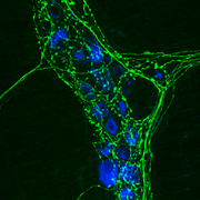
are the most common) which is detected in a fluorescence or confocal microscope. The location of fluorescence will vary according to the target molecule, external for membrane proteins, and internal for cytoplasmic proteins. In this way immunofluorescence
Immunofluorescence
Immunofluorescence is a technique used for light microscopy with a fluorescence microscope and is used primarily on biological samples. This technique uses the specificity of antibodies to their antigen to target fluorescent dyes to specific biomolecule targets within a cell, and therefore allows...
is a powerful technique when combined with confocal microscopy
Confocal microscopy
Confocal microscopy is an optical imaging technique used to increase optical resolution and contrast of a micrograph by using point illumination and a spatial pinhole to eliminate out-of-focus light in specimens that are thicker than the focal plane. It enables the reconstruction of...
for studying the location of proteins and dynamic processes (exocytosis
Exocytosis
Exocytosis , also known as 'The peni-cytosis', is the durable process by which a cell directs the contents of secretory vesicles out of the cell membrane...
, endocytosis
Endocytosis
Endocytosis is a process by which cells absorb molecules by engulfing them. It is used by all cells of the body because most substances important to them are large polar molecules that cannot pass through the hydrophobic plasma or cell membrane...
, etc.).