
Hyphema
Encyclopedia
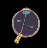
Anterior chamber
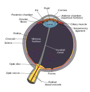
The anterior chamber is the fluid-filled space inside the eye between the iris and the cornea's innermost surface, the endothelium. Aqueous humor is the fluid that fills the anterior chamber. Hyphema and glaucoma are two main pathologies in this area. In hyphema, blood fills the anterior chamber...
) chamber of the eye
Human eye
The human eye is an organ which reacts to light for several purposes. As a conscious sense organ, the eye allows vision. Rod and cone cells in the retina allow conscious light perception and vision including color differentiation and the perception of depth...
. It may appear as a reddish tinge, or it may appear as a small pool of blood at the bottom of the iris or in the cornea.
Causes
Hyphemas are frequently caused by injury (blunt trauma) and may partially or completely block vision.Presentation and prognosis
Hyphemas may resolve by themselves, they may require medical treatment, or they may result in permanent visual impairment.A long-standing hyphema may result in hemosiderosis
Hemosiderosis
Idiopathic pulmonary haemosiderosis is a lung disease of unknown cause that is characterized by alveolar capillary bleeding and accumulation of haemosiderin in the lungs...
and heterochromia
Heterochromia
In anatomy, heterochromia refers to a difference in coloration, usually of the iris but also of hair or skin. Heterochromia is a result of the relative excess or lack of melanin...
.http://www.optometry.co.uk/files/b9ef5756eeb28a9f1aca8872fd3f9c07_swann19990129.pdf Blood accumulation may also cause an elevation of the intraocular pressure
Intraocular pressure
Intraocular pressure is the fluid pressure inside the eye. Tonometry is the method eye care professionals use to determine this. IOP is an important aspect in the evaluation of patients at risk from glaucoma...
.
Treatment
Treatment of hyphema consists of 4 major tenets:1. Light activity or even bedrest (to prevent a rebleed into the anterior chamber, which may cause obstruction of vision, or a painful rise in pressure)
2. Elevation of the head of the bed by approximately 45 degrees (so that the hyphema can settle out inferiorly and avoid obstruction of vision, as well as to facilitate resolution)
3. Wearing of an eye shield at night time (to prevent accidental rubbing of the eyes during sleep, which can precipitate a rebleed)
4. Avoidance of pain medications such as aspirin or ibuprofen (which thin the blood and increase the risk of a rebleed - instead, acetaminophen can be used for pain control).
It is controversial among ophthalmologists whether a steroid medication or a dilating drop (mydriatic) should be used in treatment of hyphema. Steroids aim to reduce the amount of inflammation, but are not without side effects. Dilating drops aim to increase comfort from the traumatized iris as well as reduce bleeding, but can also cause the pupil to be fixed in a dilated state via posterior synechiae (adhesions).
The vast majority of hyphemas resolve on their own without complication.
Some cases of non-resolving hyphemas, or hyphemas that are associated with high pressure, may require surgery to clean out the anterior chamber and prevent corneal blood staining.