
Gastroschisis
Encyclopedia
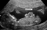
Gastroschisis represents a congenital defect characterized by a defect in the anterior abdominal wall through which the abdominal contents freely protrude. There is no overlying sac and the size of the defect is usually less than 4 cm. The abdominal wall defect is located at the junction of the umbilicus and normal skin, and is almost always to the right of the umbilicus.
Omphalocele
is another congenital birth defect, but it involves the umbilical cord itself, and the organs remain enclosed in visceral peritoneum. With omphalocele the defect is usually much larger than in gastroschisis.
A change in paternity (childbearing with different fathers) has been implicated as a risk factor in a recent study, suggesting that the immune system
of the mother may play a role in the development of gastroschisis.
manner. It may begin as a sporadic mutation
, can be associated with non-genetic congenital disorders, but has also been observed to be autosomal dominant.
 Genetic counseling
and further genetic testing
, such as amniocentesis
, may be offered during the pregnancy, as this and other abdominal wall defects are associated with genetic disorders. If there are no additional genetic problems or birth defects, surgery soon after birth can often repair the opening.
The first hypothesis does not explain why the mesoderm defect would occur in such a specific small area. The second hypothesis does not explain the low percentage of associated abnormality compared with omphalocele. The third hypothesis was criticized due to no vascular supplement of anterior abdominal wall by umbilical vein. The fourth hypothesis was commonly accepted, but it was later shown that the right vitelline artery (right omphalomesenteric artery) did not supply the anterior abdominal wall in this area. More evidence is needed to support the fifth hypothesis.
The general procedure for gastroschisis is to simply tuck the protruding organs back into the opening and apply a belly band pressure until the wound heals itself. New advances have been pioneered in repairing the protruding bowel by placing a protective "silo" around the intestine outside the abdomen, then slowly pressuring the herniated intestine into the abdominal cavity. This new procedure allows for the bowel to return to its intended shape and location without further traumatizing the infant's viscera with undue internal pressure. The main cause for lengthy recovery periods in patients is the time taken for the infants' bowel function to return to normal.
The morbidity is closely related to the presence of other malformations and complications of the wound or the intestine. Patients frequently require more than one surgery.
It has been reported that the incidence of gastroschisis has increased in recent years.
Omphalocele
Omphalocele
An omphalocele is a type of abdominal wall defect in which the intestines, liver, and occasionally other organs remain outside of the abdomen in a sac because of a defect in the development of the muscles of the abdominal wall.-Presentation:The sac, which is formed from an outpouching of...
is another congenital birth defect, but it involves the umbilical cord itself, and the organs remain enclosed in visceral peritoneum. With omphalocele the defect is usually much larger than in gastroschisis.
Causes
High-risk pregnancies such as those complicated by infection, young maternal age, smoking, drug abuse, or anything that contributes to low birth weight can increase the incidence of gastroschisis, which is more frequent in newborns who are small for gestational age. Whether the intrauterine growth retardation can facilitate the apparition of gastroschisis or the abdominal wall defect impairs fetal growth is not clear yet.A change in paternity (childbearing with different fathers) has been implicated as a risk factor in a recent study, suggesting that the immune system
Immune system
An immune system is a system of biological structures and processes within an organism that protects against disease by identifying and killing pathogens and tumor cells. It detects a wide variety of agents, from viruses to parasitic worms, and needs to distinguish them from the organism's own...
of the mother may play a role in the development of gastroschisis.
Genetics
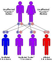
Gastroschisis as a stand-alone congenital defect is usually inherited in an autosomal recessiveRecessive
In genetics, the term "recessive gene" refers to an allele that causes a phenotype that is only seen in a homozygous genotype and never in a heterozygous genotype. Every person has two copies of every gene on autosomal chromosomes, one from mother and one from father...
manner. It may begin as a sporadic mutation
Mutation
In molecular biology and genetics, mutations are changes in a genomic sequence: the DNA sequence of a cell's genome or the DNA or RNA sequence of a virus. They can be defined as sudden and spontaneous changes in the cell. Mutations are caused by radiation, viruses, transposons and mutagenic...
, can be associated with non-genetic congenital disorders, but has also been observed to be autosomal dominant.
Genetic counseling
Genetic counseling or traveling is the process by which patients or relatives, at risk of an inherited disorder, are advised of the consequences and nature of the disorder, the probability of developing or transmitting it, and the options open to them in management and family planning...
and further genetic testing
Genetic testing
Genetic testing is among the newest and most sophisticated of techniques used to test for genetic disorders which involves direct examination of the DNA molecule itself. Other genetic tests include biochemical tests for such gene products as enzymes and other proteins and for microscopic...
, such as amniocentesis
Amniocentesis
Amniocentesis is a medical procedure used in prenatal diagnosis of chromosomal abnormalities and fetal infections, in which a small amount of amniotic fluid, which contains fetal tissues, is sampled from the amnion or amniotic sac surrounding a developing fetus, and the fetal DNA is examined for...
, may be offered during the pregnancy, as this and other abdominal wall defects are associated with genetic disorders. If there are no additional genetic problems or birth defects, surgery soon after birth can often repair the opening.
Pathophysiology
At least six hypotheses have been proposed:- Failure of mesoderm to form in the body wall
- Rupture of the amnion around the umbilical ring with subsequent herniation of bowel
- Abnormal involution of the right umbilical vein leading to weakening of the body wall and gut herniation
- Disruption of the right vitelline (yolk sac) artery with subsequent body wall damage and gut herniation
- Abnormal folding of the body wall results in a ventral body wall defect through which the gut herniates
- Failure to incorporate the yolk sac and related vitelline structures into the yolk sac
The first hypothesis does not explain why the mesoderm defect would occur in such a specific small area. The second hypothesis does not explain the low percentage of associated abnormality compared with omphalocele. The third hypothesis was criticized due to no vascular supplement of anterior abdominal wall by umbilical vein. The fourth hypothesis was commonly accepted, but it was later shown that the right vitelline artery (right omphalomesenteric artery) did not supply the anterior abdominal wall in this area. More evidence is needed to support the fifth hypothesis.
Embryology
During the fourth week of development, the lateral body folds move ventrally and fuse in the midline to form the anterior body wall. Incomplete fusion results in a defect that allows abdominal viscera to protrude through the abdominal wall. The bowel typically herniates through the rectus muscle, lying to the right of the umbilicus.Prognosis
Current advances in surgical techniques and intensive care management for neonates have increased the survival rate to 90%, in adequate settings. The possibility of prenatal diagnosis either through echosonogram or any other method available allows the mother to be referred to an adequate center where a caesarean section or induced natural birth can be performed before term (as natural birth is recommended and just as safe as with a normal baby), preferably within 2 weeks of term, and allow the immediate surgery to be performed on the newborn.The general procedure for gastroschisis is to simply tuck the protruding organs back into the opening and apply a belly band pressure until the wound heals itself. New advances have been pioneered in repairing the protruding bowel by placing a protective "silo" around the intestine outside the abdomen, then slowly pressuring the herniated intestine into the abdominal cavity. This new procedure allows for the bowel to return to its intended shape and location without further traumatizing the infant's viscera with undue internal pressure. The main cause for lengthy recovery periods in patients is the time taken for the infants' bowel function to return to normal.
The morbidity is closely related to the presence of other malformations and complications of the wound or the intestine. Patients frequently require more than one surgery.
Epidemiology
The malformation is slightly more frequent in males than females. The frequency of gastroschisis is associated with young maternal age, and low number of gestations. This abnormality is seen in ratio of 1:10,000 and is usually detected before birth.It has been reported that the incidence of gastroschisis has increased in recent years.
External links
- Fetal Treatment Program - Providence, Rhode Island at Brown UniversityBrown UniversityBrown University is a private, Ivy League university located in Providence, Rhode Island, United States. Founded in 1764 prior to American independence from the British Empire as the College in the English Colony of Rhode Island and Providence Plantations early in the reign of King George III ,...
- Fetal Treatment Center: Gastroschisis at UCSFUniversity of California, San FranciscoThe University of California, San Francisco is one of the world's leading centers of health sciences research, patient care, and education. UCSF's medical, pharmacy, dentistry, nursing, and graduate schools are among the top health science professional schools in the world...
Medical Center - Gastroschisis support, resources, birth stories http://www.gastroschisis.co.uk/gastroschisis/ gastroschisis.co.uk
- The Gastroschisis Support Network www.southenglandgastroschisis.webs.com
- Imaging Gastroschisis Ultrasound, MR, CT
- Gastroschisis Evaluation, Repair and Treatment Options Center for Fetal Diagnosis and Treatment at The Children's Hospital of Philadelphia