

Eye development
Encyclopedia
Eye
Eyes are organs that detect light and convert it into electro-chemical impulses in neurons. The simplest photoreceptors in conscious vision connect light to movement...
develops from the neural tube
Neural tube
In the developing vertebrate, the neural tube is the embryo's precursor to the central nervous system, which comprises the brain and spinal cord...
, the epidermis, and the periocular mesenchyme, which receives contributions from both the neural crest
Neural crest
Neural crest cells are a transient, multipotent, migratory cell population unique to vertebrates that gives rise to a diverse cell lineage including melanocytes, craniofacial cartilage and bone, smooth muscle, peripheral and enteric neurons and glia....
and mesoderm
Mesoderm
In all bilaterian animals, the mesoderm is one of the three primary germ cell layers in the very early embryo. The other two layers are the ectoderm and endoderm , with the mesoderm as the middle layer between them.The mesoderm forms mesenchyme , mesothelium, non-epithelial blood corpuscles and...
lineages.
Sequential inductions
This development is an example of sequential inductions where the organ is formed from three different tissues.Neural tube
First, there is an outpocketing of the neural tube called optic vesiclesOptic vesicles
The eyes begin to develop as a pair of diverticula from the lateral aspects of the forebrain. These diverticula make their appearance before the closure of the anterior end of the neural tube; after the closure of the tube they are known as the optic vesicles....
. Development of the optic vesicles starts in the 3-week embryo, from a progressively deepening groove in the neural plate called the optic sulcus. As this expands, the rostral neuropore (the exit of the brain cavity out of the embryo) closes and the optic sulcus and the neural plate becomes the optic vesicle.
Epidermis
The optic vesicles come into contact with the epithelum and induce the epidermis. The epithelium thickens to form the lens placodeLens placode
The Lens placode is a thickened portion of ectoderm which serves as the precursor to the lens.SOX2 and Pou2f1 are involved in its development.-External links:* http://cwx.prenhall.com/bookbind/pubbooks/martini10/chapter18/custom3/deluxe-content.html...
.
The lens differentiates and invaginates until it pinches off from the epithelium. The lens acts as an inducer back to the optic vesicle to transform it into the optic cup
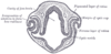
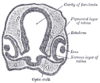
Optic cup
During embryonic development of the eye, the outer wall of the bulb of the optic vesicles becomes thickened and invaginated, and the bulb is thus converted into a cup, the optic cup , consisting of two strata of cells)...
and back to the epidermis to transform it into the cornea
Cornea
The cornea is the transparent front part of the eye that covers the iris, pupil, and anterior chamber. Together with the lens, the cornea refracts light, with the cornea accounting for approximately two-thirds of the eye's total optical power. In humans, the refractive power of the cornea is...
.
The optic cup then delaminates into two layers: The neural retina
Retina
The vertebrate retina is a light-sensitive tissue lining the inner surface of the eye. The optics of the eye create an image of the visual world on the retina, which serves much the same function as the film in a camera. Light striking the retina initiates a cascade of chemical and electrical...
and the retinal pigment epithelium.
Periocular mesenchyme
The periocular mesenchyme migrates in during the formation of the optic cup and is critical for the induction of the retinal pigment epithelium and the optic nerveOptic nerve
The optic nerve, also called cranial nerve 2, transmits visual information from the retina to the brain. Derived from the embryonic retinal ganglion cell, a diverticulum located in the diencephalon, the optic nerve doesn't regenerate after transection.-Anatomy:The optic nerve is the second of...
. The mesenchyme contributes to the cornea, iris
Iris (anatomy)
The iris is a thin, circular structure in the eye, responsible for controlling the diameter and size of the pupils and thus the amount of light reaching the retina. "Eye color" is the color of the iris, which can be green, blue, or brown. In some cases it can be hazel , grey, violet, or even pink...
, ciliary body
Ciliary body
The ciliary body is the circumferential tissue inside the eye composed of the ciliary muscle and ciliary processes. It is triangular in horizontal section and is coated by a double layer, the ciliary epithelium. This epithelium produces the aqueous humor. The inner layer is transparent and covers...
, sclera
Sclera
The sclera , also known as the white or white of the eye, is the opaque , fibrous, protective, outer layer of the eye containing collagen and elastic fiber. In the development of the embryo, the sclera is derived from the neural crest...
and blood vessels of the eye.
There is some evidence that LMX1B
LMX1B
LIM homeobox transcription factor 1-beta, also known as LMX1B, is a protein which in humans is encoded by the LMX1B gene.- Function :...
plays a role in periocular mesenchymal survival.
Developmental cascade
According to Liem et al., the organogenesis of the eye is pointed out as an example of a developmental cascade of inductions. The eye is essentially a derivative of the ectoderm from the somatic ectoderm and neural tube, with a succession of inductions by the chordamesoderm.Chordamesoderm induces the anterior portion of the neural tube to form the precursors of the synapomorphic tripartite brain of vertebrates, and it will form a bulge called the diencephalon. Further induction by the chordamesoderm will form a protrusion: the optic visicle. This visicle will be subsequently invaginated by means of further inductions from the chordamesoderm. The optic visicle will then induce the ectoderm that thickens (lens placode) and further invaginates to a point that detaches from the ectoderm and forms a neurogenic placode by itself. The lens placode is affected by the chordamesoderm making it to invaginate and forms the optic cup composed by an outer layer of neural retina and inner layer the pigmented retina that will unite and form the optic stalk. The pigmented retina is formed by rods and cones and composed of small cilia typical of the ependymal epithelium of the neural tube. Some cells in the lens vesicle will be fated to form the cornea and the lens vesicle will develop completely to form the definitive lens. Iris is formed from the optic cup cells.
Responsivity of head epidermis
Only the epidermis in the head is competent to respond to the signal from the optic vesicles. Both the optic vesicle and the head epidermis are required for eye development. The competence of the head epidermis to respond to the optic vesicle signals comes from the expression of Pax6PAX6
Paired box protein Pax-6 also known as aniridia type II protein or oculorhombin is a protein that in humans is encoded by the PAX6 gene.- Function :PAX6 is a member of the Pax gene family...
in the epidermis. Pax6 is necessary and sufficient for eye induction. This competenece is acquired gradually during gastrulation
Gastrulation
Gastrulation is a phase early in the embryonic development of most animals, during which the single-layered blastula is reorganized into a trilaminar structure known as the gastrula. These three germ layers are known as the ectoderm, mesoderm, and endoderm.Gastrulation takes place after cleavage...
and neurulation
Neurulation
Neurulation is the stage of organogenesis in vertebrate embryos, during which the neural tube is transformed into the primitive structures that will later develop into the central nervous system....
from interactions with the endoderm
Endoderm
Endoderm is one of the three primary germ cell layers in the very early embryo. The other two layers are the ectoderm and mesoderm , with the endoderm as the intermost layer...
, mesoderm
Mesoderm
In all bilaterian animals, the mesoderm is one of the three primary germ cell layers in the very early embryo. The other two layers are the ectoderm and endoderm , with the mesoderm as the middle layer between them.The mesoderm forms mesenchyme , mesothelium, non-epithelial blood corpuscles and...
, and neural plate
Neural plate
In human embryology, formation of neural plate is the first step of neurulation. It is created by a flat thickening opposite to the primitive streak of the ectoderm.-Development:...
.
Regulation and inhibition
Sonic hedgehogSonic hedgehog
Sonic hedgehog homolog is one of three proteins in the mammalian signaling pathway family called hedgehog, the others being desert hedgehog and Indian hedgehog . SHH is the best studied ligand of the hedgehog signaling pathway. It plays a key role in regulating vertebrate organogenesis, such as...
reduces the expression of Pax6. When Shh is inhibited during development, the domain of expression for Pax6 is expanded and the eyes fail to separate causing cyclopia
Cyclopia
Cyclopia is a rare form of holoprosencephaly and is a congenital disorder characterized by the failure of the embryonic prosencephalon to properly divide the orbits of the eye into two cavities...
. Overexpression of Shh causes a loss of eye structures.
Retinoic acid
Retinoic acid
Retinoic acid is a metabolite of vitamin A that mediates the functions of vitamin A required for growth and development. Retinoic acid is required in chordate animals which includes all higher animals from fishes to humans...
generated from vitamin A
Vitamin A
Vitamin A is a vitamin that is needed by the retina of the eye in the form of a specific metabolite, the light-absorbing molecule retinal, that is necessary for both low-light and color vision...
in the retina plays an essential role in eye development as a secreted paracrine signal which restricts invasion of perioptic mesenchyme around the optic cup. Vitamin A deficiency
Vitamin A deficiency
Vitamin A deficiency is a lack of vitamin A in humans. It is common in developing countries but rarely seen in developed countries. Night blindness is one of the first signs of vitamin A deficiency. Xerophthalmia and complete blindness can also occur since Vitamin A has a major role in...
during embryogenesis results in anterior segment
Anterior segment
The anterior segment is the front third of the eye that includes the structures in front of the vitreous humour: the cornea, iris, ciliary body, and lens.Within the anterior segment are two fluid-filled spaces:...
defects (particularly cornea and eyelids) that lead to vision loss or blindness.