
Epiretinal membrane
Encyclopedia

Posterior vitreous detachment
A posterior vitreous detachment is a condition of the eye in which the vitreous humour separates from the retina.Broadly speaking, the condition is common for older adults and over 75% of those over the age of 65 develop it. Although less common among people in their 40s or 50s, the condition is...
(PVD). PVD can create minor damage to the retina, stimulating exudate, inflammation, and leucocyte
White blood cell
White blood cells, or leukocytes , are cells of the immune system involved in defending the body against both infectious disease and foreign materials. Five different and diverse types of leukocytes exist, but they are all produced and derived from a multipotent cell in the bone marrow known as a...
response. These cells can form a transparent layer gradually and, like all scar tissue
Scar tissue
Scar tissue can refer to:*Granulation tissue, a product of healing in major wounds*The tissue of a scar*"Scar Tissue", a Red Hot Chili Peppers song*Scar Tissue , the autobiography of Anthony Kiedis, lead singer of the Red Hot Chili Peppers...
, tighten to create tension on the retina which may bulge and pucker (e.g. macular pucker), or even cause swelling or Macular edema
Macular edema
Macular edema occurs when fluid and protein deposits collect on or under the macula of the eye and causes it to thicken and swell. The swelling may distort a person's central vision, as the macula is near the center of the retina at the back of the eyeball...
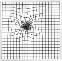
. Often this results in distortions of vision that are clearly visible as bowing and blurring when looking at lines on chart paper (or an Amsler grid
Amsler grid
The Amsler grid, used since 1945, is a grid of horizontal and vertical lines used to monitor a person's central visual field. The grid was developed by Marc Amsler, a Swiss ophthalmologist. It is a diagnostic tool that aids in the detection of visual disturbances caused by changes in the retina,...
) within the macula
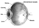
Macula
The macula or macula lutea is an oval-shaped highly pigmented yellow spot near the center of the retina of the human eye. It has a diameter of around 5 mm and is often histologically defined as having two or more layers of ganglion cells...
r area, or central 1.0 degree of visual arc. Usually it occurs in one eye first, and may cause binocular diplopia
Diplopia
Diplopia, commonly known as double vision, is the simultaneous perception of two images of a single object that may be displaced horizontally, vertically, or diagonally in relation to each other...
or double vision
Diplopia
Diplopia, commonly known as double vision, is the simultaneous perception of two images of a single object that may be displaced horizontally, vertically, or diagonally in relation to each other...
if the image from eye is too different from the image of the other eye. The distortions can make objects look different in size (usually larger = macropsia
Macropsia
Macropsia is a neurological condition affecting human visual perception, in which objects within an affected section of the visual field appear larger than normal, causing the subject to feel smaller. Macropsia, along with its opposite condition, micropsia, can be categorized under dysmetropsia...
), especially in the central portion of the visual field, creating a localized or field dependent aniseikonia
Aniseikonia
Aniseikonia is an ocular condition where there is a significant difference in the perceived size of images. It can occur as an overall difference between the two eyes, or as a difference in a particular meridian.-Causes:Retinal image size is determined by many factors...
that cannot be fully corrected optically with glasses. Partial correction often improves the binocular vision considerably though. In the young (under 50 years of age), these cells occasionally pull free and disintegrate on their own; but in the majority of sufferers (over 60 years of age) the condition is permanent. The underlying photoreceptor cells, rod cells and cone cells, are usually not damaged unless the membrane becomes quite thick and hard; so usually there is no macular degeneration
Macular degeneration
Age-related macular degeneration is a medical condition which usually affects older adults and results in a loss of vision in the center of the visual field because of damage to the retina. It occurs in “dry” and “wet” forms. It is a major cause of blindness and visual impairment in older adults...
.
Surgery for epiretinal membrane
Surgeons can remove or peel the membrane through the scleraSclera
The sclera , also known as the white or white of the eye, is the opaque , fibrous, protective, outer layer of the eye containing collagen and elastic fiber. In the development of the embryo, the sclera is derived from the neural crest...
and improve vision by 2 or more Snellen lines. Usually the vitreous is replaced at the same time with clear fluid, in a vitrectomy
Vitrectomy
Vitrectomy is a surgery to remove some or all of the vitreous humor from the eye. Anterior vitrectomy entails removing small portions of the vitreous from the front structures of the eye—often because these are tangled in an intraocular lens or other structures...
. Surgery is not usually recommended unless the distortions are severe enough to interfere with daily living, since there are the usual hazards of surgery, infections, and a possibility of retinal detachment
Retinal detachment
Retinal detachment is a disorder of the eye in which the retina peels away from its underlying layer of support tissue. Initial detachment may be localized, but without rapid treatment the entire retina may detach, leading to vision loss and blindness. It is a medical emergency.The retina is a...
. More common complications are high intraocular pressure, bleeding in the eye, and cataract
Cataract
A cataract is a clouding that develops in the crystalline lens of the eye or in its envelope, varying in degree from slight to complete opacity and obstructing the passage of light...
s, which are the most frequent complication of vitrectomy surgery. Many patients will develop a cataract within the first few years after surgery. In fact, the visual distortions and diplopia
Diplopia
Diplopia, commonly known as double vision, is the simultaneous perception of two images of a single object that may be displaced horizontally, vertically, or diagonally in relation to each other...
created by cataracts may sometimes be confused with epiretinal membrane.
Prevention
There is no good evidence for any preventative actions, since it appears this is a natural response to aging changes in the vitreous. posterior vitreous detachmentPosterior vitreous detachment
A posterior vitreous detachment is a condition of the eye in which the vitreous humour separates from the retina.Broadly speaking, the condition is common for older adults and over 75% of those over the age of 65 develop it. Although less common among people in their 40s or 50s, the condition is...
(PVD) has been estimated to occur in over 75 per cent of the population over age 65, that PVD is essentially a harmless condition (although with some disturbing symptoms), and that it does not normally threaten sight. However, since epiretinal membrane appears to be a protective response to PVD, where inflammation
Inflammation
Inflammation is part of the complex biological response of vascular tissues to harmful stimuli, such as pathogens, damaged cells, or irritants. Inflammation is a protective attempt by the organism to remove the injurious stimuli and to initiate the healing process...
, exudative fluid, and scar tissue
Scar tissue
Scar tissue can refer to:*Granulation tissue, a product of healing in major wounds*The tissue of a scar*"Scar Tissue", a Red Hot Chili Peppers song*Scar Tissue , the autobiography of Anthony Kiedis, lead singer of the Red Hot Chili Peppers...
is formed, it is possible that NSAIDS may reduce the inflammation response. Usually there are flashing light experiences and the emergence of floater
Floater
Floaters are deposits of various size, shape, consistency, refractive index, and motility within the eye's vitreous humour, which is normally transparent. At young age the vitreous is perfectly transparent, but during life imperfections gradually develop. The common type of floater, which is...
s in the eye that herald changes in the vitreous
Vitreous humour
The vitreous humour or vitreous humor is the clear gel that fills the space between the lens and the retina of the eyeball of humans and other vertebrates...
before the epiretinal membrane forms.
Scientific background
This ocular pathology was first described by Iwanoff in 1865, and it has been shown to occur in about 7% of the population. It can occur more frequently in the older population with postmortem studies showing it in 2% of those aged 50 years and 20% in those aged 75 years.The source of the cells in epiretinal membranes (ERM) has been found to comprise glial cells, retinal pigment epithelial (RPE) cells, macrophages, fibrocytes, and collagen cells. These cells are found in varying proportions. Those from retinal breaks, previous retinal detachments, or cryopexy are composed mainly of dispersed RPE cells, while cells of glial origin predominate in idiopathic pathology. Laminocytes are the fundamental cell type in idiopathic ERMs. These cells are frequently found in small and dispersed numbers in eyes containing a PVD. The presence of retinal pigment cells invariable indicates proliferative retinopathy and is only seen in association with a retinal detachment or tear.
The incidence of associated PVD range from 75-93%, and PVD is present in virtually all eyes with retinal breaks or retinal detachments and subsequent ERM formation. PVD can lead to retinal breaks that may liberate RPE cells that initiate membrane formation. Small breaks in the internal limiting membrane (ILM) after PVD also may provide retinal astrocytes access to the vitreous cavity, where they may subsequently proliferate. Many ERM also have ILM fragments that may be peeled separately. Finally, vitreous hemorrhage, inflammation, or both associated with a PVD also may stimulate ERM formation.
Both sexes appear to be affected equally frequently.
Synonyms
Macular pucker, epimacular membrane, preretinal membrane, cellophane maculopathy, retina wrinkle, surface wrinkling retinopathy, premacular fibrosis, and internal limiting membrane disease.External links
- Macular Pucker Resource Guide from the National Eye Institute (NEI).
- Treatment of Epiretinal Membrane, Mayo ClinicMayo ClinicMayo Clinic is a not-for-profit medical practice and medical research group specializing in treating difficult patients . Patients are referred to Mayo Clinic from across the U.S. and the world, and it is known for innovative and effective treatments. Mayo Clinic is known for being at the top of...