
Development of the gonads
Encyclopedia
The prenatal development of the gonads is a part of the development of reproductive system and ultimately forms the testes in males and ovaries in females. They initially develop from the mesothelial layer of the peritoneum.
The ovary is differentiated into a central part, the medulla of ovary, covered by a surface layer, the germinal epithelium. The immature ova originate from cells from the dorsal endoderm
of the yolk sac
. Once they have reached the gonadal ridge they are called oogonia. Development proceeds and the oogonia become fully surrounded by a layer of connective tissue cells (pre-granulosa cells) In this way, the rudiments of the ovarian follicles are formed.
In the testis, a network of tubules fuse to create the seminiferous tubules
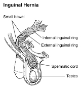
. Via the rete testis, the seminiferous tubules become connected with outgrowths from the mesonephros, which form the efferent ducts of the testis. The descent of the testes consists of the opening of a connection from the testis to its final location at the anterior abdominal wall, followed by the development of the gubernaculum, which subsequently pulls and translocates the testis down into the developing scrotum. Ultimately, the passageway closes behind the testis. A failure in this process causes indirect inguinal hernia
.
is essentially the same in the two sexes, and consists in a thickening of the mesothelial layer of the peritoneum
. The thick plate of epithelium extends deeply, pushing before it the mesoderm and forming a distinct projection. This is termed the gonadal ridge
. The gonadal ridge, in turn, develops into a gonad. This is a testis in the male and an ovary
in the female.
At first, the mesonephros
and gonadal ridge are continuous, but as the embryo grows the gonadal ridge gradually becomes pinched off from the mesonephros. However, some cells of mesonephric origin join the gonadal ridge. Furthermore, the gonadal ridge still remains connected to the remnant of that body by a fold of peritoneum, namely the mesorchium
or mesovarium
.
About the seventh week the distinction of sex in the gonadal ridge begins to be perceptible.
is thus formed mainly from the genital ridge and partly from the mesonephros. Later the mass is differentiated into a central part, the medulla of ovary
, covered by a surface layer, the germinal epithelium
. Between the cells of the germinal epithelium a number of larger cells, the immature ova, are found. The immature ova, in turn, are carried into the stroma beneath by bud-like ingrowths (genital cords of the germinal epithelium). The surface germinal epithelium ultimately forms the permanent epithelial covering of this organ. Furthermore, it soon loses its connection with the central mass. Instead, the tunica albuginea of the ovaries develops between them.
of the yolk sac
migrate along the hindgut to the gonadal ridge. These primordial germ cells (PGCs) multiply by mitosis and once they have reached the gonadal ridge they are called oogonia (diploid stem cells of the ovary).
Once oogonia enter this area they attempt to associate with the other somatic cells, derived from both the peritoneum and mesonephros.
Development proceeds and the oogonia become fully surrounded by a layer of connective tissue cells (pre-granulosa cells) in an irregular manner. In this way, the rudiments of the ovarian follicles are formed.
In the 1970’s, evidence emerged that the first cells to make contact with the oogonia were of mesonephric origin.
It was suggested that mesonephric cells already closely associated with the oogonia proliferated throughout development to form the granulosa cell layer.
Recently this hypothesis has been challenged with some thorough histology. Sawyer et al. hypothesised that in sheep most of the granulosa cells develop from cells of the mesothelium
(i.e., epithelial cells from the presumptive surface epithelium of the ovary).
in the female, which effects a considerable change in the position of the ovary, though not so extensive a change as in that of the testis. The gubernaculum in the female lies in contact with the fundus of the uterus and adheres to this organ, and thus the ovary can only descend as far as to this level. The part of the gubernaculum between the ovary and the uterus ultimately becomes the proper ovarian ligament
, while the part between the uterus and the labium majus forms the round ligament of the uterus. A pouch of peritoneum analogous to the processus vaginalis
in the male accompanies it along the inguinal canal: it is called the canal of Nuck
.
, thus excluding the surface epithelium from any part in the formation of the tissue of the testis. The cords of the central mass run together toward the future hilum
and form a network which ultimately becomes the rete testis
. On the other hand, the seminiferous tubules
are developed from the cords distal to the hilum, and between them connective-tissue septa extend. Via the rete testis, the seminiferous tubules become connected with outgrowths from the mesonephros, which form the efferent ducts of the testis.
, to the mesonephros. From the front of the mesonephros a fold of peritoneum termed the inguinal fold grows forward to meet and fuse with a peritoneal fold, the inguinal crest, which grows backward from the antero-lateral abdominal wall. The testis thus acquires an indirect connection with the anterior abdominal wall. At the same time, a portion of the peritoneal cavity lateral to these fused folds is marked off as the future saccus vaginalis.
, makes its appearance. This is at first a slender band, extending from that part of the skin of the groin
which afterward forms the scrotum
through the inguinal canal
to the body and epididymis of the testis. As development advances, the peritoneum enclosing the gubernaculum forms two folds, one above the testis and the other below it. The one above the testis is the plica vascularis, and contains the upper part of the gubernaculum, and ultimately also the internal spermatic vessels; the one below, the plica gubernatrix, contains the lower part of the gubernaculum.
The gubernaculum grows into a thick cord. It ends below at the abdominal inguinal ring in a tube of peritoneum, the saccus vaginalis, which protrudes itself down the inguinal canal. By the fifth month the lower part of the gubernaculum still is a thick cord, while the upper part has disappeared. The lower part now consists of a central core of smooth muscle
fibers, surrounded by a firm layer of striated muscle
elements, connected, behind the peritoneum, with the abdominal wall.
of the obliquus externus, which form respectively the cremaster muscle
and the external spermatic fascia
. The saccus vaginalis forms a gradually elongating pouch, which eventually reaches the bottom of the scrotum, and behind this pouch the testis is drawn by the growth of the body of the fetus, for the gubernaculum does not grow proportionately with the growth of other parts, and therefore the testis, being attached by the gubernaculum to the bottom of the scrotum, is prevented from rising as the body grows, and is instead drawn first into the inguinal canal and eventually into the scrotum. It seems certain also that the gubernacular cord becomes shortened as development proceeds, and this assists in causing the testis to reach the bottom of the scrotum.
 By the end of the eighth month the testis has reached the scrotum, preceded by the saccus vaginalis, which communicates by its upper extremity with the peritoneal cavity. Just before birth the upper part of the saccus vaginalis, at the internal inguinal ring, usually becomes closed, and this obliteration extends gradually downward to within a short distance of the testis. The process of peritoneum surrounding the testis is now entirely cut off from the general peritoneal cavity and constitutes the tunica vaginalis.
If the internal inguinal ring doesn't close properly, then there is a risk that other contents of the abdominal cavity protrudes through the passageway and cause indirect inguinal hernia
.
The ovary is differentiated into a central part, the medulla of ovary, covered by a surface layer, the germinal epithelium. The immature ova originate from cells from the dorsal endoderm
Endoderm
Endoderm is one of the three primary germ cell layers in the very early embryo. The other two layers are the ectoderm and mesoderm , with the endoderm as the intermost layer...
of the yolk sac
Yolk sac
The yolk sac is a membranous sac attached to an embryo, providing early nourishment in the form of yolk in bony fishes, sharks, reptiles, birds, and primitive mammals...
. Once they have reached the gonadal ridge they are called oogonia. Development proceeds and the oogonia become fully surrounded by a layer of connective tissue cells (pre-granulosa cells) In this way, the rudiments of the ovarian follicles are formed.
In the testis, a network of tubules fuse to create the seminiferous tubules
Seminiferous tubules
Seminiferous tubules are located in the testes, and are the specific location of meiosis, and the subsequent creation of gametes, namely spermatozoa....
. Via the rete testis, the seminiferous tubules become connected with outgrowths from the mesonephros, which form the efferent ducts of the testis. The descent of the testes consists of the opening of a connection from the testis to its final location at the anterior abdominal wall, followed by the development of the gubernaculum, which subsequently pulls and translocates the testis down into the developing scrotum. Ultimately, the passageway closes behind the testis. A failure in this process causes indirect inguinal hernia
Indirect inguinal hernia
An indirect inguinal hernia is an inguinal hernia that results from the failure of embryonic closure of the deep inguinal ring after the testicle has passed through it. Like other inguinal hernias, it protrudes through the superficial inguinal ring...
.
Before differentiation
The first appearance of the gonadGonad
The gonad is the organ that makes gametes. The gonads in males are the testes and the gonads in females are the ovaries. The product, gametes, are haploid germ cells. For example, spermatozoon and egg cells are gametes...
is essentially the same in the two sexes, and consists in a thickening of the mesothelial layer of the peritoneum
Peritoneum
The peritoneum is the serous membrane that forms the lining of the abdominal cavity or the coelom — it covers most of the intra-abdominal organs — in amniotes and some invertebrates...
. The thick plate of epithelium extends deeply, pushing before it the mesoderm and forming a distinct projection. This is termed the gonadal ridge
Gonadal ridge
In embryology, the gonadal ridge is the precursor to the gonads. The gonadal ridge initially consists mainly of mesenchyme and cells of underlying mesonephric origin. Once oogonia enter this area they attempt to associate with these somatic cells...
. The gonadal ridge, in turn, develops into a gonad. This is a testis in the male and an ovary
Ovary
The ovary is an ovum-producing reproductive organ, often found in pairs as part of the vertebrate female reproductive system. Ovaries in anatomically female individuals are analogous to testes in anatomically male individuals, in that they are both gonads and endocrine glands.-Human anatomy:Ovaries...
in the female.
At first, the mesonephros
Mesonephros
The mesonephros is one of three excretory organs that develop in vertebrates. It serves as the main excretory organ of aquatic vertebrates and as a temporary kidney in reptiles, birds, and mammals. The mesonephros is included in the Wolffian body after Caspar Friedrich Wolff who described it in 1759...
and gonadal ridge are continuous, but as the embryo grows the gonadal ridge gradually becomes pinched off from the mesonephros. However, some cells of mesonephric origin join the gonadal ridge. Furthermore, the gonadal ridge still remains connected to the remnant of that body by a fold of peritoneum, namely the mesorchium
Mesorchium
The testes, at an early period of fetal life, are placed at the back part of the abdominal cavity, behind the peritoneum, and each is attached by a peritoneal fold, the mesorchium, to the mesonephros.-See also:* mesentery* mesovarium...
or mesovarium
Mesovarium
The mesovarium is the portion of the broad ligament of the uterus that covers the ovaries.At first the mesonephros and genital ridge are suspended by a common mesentery, but as the embryo grows the genital ridge gradually becomes pinched off from the mesonephros, with which it is at first...
.
About the seventh week the distinction of sex in the gonadal ridge begins to be perceptible.
The Ovary
The ovaryOvary
The ovary is an ovum-producing reproductive organ, often found in pairs as part of the vertebrate female reproductive system. Ovaries in anatomically female individuals are analogous to testes in anatomically male individuals, in that they are both gonads and endocrine glands.-Human anatomy:Ovaries...
is thus formed mainly from the genital ridge and partly from the mesonephros. Later the mass is differentiated into a central part, the medulla of ovary
Medulla of ovary
The medulla of ovary is a highly vascular stroma in the center of the ovary. It forms from embryonic mesenchyme and contains blood vessels, lymphatic vessels, and nerves....
, covered by a surface layer, the germinal epithelium
Germinal epithelium (female)
The surface of the ovary is covered by a layer of simple cuboidal cells which constitutes the germinal epithelium of Waldeyer.These cells are derived from the mesoderm during embryonic development and are closely related to the mesothelium of the peritoneum...
. Between the cells of the germinal epithelium a number of larger cells, the immature ova, are found. The immature ova, in turn, are carried into the stroma beneath by bud-like ingrowths (genital cords of the germinal epithelium). The surface germinal epithelium ultimately forms the permanent epithelial covering of this organ. Furthermore, it soon loses its connection with the central mass. Instead, the tunica albuginea of the ovaries develops between them.
Origin of ova
During early embryonic development, cells from the dorsal endodermEndoderm
Endoderm is one of the three primary germ cell layers in the very early embryo. The other two layers are the ectoderm and mesoderm , with the endoderm as the intermost layer...
of the yolk sac
Yolk sac
The yolk sac is a membranous sac attached to an embryo, providing early nourishment in the form of yolk in bony fishes, sharks, reptiles, birds, and primitive mammals...
migrate along the hindgut to the gonadal ridge. These primordial germ cells (PGCs) multiply by mitosis and once they have reached the gonadal ridge they are called oogonia (diploid stem cells of the ovary).
Once oogonia enter this area they attempt to associate with the other somatic cells, derived from both the peritoneum and mesonephros.
Development proceeds and the oogonia become fully surrounded by a layer of connective tissue cells (pre-granulosa cells) in an irregular manner. In this way, the rudiments of the ovarian follicles are formed.
Origin of granulosa cells
The embryological origin of granulosa cells remains controversial.In the 1970’s, evidence emerged that the first cells to make contact with the oogonia were of mesonephric origin.
It was suggested that mesonephric cells already closely associated with the oogonia proliferated throughout development to form the granulosa cell layer.
Recently this hypothesis has been challenged with some thorough histology. Sawyer et al. hypothesised that in sheep most of the granulosa cells develop from cells of the mesothelium
Mesothelium
The mesothelium is a membrane that forms the lining of several body cavities: the pleura , peritoneum and pericardium . Mesothelial tissue also surrounds the male internal reproductive organs and covers the internal reproductive organs of women...
(i.e., epithelial cells from the presumptive surface epithelium of the ovary).
Descent of the Ovaries
Just as in the male, there is a gubernaculumGubernaculum
The paired Gubernacula are embryonic structures which begin as undifferentiated mesenchyme attaching to the caudal end of the gonads .-Function during development:...
in the female, which effects a considerable change in the position of the ovary, though not so extensive a change as in that of the testis. The gubernaculum in the female lies in contact with the fundus of the uterus and adheres to this organ, and thus the ovary can only descend as far as to this level. The part of the gubernaculum between the ovary and the uterus ultimately becomes the proper ovarian ligament
Ovarian ligament
The ovarian ligament is a fibrous ligament that connects the ovary to the lateral surface of the uterus....
, while the part between the uterus and the labium majus forms the round ligament of the uterus. A pouch of peritoneum analogous to the processus vaginalis
Processus vaginalis
The processus vaginalis is an embryonic developmental outpouching of the peritoneum.It is present from around the 12th week of gestation, and commences as a peritoneal outpouching.-Gender differences:...
in the male accompanies it along the inguinal canal: it is called the canal of Nuck
Canal of Nuck
The canal of Nuck, described by Anton Nuck in 1691, is an abnormal patent pouch of peritoneum extending into the labia majora of women. It is analogous to the processus vaginalis in males ....
.
Pathology
In rare cases the gubernaculum may fail to contract adhesions to the uterus, and then the ovary descends through the inguinal canal into the labium majus, and under these circumstances its position resembles that of the testis.The testis
The testis is developed in much the same way as the ovary, originating from mesothelium as well as mesonephros. Like the ovary, in its earliest stages it consists of a central mass covered by a surface epithelium. In the central mass a series of cords appear, and the periphery of the mass is converted into the tunica albugineaTunica albuginea (testicles)
The Tunica Albuginea is the fibrous covering of the testis.It is a dense blue-white membrane, composed of bundles of white fibrous tissue which interlace in every direction....
, thus excluding the surface epithelium from any part in the formation of the tissue of the testis. The cords of the central mass run together toward the future hilum
Hilum (anatomy)
In human anatomy, the hilum is a depression or fissure where structures such as blood vessels and nerves enter an organ.-Examples of hila:* Hilum of kidney, admits the renal artery, vein, ureter, and nerves...
and form a network which ultimately becomes the rete testis
Rete testis
Rete testis is an anastomosing network of delicate tubules located in the hilum of the testicle that carries sperm from the seminiferous tubules to the vasa efferentia....
. On the other hand, the seminiferous tubules
Seminiferous tubules
Seminiferous tubules are located in the testes, and are the specific location of meiosis, and the subsequent creation of gametes, namely spermatozoa....
are developed from the cords distal to the hilum, and between them connective-tissue septa extend. Via the rete testis, the seminiferous tubules become connected with outgrowths from the mesonephros, which form the efferent ducts of the testis.
Descent of the Testes
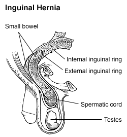
The descent of the testes consists of the opening of a connection from the testis to its final location at the anterior abdominal wall, followed by the development of the gubernaculum, which subsequently pulls and translocates the testis down into the developing scrotum. Ultimately, the passageway closes behind the testis.Opening of connection
At an early period of fetal life the testes are placed at the back part of the abdominal cavity, behind the peritoneum, and each is attached by a peritoneal fold, the mesorchiumMesorchium
The testes, at an early period of fetal life, are placed at the back part of the abdominal cavity, behind the peritoneum, and each is attached by a peritoneal fold, the mesorchium, to the mesonephros.-See also:* mesentery* mesovarium...
, to the mesonephros. From the front of the mesonephros a fold of peritoneum termed the inguinal fold grows forward to meet and fuse with a peritoneal fold, the inguinal crest, which grows backward from the antero-lateral abdominal wall. The testis thus acquires an indirect connection with the anterior abdominal wall. At the same time, a portion of the peritoneal cavity lateral to these fused folds is marked off as the future saccus vaginalis.
Development of gubernaculum
Also, in the inguinal crest a structure, the gubernaculum testisGubernaculum testis
In the inguinal crest a peculiar structure, the gubernaculum testis, makes its appearance. This is at first a slender band, extending from that part of the skin of the groin which afterward forms the scrotum through the inguinal canal to the body and epididymis of the testis.-External links:*...
, makes its appearance. This is at first a slender band, extending from that part of the skin of the groin
Groin
In human anatomy, the groin areas are the two creases at the junction of the torso with the legs, on either side of the pubic area. This is also known as the medial compartment of the thigh. A pulled groin muscle usually refers to a painful injury sustained by straining the hip adductor muscles...
which afterward forms the scrotum
Scrotum
In some male mammals the scrotum is a dual-chambered protuberance of skin and muscle containing the testicles and divided by a septum. It is an extension of the perineum, and is located between the penis and anus. In humans and some other mammals, the base of the scrotum becomes covered with curly...
through the inguinal canal
Inguinal canal
The inguinal canal is a passage in the anterior abdominal wall which in men conveys the spermatic cord and in women the round ligament. The inguinal canal is larger and more prominent in men.-Site:...
to the body and epididymis of the testis. As development advances, the peritoneum enclosing the gubernaculum forms two folds, one above the testis and the other below it. The one above the testis is the plica vascularis, and contains the upper part of the gubernaculum, and ultimately also the internal spermatic vessels; the one below, the plica gubernatrix, contains the lower part of the gubernaculum.
The gubernaculum grows into a thick cord. It ends below at the abdominal inguinal ring in a tube of peritoneum, the saccus vaginalis, which protrudes itself down the inguinal canal. By the fifth month the lower part of the gubernaculum still is a thick cord, while the upper part has disappeared. The lower part now consists of a central core of smooth muscle
Smooth muscle
Smooth muscle is an involuntary non-striated muscle. It is divided into two sub-groups; the single-unit and multiunit smooth muscle. Within single-unit smooth muscle tissues, the autonomic nervous system innervates a single cell within a sheet or bundle and the action potential is propagated by...
fibers, surrounded by a firm layer of striated muscle
Striated muscle
Striated muscle tissue is a form of fibers that are combined into parallel fibers. More specifically, it can refer to:* Cardiac muscle .* Skeletal muscle* Branchiomeric muscles...
elements, connected, behind the peritoneum, with the abdominal wall.
Translocation
As the testes develops, the main portion of the lower end of the gubernaculum is carried, following the skin to which it is attached, to the bottom of this pouch. Other bands are carried to the medial side of the thigh and to the perineum. The tube of peritoneum constituting the saccus vaginalis projects itself downward into the inguinal canal, and emerges at the external inguinal ring, pushing before it a part of the obliquus internus and the aponeurosisAponeurosis
Aponeuroses are layers of flat broad tendons. They have a shiny, whitish-silvery color, are histologically similar to tendons, and are very sparingly supplied with blood vessels and nerves. When dissected, aponeuroses are papery, and peel off by sections...
of the obliquus externus, which form respectively the cremaster muscle
Cremaster muscle
-Contraction:Its function is to raise and lower the testes in order to regulate the temperature of the testes and promote spermatogenesis. Contraction may also occur during arousal which can prevent injury to the testicles during sex....
and the external spermatic fascia
External spermatic fascia
The external spermatic fascia is a thin membrane, prolonged downward around the surface of the spermatic cord and testis. It is separated from the dartos tunic by loose areolar tissue...
. The saccus vaginalis forms a gradually elongating pouch, which eventually reaches the bottom of the scrotum, and behind this pouch the testis is drawn by the growth of the body of the fetus, for the gubernaculum does not grow proportionately with the growth of other parts, and therefore the testis, being attached by the gubernaculum to the bottom of the scrotum, is prevented from rising as the body grows, and is instead drawn first into the inguinal canal and eventually into the scrotum. It seems certain also that the gubernacular cord becomes shortened as development proceeds, and this assists in causing the testis to reach the bottom of the scrotum.
Closing of connection
Pathology
If the internal inguinal ring doesn't close properly, then there is a risk that other contents of the abdominal cavity protrudes through the passageway and cause indirect inguinal hernia
Indirect inguinal hernia
An indirect inguinal hernia is an inguinal hernia that results from the failure of embryonic closure of the deep inguinal ring after the testicle has passed through it. Like other inguinal hernias, it protrudes through the superficial inguinal ring...
.