
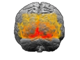
Brodmann area 19
Encyclopedia
Human
Brodmann area 19, or BA19, is part of the occipital lobeOccipital lobe
The occipital lobe is the visual processing center of the mammalian brain containing most of the anatomical region of the visual cortex. The primary visual cortex is Brodmann area 17, commonly called V1...
cortex
Cerebral cortex
The cerebral cortex is a sheet of neural tissue that is outermost to the cerebrum of the mammalian brain. It plays a key role in memory, attention, perceptual awareness, thought, language, and consciousness. It is constituted of up to six horizontal layers, each of which has a different...
in the human brain
Human brain
The human brain has the same general structure as the brains of other mammals, but is over three times larger than the brain of a typical mammal with an equivalent body size. Estimates for the number of neurons in the human brain range from 80 to 120 billion...
. Along with area 18, it comprises the extrastriate (or peristriate) cortex. In normally-sighted humans, extrastriate cortex
Extrastriate cortex
The extrastriate cortex is the region of the occipital cortex of the mammalian brain located next to the primary visual cortex, which is also named striate cortex because of its appeareance in the microscope. The extrastriate cortex encompasses multiple functional areas, including V3, V4, V5/MT...
is a visual association area, with feature-extracting, shape recognition, attentional, and multimodal integrating functions.
This area is also known as peristriate area 19, and it refers to a subdivision of the cytoarchitecturally defined occipital region of cerebral cortex
Cerebral cortex
The cerebral cortex is a sheet of neural tissue that is outermost to the cerebrum of the mammalian brain. It plays a key role in memory, attention, perceptual awareness, thought, language, and consciousness. It is constituted of up to six horizontal layers, each of which has a different...
. In the human it is located in parts of the lingual gyrus
Lingual gyrus
The lingual gyrus of the occipital lobe lies between the calcarine sulcus and the posterior part of the collateral sulcus; behind, it reaches the occipital pole; in front, it is continued on to the tentorial surface of the temporal lobe, and joins the parahippocampal gyrus...
, the cuneus
Cuneus
The cuneus is a portion of the human brain in the occipital lobe.The cuneus receives visual information from the contralateral superior retina representing the inferior visual field. It is most known for its involvement in basic visual processing. Pyramidal cells in the cuneus project to...
, the lateral occipital gyrus (H) and the superior occipital gyrus (H) of the occipital lobe
Occipital lobe
The occipital lobe is the visual processing center of the mammalian brain containing most of the anatomical region of the visual cortex. The primary visual cortex is Brodmann area 17, commonly called V1...
where it is bounded approximately by the parieto-occipital sulcus. Cytoarchitecturally it is bounded on one side by the parastriate area 18 which it surrounds. Rostrally it is bounded by the angular area 39 (H) and the occipitotemporal area 37 (H) (Brodmann-1909).
Guenon
Brodmann area 19-1909 is a subdivision of the cerebral cortex of the guenonGuenon
The guenons are the genus Cercopithecus of Old World monkeys. Not all the members of this genus have the word "guenon" in their common names, and because of changes in scientific classification, some monkeys in other genera may have common names that do include the word "guenon"...
defined on the basis of cytoarchitecture. It is cytoarchitecturally homologous
Homology (biology)
Homology forms the basis of organization for comparative biology. In 1843, Richard Owen defined homology as "the same organ in different animals under every variety of form and function". Organs as different as a bat's wing, a seal's flipper, a cat's paw and a human hand have a common underlying...
to the peristriate area 19 of the human (Brodmann-1909). Distinctive features (Brodmann-1905): Compared to Brodmann area 18
Brodmann area 18
-Human:Brodmann area 18, or BA18, is part of the occipital cortex in the human brain. It accounts for the bulk of the volume of the occipital lobe....
-1909, the pyramidal cell
Pyramidal cell
Pyramidal neurons are a type of neuron found in areas of the brain including cerebral cortex, the hippocampus, and in the amygdala. Pyramidal neurons are the primary excitation units of the mammalian prefrontal cortex and the corticospinal tract. Pyramidal neurons were first discovered and...
s of sublayer 3b of the external pyramidal layer (III) are not as densely distributed, the layer is not as narrow, and its boundary with the internal granular layer
Granular layer
The term granular layer may refer to:*the granular layer of Tomes, seen in dentin of the teeth. When dry section of the root dentin of teeth are visualized under transmitted light, a granular layer is seen adjacent to cementum.It is believed to be caused by coalescing & looping of terminal portion...
(IV) is not as distinct; the cells in sublayer 3b are concentrated at its outer boundary leaving a narrow clear zone with no large pyramidal cells adjacent to layer IV; the granule cell
Granule cell
In neuroscience, granule cells refer to tiny neurons that are around 10 micrometres in diameter. Granule cells are found within the granular layer of the cerebellum , the dentate gyrus of the...
s of layer IV are less densely distributed and are intermixed with larger polymorphic cells so that, while the layer is still quite dark and prominent, it is somewhat widened and not as self-contained; the internal pyramidal layer (V) is characterized by large pyramidal ganglion cell
Ganglion cell
A retinal ganglion cell is a type of neuron located near the inner surface of the retina of the eye. It receives visual information from photoreceptors via two intermediate neuron types: bipolar cells and amacrine cells...
s, most in small groups, a pattern not seen in area 18; the cells in the multiform layer (VI) are clearly larger than in area 18; overall area 19 is somewhat thicker and less densely populated than area 18.
Function
Area 19 is a histologically delineated band anterolaterally abutting visual area 18. Single-cell electrophysiological recordings from area 19 in the catCat
The cat , also known as the domestic cat or housecat to distinguish it from other felids and felines, is a small, usually furry, domesticated, carnivorous mammal that is valued by humans for its companionship and for its ability to hunt vermin and household pests...
suggest sensitivity to motion-delineated forms; recordings from primate
Primate
A primate is a mammal of the order Primates , which contains prosimians and simians. Primates arose from ancestors that lived in the trees of tropical forests; many primate characteristics represent adaptations to life in this challenging three-dimensional environment...
s have yielded varying results, indicating that this area may be a heterogeneous collection of visual areas, with multiple incomplete representations of the visual scene.
In humans, this band putatively contains regions of the visual areas designated V3, V4, V5 (also known as the middle temporal area, or MT) and V6 (also known as dorsomedial area
Dorsomedial area
The Dorsomedial area, also known as DM or V6, is a subdivision of the visual cortex of primates first described by John Allman and Jon Kaas in 1975...
) in the primate. Functional magnetic resonance imaging
Functional magnetic resonance imaging
Functional magnetic resonance imaging or functional MRI is a type of specialized MRI scan used to measure the hemodynamic response related to neural activity in the brain or spinal cord of humans or other animals. It is one of the most recently developed forms of neuroimaging...
shows the existence of various retinotopic maps within area 19. In general, the diverse fields that comprise area 19 have reciprocal connections with areas 17 and 18, as well as posterior parietal and inferior temporal association areas.
Area 19 has been noted to receive inputs from the retina
Retina
The vertebrate retina is a light-sensitive tissue lining the inner surface of the eye. The optics of the eye create an image of the visual world on the retina, which serves much the same function as the film in a camera. Light striking the retina initiates a cascade of chemical and electrical...
via the superior colliculus
Superior colliculus
The optic tectum or simply tectum is a paired structure that forms a major component of the vertebrate midbrain. In mammals this structure is more commonly called the superior colliculus , but, even in mammals, the adjective tectal is commonly used. The tectum is a layered structure, with a...
and pulvinar
Pulvinar
The pulvinar nuclei are a collection of nuclei located in the pulvinar thalamus. The pulvinar part is the most posterior region of the thalamus....
, and may contribute to the phenomenon of blindsight
Blindsight
Blindsight is a phenomenon in which people who are perceptually blind in a certain area of their visual field demonstrate some response to visual stimuli...
. In patients blind from a young age, the area has been found to be activated by somatosensory stimuli.
Because of these findings, it is thought that area 19 is the differentiation point of the two visual streams, of the 'what' and 'where' visual pathways. The dorsal region may contain motion-sensitive neurons, and ventral areas may be specialised for object recognition.