
Brodmann area 18
Encyclopedia
Human
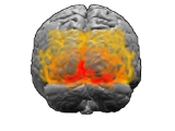
Brodmann area 18, or BA18, is part of the occipitalOccipital lobe
The occipital lobe is the visual processing center of the mammalian brain containing most of the anatomical region of the visual cortex. The primary visual cortex is Brodmann area 17, commonly called V1...
cortex
Cerebral cortex
The cerebral cortex is a sheet of neural tissue that is outermost to the cerebrum of the mammalian brain. It plays a key role in memory, attention, perceptual awareness, thought, language, and consciousness. It is constituted of up to six horizontal layers, each of which has a different...
in the human brain
Human brain
The human brain has the same general structure as the brains of other mammals, but is over three times larger than the brain of a typical mammal with an equivalent body size. Estimates for the number of neurons in the human brain range from 80 to 120 billion...
. It accounts for the bulk of the volume of the occipital lobe.
This area is also known as parastriate area 18. It is a subdivision of the cytoarchitecturally defined occipital region of cerebral cortex
Cerebral cortex
The cerebral cortex is a sheet of neural tissue that is outermost to the cerebrum of the mammalian brain. It plays a key role in memory, attention, perceptual awareness, thought, language, and consciousness. It is constituted of up to six horizontal layers, each of which has a different...
. In the human it is located in parts of the cuneus
Cuneus
The cuneus is a portion of the human brain in the occipital lobe.The cuneus receives visual information from the contralateral superior retina representing the inferior visual field. It is most known for its involvement in basic visual processing. Pyramidal cells in the cuneus project to...
, the lingual gyrus
Lingual gyrus
The lingual gyrus of the occipital lobe lies between the calcarine sulcus and the posterior part of the collateral sulcus; behind, it reaches the occipital pole; in front, it is continued on to the tentorial surface of the temporal lobe, and joins the parahippocampal gyrus...
and the lateral occipital gyrus (H) of the occipital lobe
Occipital lobe
The occipital lobe is the visual processing center of the mammalian brain containing most of the anatomical region of the visual cortex. The primary visual cortex is Brodmann area 17, commonly called V1...
. Cytoarchitecturally it is bounded on one side by the striate area 17, from which it is distinguished by absence of a band of Gennari, and on the other by the peristriate area 19 (Brodmann-1909).
Guenon
Brodmann area 18 is a subdivision of the cerebral cortex of the guenonGuenon
The guenons are the genus Cercopithecus of Old World monkeys. Not all the members of this genus have the word "guenon" in their common names, and because of changes in scientific classification, some monkeys in other genera may have common names that do include the word "guenon"...
defined on the basis of cytoarchitecture. It is topographically and cytoarchitecturally homologous
Homology (biology)
Homology forms the basis of organization for comparative biology. In 1843, Richard Owen defined homology as "the same organ in different animals under every variety of form and function". Organs as different as a bat's wing, a seal's flipper, a cat's paw and a human hand have a common underlying...
to parastriate area 18 of the human (Brodmann-1909). Distinctive features (Brodmann-1905): a wide, dense internal granular cell layer (IV); a distinct sublayer 3b of closely packed large pyramidal cell
Pyramidal cell
Pyramidal neurons are a type of neuron found in areas of the brain including cerebral cortex, the hippocampus, and in the amygdala. Pyramidal neurons are the primary excitation units of the mammalian prefrontal cortex and the corticospinal tract. Pyramidal neurons were first discovered and...
s positioned in the external pyramidal layer (III) directly above layer IV; an almost cell free, narrow internal pyramidal layer (V) with no larger ganglion cell
Ganglion cell
A retinal ganglion cell is a type of neuron located near the inner surface of the retina of the eye. It receives visual information from photoreceptors via two intermediate neuron types: bipolar cells and amacrine cells...
s; a likewise very narrow, dense multiform layer (VI) composed of small polymorphic cells that form a distinct boundary with the underlying subcortical white matter
White matter
White matter is one of the two components of the central nervous system and consists mostly of myelinated axons. White matter tissue of the freshly cut brain appears pinkish white to the naked eye because myelin is composed largely of lipid tissue veined with capillaries. Its white color is due to...
. Like area 17 of Brodmann-1905, area 18 is relatively thin; the three deep layers are thin relative to the three outer layers; distinct boundaries between layers; abundance of granule cell
Granule cell
In neuroscience, granule cells refer to tiny neurons that are around 10 micrometres in diameter. Granule cells are found within the granular layer of the cerebellum , the dentate gyrus of the...
s; narrow layer VI; and sharp boundary between cortex and subcortical white matter.
External links
- For Neuroanatomy of the parastriate area 18 visit BrainInfo
- For Neuroanatomy of Brodmann area 18 visit BrainInfo
See also
- Brodmann areaBrodmann areaA Brodmann area is a region of the cerebral cortex defined based on its cytoarchitectonics, or structure and organization of cells.-History:...
- List of regions in the human brain