

Aortic valve area calculation
Encyclopedia
Aortic valve area calculation is an indirect method of determining the area of the aortic valve
. The calculated aortic valve orifice area is currently one of the measures for evaluating the severity of aortic stenosis
. A valve area of less than 0.8 cm² is considered to be severe aortic stenosis.
There are many ways to calculate the valve area of aortic stenosis. The most commonly used methods involve measurements taken during echocardiography
. For interpretation of these values, the area is generally divided by the body surface area
, to arrive at the patient's optimal aortic valve orifice area.
, when the valve is supposed to be open. While this method directly measures the valve area, the image may be difficult to obtain due to artifacts during echocardiography, and the measurements are dependent on the technician who has to manually trace the perimeter of the open aortic valve. Because of these reasons, planimetry of aortic valve is not routinely performed.

The weakest aspect of this calculation is the variability in measurement of LVOT area, because it involves squaring the LVOT dimension. Therefore, it is crucial for the sonographer to take great care in measuring the LVOT diameter.
The measurement using echocardiogram may be inaccurate in cases of Aortic subvalvular stenosis, because there is not a uniform diameter, as assumed during echocardiogram. The viewpoint of the echocardiogram may distort the true diameter of the LVOT in some patients. For verification purposes of the obtained valve area using echocardiogram and doppler measures, especially if the obtained valve area is in the range requiring surgery and cardiac output is low, the Gold standard of left heart catheterization for true hemodynamics should be obtained for validation using the Gorlin formula, so patient does not go for unneeded surgery.
(measured in liters/minute) and dividing it by the heart rate (to give output per cardiac cycle
) and then dividing it by the systolic ejection period measured in seconds per beat (to give flow per ventricular contraction).

The Gorlin equation is related to flow across the valve. Because of this, the valve area may be erroneously calculated as stenotic if the flow across the valve is low (ie: if the cardiac output is low). The measurement of the true gradient is accomplished by temporarily increasing the cardiac output by the infusion of positive inotropic agents, such as dobutamine
.
 . The resulting simplified formula is:
Aortic valve
The aortic valve is one of the valves of the heart. It is normally tricuspid , although in 1% of the population it is found to be congenitally bicuspid . It lies between the left ventricle and the aorta....
. The calculated aortic valve orifice area is currently one of the measures for evaluating the severity of aortic stenosis
Aortic valve stenosis
Aortic valve stenosis is a disease of the heart valves in which the opening of the aortic valve is narrowed. The aortic valve is the valve between the left ventricle of the heart and the aorta, which is the largest artery in the body and carries the entire output of blood.-Pathophysiology:The...
. A valve area of less than 0.8 cm² is considered to be severe aortic stenosis.
There are many ways to calculate the valve area of aortic stenosis. The most commonly used methods involve measurements taken during echocardiography
Echocardiography
An echocardiogram, often referred to in the medical community as a cardiac ECHO or simply an ECHO, is a sonogram of the heart . Also known as a cardiac ultrasound, it uses standard ultrasound techniques to image two-dimensional slices of the heart...
. For interpretation of these values, the area is generally divided by the body surface area
Body surface area
In physiology and medicine, the body surface area is the measured or calculated surface of a human body. For many clinical purposes BSA is a better indicator of metabolic mass than body weight because it is less affected by abnormal adipose mass...
, to arrive at the patient's optimal aortic valve orifice area.
Planimetry
Planimetry is the tracing out of the opening of the aortic valve in a still image obtained during echocardiographic acquisition during ventricular systoleSystole (medicine)
Systole is the contraction of the heart. Used alone, it usually means the contraction of the left ventricle.In all mammals, the heart has 4 chambers. The left and right ventricles pump together. The atria and ventricles pump in sequence...
, when the valve is supposed to be open. While this method directly measures the valve area, the image may be difficult to obtain due to artifacts during echocardiography, and the measurements are dependent on the technician who has to manually trace the perimeter of the open aortic valve. Because of these reasons, planimetry of aortic valve is not routinely performed.
The continuity equation
The continuity equation states that the flow in one area must equal the flow in a second area if there are no shunts in between the two areas. In practical terms, the flow from the left ventricular outflow tract (LVOT) is compared to the flow at the level of the aortic valve. Using Echocardiography, the aortic valve area calculated using the time velocity integral (TVI) is most accurate method and is the preferred method. The flow through the LVOT, or LV Stroke Volume (cm3 or cc), can be calculated by measuring the LVOT diameter (cm), squaring that value, multiplying the value by 0.78540 giving cross sectional area of the LVOT (cm2)and multiplying that value by the LVOT TVI (cm), measured on the spectral Doppler display using pulsed-wave Doppler. From these, it is easy to calculate the area (cm2) of the aortic valve by simply dividing the LV Stroke Volume (cm3) by the AV TVI (cm) measured on the spectral Doppler display using continuous-wave Doppler.| Example: An individual undergoes transthoracic echocardiography for the evaluation of a systolic ejection murmur with delayed carotid upstroke noted on physical examination. During echocardiography, the following measurements were made: LVOT diameter of 2 cm, LVOT TVI of 24 cm, and an Aortic Valve TVI of 50 cm. What is the aortic valve area? | ||
| Answer: An LVOT diameter of 2 cm gives a LVOT cross-sectional area of, 2 * 2 * 0.78540 = 3.14 cm2. To calculate stroke volume, multiply the cross-sectional area of 3.14 cm2 by the LVOT TVI 24 cm. This gives an LV Stroke Volume of 3.14 * 24 = 75.40 cc. Divide the LV Stroke Volume, 75.40 cc by the Aortic Valve TVI, 50 cm and this gives an aortic valve area of 75.40 / 50 = 1.51 cm2. |
The weakest aspect of this calculation is the variability in measurement of LVOT area, because it involves squaring the LVOT dimension. Therefore, it is crucial for the sonographer to take great care in measuring the LVOT diameter.
The measurement using echocardiogram may be inaccurate in cases of Aortic subvalvular stenosis, because there is not a uniform diameter, as assumed during echocardiogram. The viewpoint of the echocardiogram may distort the true diameter of the LVOT in some patients. For verification purposes of the obtained valve area using echocardiogram and doppler measures, especially if the obtained valve area is in the range requiring surgery and cardiac output is low, the Gold standard of left heart catheterization for true hemodynamics should be obtained for validation using the Gorlin formula, so patient does not go for unneeded surgery.
The Gorlin equation
The Gorlin equation states that the aortic valve area is equal to the flow through the aortic valve during ventricular systole divided by the systolic pressure gradient across the valve times a constant. The flow across the aortic valve is calculated by taking the cardiac outputCardiac output
Cardiac output is the volume of blood being pumped by the heart, in particular by a left or right ventricle in the time interval of one minute. CO may be measured in many ways, for example dm3/min...
(measured in liters/minute) and dividing it by the heart rate (to give output per cardiac cycle
Cardiac cycle
The cardiac cycle is a term referring to all or any of the events related to the flow or blood pressure that occurs from the beginning of one heartbeat to the beginning of the next. The frequency of the cardiac cycle is described by the heart rate. Each beat of the heart involves five major stages...
) and then dividing it by the systolic ejection period measured in seconds per beat (to give flow per ventricular contraction).
The Gorlin equation is related to flow across the valve. Because of this, the valve area may be erroneously calculated as stenotic if the flow across the valve is low (ie: if the cardiac output is low). The measurement of the true gradient is accomplished by temporarily increasing the cardiac output by the infusion of positive inotropic agents, such as dobutamine
Dobutamine
Dobutamine is a sympathomimetic drug used in the treatment of heart failure and cardiogenic shock. Its primary mechanism is direct stimulation of β1 receptors of the sympathetic nervous system. Dobutamine was developed by a laboratory led by Drs...
.
| Example: An individual undergoes left and right heart cardiac catheterization as part of the evaluation of aortic stenosis. The following hemodynamic parameters were measured. With a heart rate of 80 beats/minute and a systolic ejection period of 0.33 seconds, the cardiac output was 5 liters/minute. During simultaneous measurement of pressures in the left ventricle and aorta (with the use of one catheter in the left ventricle and a second in the ascending aorta), the mean systolic pressure gradient was measured at 50 mmHg. What is the valve area as measured by the Gorlin equation? | ||
Answer: 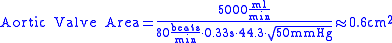 |
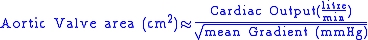
The Hakki equation
The Hakki equation is a simplification of the Gorlin equation, relying on the observation that in most cases, the numerical value of. The resulting simplified formula is:| Example: An individual undergoes left and right cardiac catheterization for the evaluation of aortic stenosis. Measurements includes an aortic pressure of 120/60, LV pressure of 170/15, cardiac output of 3.5 liters/minute. What is the aortic valve area? | ||
Answer: The peak gradient between the LV and aorta is 50 mmHg. This gives |