
Aortic coarctation
Encyclopedia

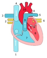
Aorta
The aorta is the largest artery in the body, originating from the left ventricle of the heart and extending down to the abdomen, where it branches off into two smaller arteries...
narrows in the area where the ductus arteriosus
Ductus arteriosus
In the developing fetus, the ductus arteriosus , also called the ductus Botalli, is a shunt connecting the pulmonary artery to the aortic arch. It allows most of the blood from the right ventricle to bypass the fetus's fluid-filled lungs. Upon closure at birth, it becomes the ligamentum arteriosum...
(ligamentum arteriosum
Ligamentum arteriosum
The ligamentum arteriosum is a small ligament attached to the superior surface of the pulmonary trunk and the inferior surface of the aortic arch...
after regression) inserts.
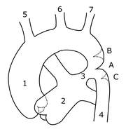
Types
There are three types:- Preductal coarctation: The narrowing is proximal to the ductus arteriosusDuctus arteriosusIn the developing fetus, the ductus arteriosus , also called the ductus Botalli, is a shunt connecting the pulmonary artery to the aortic arch. It allows most of the blood from the right ventricle to bypass the fetus's fluid-filled lungs. Upon closure at birth, it becomes the ligamentum arteriosum...
. Blood flow to the aorta that is distal to the narrowing is dependent on the ductus arteriosus; therefore severe coarctation can be life-threatening. Preductal coarctation results when an intracardiac anomaly during fetal life decreases blood flow through the left side of the heart, leading to hypoplastic development of the aorta. This is the type seen in approximately 5% of infants with Turner SyndromeTurner syndromeTurner syndrome or Ullrich-Turner syndrome encompasses several conditions in human females, of which monosomy X is most common. It is a chromosomal abnormality in which all or part of one of the sex chromosomes is absent...
. - Ductal coarctation: The narrowing occurs at the insertion of the ductus arteriosus. This kind usually appears when the ductus arteriosus closes.
- Postductal coarctation: The narrowing is distal to the insertion of the ductus arteriosus. Even with an open ductus arteriosus blood flow to the lower body can be impaired. This type is most common in adults. It is associated with notching of the ribsNotching of the ribsNotching of the ribs is a radiological finding where the surface of the rib is deformed. It can be characterized as unilateral or bilateral...
(because of collateral circulation), hypertension in the upper extremities, and weak pulses in the lower extremities. Postductal coarctation is most likely the result of the extension of a muscular artery (ductus arteriosus) into an elastic artery (aorta) during fetal life, where the contraction and fibrosis of the ductus arteriosus upon birth subsequently narrows the aortic lumen.
Signs and symptoms
In mild cases, children may show no signs or symptoms at first and their condition may not be diagnosed until later in life.Some children born with coarctation of the aorta have other heart defects, too, such as aortic stenosis, ventricular septal defect, patent ductus arteriosus or mitral valve abnormalities.
Coarctation is about twice as common in boys as it is in girls. It’s common in girls who have Turner syndrome.
Symptoms may be absent with mild narrowings (coarctation). When present, they include: difficulty breathing, poor appetite or trouble feeding, failure to thrive. Later on, children may develop symptoms related to problems with blood flow and an enlarged heart. They may experience dizziness or shortness of breath, faint or near-fainting episodes, chest pain, abnormal tiredness or fatigue, headaches, or nosebleeds. They may have cold legs and feet or have pain in their legs with exercise (intermittent claudication
Intermittent claudication
Intermittent claudication is a clinical diagnosis given for muscle pain , classically in the calf muscle, which occurs during exercise, such as walking, and is relieved by a short period of rest.Claudication derives from the Latin verb claudicare, "to limp".-Signs:One of the hallmarks of arterial...
).
In more severe cases, where severe coarctations, babies may develop serious problems soon after birth because not enough blood can get through the aorta to the rest of their body.
Arterial hypertension in the arms with normal to low blood pressure in the lower extremities is classic. Poor peripheral pulses in the legs and a weak femoral artery pulse may be found in severe cases.
The coarctation typically occurs after the left subclavian artery
Subclavian artery
In human anatomy, the subclavian arteries are two major arteries of the upper thorax , below the clavicle . They receive blood from the top of the aorta...
. However, if situated before it, blood flow to the left arm is compromised and asynchronous or radial pulses of different "strength" may be detected (normal on the right arm, weak or delayed on the left). In these cases, a difference between the normal radial pulse in the right arm and the delayed femoral pulse in the legs (either side) may be apparent, whilst no such delay would be appreciated with palpation of both delayed left arm and either femoral pulses. On the other hand, a coarctation occurring after the left subclavian artery will produce synchronous radial pulses, but radial-femoral delay will be present under palpation in either arm (both arm pulses are normal compared to the delayed leg pulses).
Imaging and diagnosis
With imaging, resorption of the lower part of the ribs may be seen, due to increased blood flow over the neurovascular bundleNeurovascular bundle
A Neurovascular bundle is a term applied to the body nerves, arteries, veins and lymphatics that tend to travel together in the body...
that runs there. Post-stenotic dilation of the aorta results in a classic 'figure 3 sign' on x-ray
X-ray
X-radiation is a form of electromagnetic radiation. X-rays have a wavelength in the range of 0.01 to 10 nanometers, corresponding to frequencies in the range 30 petahertz to 30 exahertz and energies in the range 120 eV to 120 keV. They are shorter in wavelength than UV rays and longer than gamma...
. The characteristic bulging of the sign is caused by dilatation of the aorta due to an indrawing of the aortic wall
Aorta
The aorta is the largest artery in the body, originating from the left ventricle of the heart and extending down to the abdomen, where it branches off into two smaller arteries...
at the site of cervical rib
Cervical rib
A cervical rib is a supernumerary rib which arises from the seventh cervical vertebra. It is a congenital abnormality located above the normal first rib. A cervical rib is present in only about 1 in 500 of people; in even rarer cases, an individual may have two cervical ribs...
obstruction, with consequent post-stenotic dilation. This physiology results in the '3' image for which the sign is named. When the esophagus is filled with barium, a reverse 3 or E sign is often seen and represents a mirror image of the areas of prestenotic and poststenotic dilatation.
Coarctation of the aorta can be accurately diagnosed with magnetic resonance angiography
Magnetic Resonance Angiography
Magnetic resonance angiography is a group of techniques based on Magnetic Resonance Imaging to image blood vessels. Magnetic resonance angiography is used to generate images of the arteries in order to evaluate them for stenosis , occlusion or aneurysms...
. In teenagers and adults echocardiograms may not be conclusive. In adults with untreated coarctation blood often reaches the lower body through collaterals, e.g. internal thoracic arteries via. the subclavian arteries. Those can be seen on MR, CT or angiography. An untreated coarctation may also result in hypertrophy of the left ventricle.
coarctation of the aorta by CMR
A case of coarctation of the aorta was published in the New England Journal of Medicine
New England Journal of Medicine
The New England Journal of Medicine is an English-language peer-reviewed medical journal published by the Massachusetts Medical Society. It describes itself as the oldest continuously published medical journal in the world.-History:...
in 2007 employing chest radiography and magnetic resonance images.
A case of long-standing misdiagnosed coarctation of the aorta in an adult was described in the New York Times Magazine.
Treatment
Therapy/Treatment is conservative if asymptomatic, but may require surgical resection of the narrow segment if there is arterial hypertension. The first operations to treat coarctation were carried out by Clarence CrafoordClarence Crafoord
Clarence Crafoord was a Swedish cardiovascular surgeon, best known for performing the first successful repair of aortic coarctation on 19 October 1944, one year before Robert E...
in Sweden in 1944. In some cases angioplasty
Angioplasty
Angioplasty is the technique of mechanically widening a narrowed or obstructed blood vessel, the latter typically being a result of atherosclerosis. An empty and collapsed balloon on a guide wire, known as a balloon catheter, is passed into the narrowed locations and then inflated to a fixed size...
can be performed to dilate the narrowed artery. If the coarctation is left untreated, arterial hypertension may become permanent due to irreversible changes in some organs (such as the kidney
Kidney
The kidneys, organs with several functions, serve essential regulatory roles in most animals, including vertebrates and some invertebrates. They are essential in the urinary system and also serve homeostatic functions such as the regulation of electrolytes, maintenance of acid–base balance, and...
).
For fetuses at high risk for developing coarctation, a novel experimental treatment approach is being investigated, wherein the mother inhales 45% oxygen three times a day (3 x 3–4 hours) beyond 34 weeks of gestation. The oxygen is transferred via the placenta to the fetus and results in dilatation of the fetal lung vessels. As a consequence, the flow of blood through the fetal circulatory system increases, including that through the underdeveloped arch. In suitable fetuses, marked increases in aortic arch dimensions have been observed over treatment periods of about two to three weeks
COMPLICATIONS:1-left venticular failure 2-aortic dissection 3-cerebral hemorrhage 4-progressive aortic regurgitation or stenosis
External links
- Aortic Coarctation information from Seattle Children's Hospital Heart Center
- Diagram at kumc.edu
- Overview and diagram at umich.edu
- Down's Heart Group Specific information relating to heart conditions and Down's Syndrome.
- http://www.uniklinik-bonn.de/dzft Specific information about hyperoxygenation approach (Note: in German)
- "English version" of some (other?) information from the [same web site as the] above source
- 3D CT Angiogram Radiology of Coarctation